Received:June 30, 2015, Revised:August 3, 2015, Accepted:August 7, 2015
Corresponding to:Seung-Geun Lee, Division of Rheumatology, Department of Internal Medicine, Pusan National University Hospital, 179 Gudeok-ro, Seo-gu, Busan 49241, Korea. E-mail:[email protected]
pISSN: 2093-940X, eISSN: 2233-4718
Copyright ⓒ 2016 by The Korean College of Rheumatology. All rights reserved.
This is a Free Access article, which permits unrestricted non-commerical use, distribution, and reproduction in any medium, provided the original work is properly cited.
Discovery of Splenic Sarcoidosis Concurrent with the Diagnosis of Ovarian Cancer: A Case Report
Eun-Heui Kim1, Seung-Geun Lee1, Ki-Hyung Kim2, Young-Mi Seol3, Eun-Kyoung Park1, Dong-Wan Koo1, Na-Kyoung Hwang1, In-Sub Han1, Moon-Won Lee1, Sung-Yong Han1, Geun-Tae Kim4, Hee-Sang Tag4
1Division of Rheumatology, Department of Internal Medicine, 2Department of Obstetrics and Gynecology, 3Division of Hemato-oncology, Department of Internal Medicine, Pusan National University School of Medicine, Busan, 4Division of Rheumatology, Department of Internal Medicine, Kosin University College of Medicine, Busan, Korea
Sarcoidosis is a multisystem inflammatory disease of unknown etiology characterized by noncaseating epithelioid granuloma formation. Although the relationship between sarcoidosis and malignancy has been noted in recent decades, there are few case reports describing the concurrent diagnosis of sarcoidosis and malignancy. Herein, we describe a case of biopsy-proven splenic sarcoidosis mimicking metastasis at the time of ovarian adenocarcinoma. Imaging studies including positron-emission tomog- raphy-computed tomography were not useful for differentiating sarcoidosis from malignancy. Thus, our case highlights the im- portance of histopathological examination to rule out nonmalignant conditions before the diagnosis of metastatic disease is made. (J Rheum Dis 2016;23:130-135)
Key Words. Sarcoidosis, Ovarian neoplasms, Granuloma, Positron-emission tomography
INTRODUCTION
Sarcoidosis is a multisystem inflammatory disease of unknown etiology characterized by the development and accumulation of noncaseating epitheloid granuloma in any organ system. In most cases, the lungs are affected, but the lymph nodes, parotid gland, liver and spleen can also be involved. Diagnosis of sarcoidosis is based on compatible clinical features and histological tissue examination. However, owing to the uncertainty of its cause and the variability in the clinical manifestations of sarcoidosis, diagnosis of this disease is often difficult.
Over the past decades, the relationship between sarcoi- dosis and malignancy has been noted [1,2]; however, the conclusions are conflicting [3]. In previous studies, sar- coidosis has more often been observed in association with hematologic malignancy than with solid tumors [4].
It has been reported that malignancy developed after the diagnosis of sarcoidosis and that sarcoidosis occurred in
patients with malignant tumor [4,5]. Otherwise, case re- ports of the concurrent discovery of sarcoidosis and ma- lignancy are lacking. In the present paper, we describe a case of the splenic sarcoidosis mimicking metastasis at the time of diagnosis of ovarian adenocarcinoma.
CASE REPORT
A 66-year-old South Korean woman, gravida 4 para 1, visited to Pusan National University Hospital complain- ing of low abdominal discomfort and weight loss. She de- nied any cutaneous or ocular symptoms. The patient was a housewife with no history of smoking or occupational exposure to chemical agents. She had a history of hyper- tension and her brother died from colon cancer.
Computed tomography (CT) of the abdomen showed a 100×47 mm sized multiseptated cystic mass in the right ovary; multiple extensive lymphadenopathy including multiple 2 to 6 cm sized lymph nodes in the splenic hi-
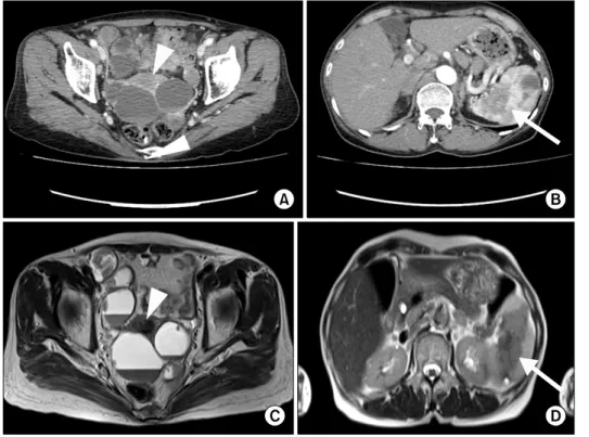
Figure 1. Abdominal computed tomography (CT; A, B) and mag- netic resonance imaging (MRI;
C, D) showed 100×47 mm sized multiseptated cystic mass (arrowheads in A and C) and multiple extensive lymphaden- opathy including multiple 2 to 6 cm sized lymph nodes. In the spleen, a low density lesion measuring approximately 12×
27 mm on CT (arrow in B) and a 62 mm sized mass lesion on MRI (arrow in D) were observed.
common iliac and left external iliac lymph node; the peri- toneal nodule; and the spleen (Figure 2). Based on these imaging findings, the diagnosis of ovarian carcinoma with multiple lymph node and splenic metastases and peri- toneal seeding was considered. To confirm the diagnosis of malignancy histologically, we planned an open hyster- ectomy and bilateral salpingo-oophorectomy, but the pa- tient refused the operation. After discussing the diag- nostic process with the patient, a percutaneous needle bi- opsy was performed on a splenic lesion that was pre- sumed to be metastatic.
Histophathologic findings revealed noncaseating gran- ulomatous inflammation without tumor cells. A poly- merase chain reaction-based assay was negative for myco- bacterium tuberculosis, leaving sarcoidosis as the prob-
min D3 56.74 pg/mL (reference, 19.6 to 54.3 pg/mL), in- tact parathyroid hormone 16.61 pg/mL (reference, 10 to 57 pg/mL), angiotensin converting enzyme (ACE) 98.8 U/L (reference, 9 to 47 U/L), CA-125 32.55 U/mL (refe- rence, 0 to 35 U/mL) and CA19-9 15.51 U/mL (reference, 0 to 39 U/mL). Lymphopenia, hypercalcemia with an in- creased 1,25-OH vitamin D3 and an elevated ACE level were recognized to be consistent with sarcoidosis. Based on the imaging, histologic and laboratory results, two types of diagnoses were considered, 1) coexisting ovarian cancer and splenic sarcoidosis or 2) sarcoidosis with ex- tensive organ involvement including ovary, lymph node and spleen without malignancy. However, the patient de- nied the possibility of malignancy and refused further di- agnostic work-up including surgical biopsy of an ovary or
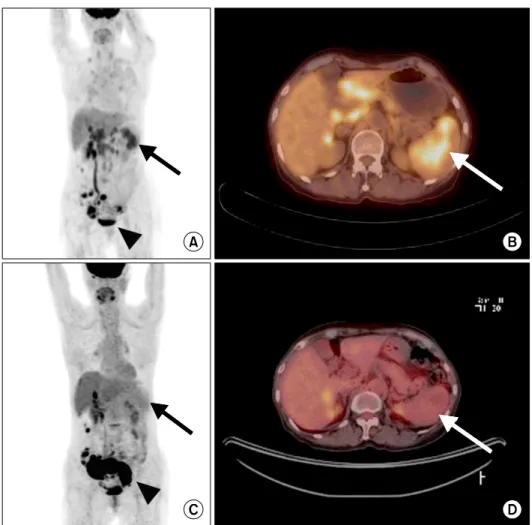
Figure 2. 18F- fluorodeoxyglucose (FDG) labeled positron-emi- ssion tomography before (A and B) and 1 month after (C and D) prednisolone therapy. The mag- nitude of 18F-FDG uptake in multiple hypermetabolic nod- ules and the multiseptated mass in the right ovary (arrowhead in A) increased after 1 month (arro- whead in C). Increased 18F-FDG uptake in spleen (arrows in A and B) nearly resolved after glu- cocorticoids treatment (arrows in C and D).
lymph node. Thus, we empirically initiated 40 mg of pre- dnisolone for presumed sarcoidosis.
After 1 month of glucocorticoids therapy, a follow-up PET-CT scan revealed the 18F-FDG uptake was dramati- cally decreased in the spleen but increased in extent and intensity in the right ovary and multiple lymph nodes in the abdominal cavity. Serum creatinine level was de- creased to 1.32 mg/dL and the calcium concentration had normalized. At that time, a diagnostic laparotomy was performed and biopsy of the omentum revealed meta- static adenocarcinoma with positive estrogen receptor staining that was morphologically and immunohisto- chemically consistent with ovarian serous carcinoma (Figure 4). The prednisolone was tapered off and the pa- tient received chemotherapy with paclitaxel and carbo- platin.
DISCUSSION
Since Brincker and Wilbek [1] reported a significantly increased risk of malignant tumor, such as lung cancer
and lymphoma in patients with sarcoidosis in 1974, the clinical and epidemiological association between sarcoi- dosis and malignancy has been increasingly recognized based on numerous case reports and registry data [2,4,5], even though other studies argued that this association is more likely to be due to selection bias and misclassi- fication [3,6]. There are 3 types of clinical settings in which sarcoidosis and malignancy are found together.
First, a hematologic malignancy or solid tumor can devel- op after the diagnosis of sarcoidosis. Several studies have shown that sarcoidosis is associated with an increased risk of malignant tumor [1,2,5]. Secondly, a sarcoid re- action, typically limited to the regional lymph nodes or the organ of tumor origin, or systemic sarcoidosis can oc- cur following the diagnosis of malignancy [7,8]. Lastly, al- though very rare, sarcoidosis can present concurrently with the diagnosis of a malignant tumor including, breast [9], colorectal [10], gastric [11] and ovarian cancer [12], as in the present case. To our knowledge, this is the first case report of splenic sarcoidosis diagnosed concurrently with ovarian adenocarcinoma in a Korean patient.
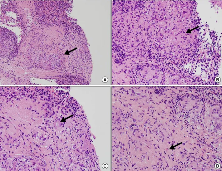
Figure 3. Microscopic findings in the spleen (hematoxylin and eosin staining). Noncaseating granulomas with aggregations of in- flammatory cells (arrow in A), multinucleated giant cells (arrow in B), asteroid body (arrow in C), and eosinophils (arrow in D) were observed. (A, B) ×200, (C, E) ×400.
Because sarcoidosis of the lymph nodes or spleen in an oncology patient is often assumed to be metastatic spread from the primary tumor, making the correct diagnosis in this situation can be challenging. Imaging studies includ- ing CT, MRI and even 18F-FDG labeled PET-CT are not usually helpful for differentiating malignant tumor from sarcoidosis. In addition, a majority of patients with sar- coidosis are asymptomatic, which can lead to misdiag- nosis or delayed diagnosis. In our case, because the mag- nitude of 18F-FDG uptake in the splenic mass on PET-CT scan was similar to that of a primary tumor and metastatic lymph node, we initially concluded it was metastatic ovarian cancer. Although our patient was found to have characteristic laboratory findings such as increased ACE and 1,25-OH vitamin D3 levels after histopathologic con- firmation of sarcoidosis, these markers are not routinely obtained as part of the assessment in patients with ovar-
ian cancer. Moreover, our patient did not have any symp- toms of sarcoidosis. Only after histological examination, we were able to make the diagnosis of splenic sarcoidosis.
Similar to our case, Mapelli et al. [12] reported 4 cases of histologically confirmed sarcoidosis of the lymph nodes mimicking metastases in gynecological malignancies in- cluding ovarian, cervical and endometrial cancer and 18F- FDG labeled PET-CT imaging was not useful for differ- entiating sarcoidosis from malignant lymph nodes. Taken together, this highlights the need for clinicians to pay spe- cial attention to the potential linkage between sarcoidosis and malignancy to avoid an erroneous diagnosis of cancer progression and inappropriate treatment.
For the treatment of sarcoidosis, the organ involvement and severity of symptoms should be considered. While patients with no or mild symptoms may not require treat- ment, symptomatic patients with multiple organ involve-
Figure 4. Microscopic findings of the omentum. (A) Hematoxylin and eosin staining showed metastatic adenocarcinoma on des- moplastic changed omentum (×200). (B) Calretinin staining was negative (×100). (C) The tumor was estrogen receptor positive, suggesting an ovarian origin (×100). (D) The tumor protein p53 was positive (×100).
ment require systemic glucocorticoids. In addition, glu- cocorticoids therapy may be considered for patients with an abnormal calcium level or neurologic, cardiac or ocular involvements. Glucocorticoids remain the initial drugs of choice for sarcoidosis, but hydroxychloroquine, metho- trexate, azathioprine and anti-tumor necrosis factor agents may also be used. In our case, we introduced 40 mg of prednisolone due to hypercalcemia and the possibility of sarcoidosis with extensive organ involvement. With prednisolone therapy, our patient’s hypercalcemia cor- rected and the size of the splenic sarcoidosis lesions dra- matically decreased. Not all cases of sarcoidosis of the lymph nodes or spleen diagnosed concurrently with a ma- lignancy required treatment in previous reports [9-12].
Thus, an appropriate therapeutic approach based on the extent of disease involvement and clinical symptoms should be considered in cases of sarcoidosis associated
with cancer.
The clinical course and prognosis of sarcoidosis asso- ciated with malignancy are not well established. There have been several reports of sarcoidosis developing or flaring after a patient received anti-neoplastic agents or biologic modifiers such as interleukin-2 [4,7,8]. The presence of a sarcoid reaction in patients with Hodgkin lymphoma and gastric carcinoma was reported to be asso- ciated with positive prognostic significance [4]. However, due to the rarity of this condition, the effect of sarcoidosis on the prognosis of malignant tumor and vice versa re- mains elusive.
Although a relationship between sarcoidosis and malig- nant tumors has been proposed over the past decades, the exact mechanisms by which sarcoidosis promotes onco- genesis is not well understood. Considering that chronic inflammation has been presumed to be the putative me-
the molecular and immunological mechanism of linking sarcoidosis and malignancy.
SUMMARY
We describe a case of the splenic sarcoidosis discovered concurrently with the diagnosis of ovarian adeno- carcinoma. The present case highlights the importance of histopathological examination in patients with suspected splenic metastases of malignant disease including ovar- ian cancer because imaging studies including PET-CT were not useful for differentiating sarcoidosis from malignancy. Added awareness of the potential association between sarcoidosis and malignant tumor is needed.
ACKNOWLEDGMENTS
We thank the late Professor Sung-Il Kim who was de- voted himself to education, research and patient care in Division of Rheumatology, Department of Internal Medi- cine, Pusan National University School of Medicine (1963 to 2011). This work was supported by clinical re- search grant form Pusan National University Hospital 2015.
CONFLICT OF INTEREST
No potential conflict of interest relevant to this article was reported.
Crit Care Med 1999;160:1668-72.
6. Seersholm N, Vestbo J, Viskum K. Risk of malignant neo- plasms in patients with pulmonary sarcoidosis. Thorax 1997;52:892-4.
7. Kim MH, Lee K, Kim KU, Park HK, Lee MK, Suh DS.
Sarcoidosis mimicking cancer metastasis following chemo- therapy for ovarian cancer. Cancer Res Treat 2013;45:
354-8.
8. Logan TF, Bensadoun ES. Increased disease activity in a pa- tient with sarcoidosis after high dose interleukin 2 treat- ment for metastatic renal cancer. Thorax 2005;60:610-1.
9. Tolaney SM, Colson YL, Gill RR, Schulte S, Duggan MM, Shulman LN, et al. Sarcoidosis mimicking metastatic breast cancer. Clin Breast Cancer 2007;7:804-10.
10. Lequoy M, Coriat R, Rouquette A, Mir O, Perkins G, Regnard JF, et al. Sarcoidosis lung nodules in colorectal can- cer follow-up: sarcoidosis or not? Am J Med 2013;126:
642-5.
11. Jiao Y, Ning J, Zhao WD, Li YL, Wu HY, Gu KS. Sarcoidosis in gastric cancer at the time of diagnosis: a case report.
Oncol Lett 2015;9:1159-62.
12. Mapelli P, Mangili G, Picchio M, Rabaiotti E, Gianolli L, Messa C, et al. Sarcoidosis mimicking metastatic gynaeco- logical malignancies: a diagnostic and therapeutic chal- lenge? Rev Esp Med Nucl Imagen Mol 2013;32:314-7.
13. Idali F, Wahlström J, Müller-Suur C, Eklund A, Grunewald J. Analysis of regulatory T cell associated forkhead box P3 expression in the lungs of patients with sarcoidosis. Clin Exp Immunol 2008;152:127-37.
14. Miyara M, Amoura Z, Parizot C, Badoual C, Dorgham K, Trad S, et al. The immune paradox of sarcoidosis and regu- latory T cells. J Exp Med 2006;203:359-70.
15. Tsiatas ML, Gyftaki R, Liacos C, Politi E, Rodolakis A, Dimopoulos MA, et al. Study of T lymphocytes infiltrating peritoneal metastases in advanced ovarian cancer: associa- tions with vascular endothelial growth factor levels and prognosis in patients receiving platinum-based chemo- therapy. Int J Gynecol Cancer 2009;19:1329-34.