• 검색 결과가 없습니다.

Outcomes and Patency of Complex Configurations of Composite Grafts Using Bilateral Internal Thoracic Arteries

N/A
N/A
Protected

Academic year: 2021

Share "Outcomes and Patency of Complex Configurations of Composite Grafts Using Bilateral Internal Thoracic Arteries"

Copied!
9
0
0

로드 중.... (전체 텍스트 보기)

전체 글

(1)

KJTCVS

The Korean Journal of Thoracic and Cardiovascular Surgery

Clinical Research

https://doi.org/10.5090/kjtcs.2020.53.2.64 pISSN: 2233-601X eISSN: 2093-6516

Korean J Thorac Cardiovasc Surg. 2020;53(2):64-72

Outcomes and Patency of Complex Configurations of Composite Grafts Using Bilateral Internal Thoracic Arteries

Beatrice Chia-Hui Shih, M.D. 1 , Suryeun Chung, M.D. 2 , Hakju Kim, M.D. 1 , Hyoung Woo Chang, M.D. 1 , Dong Jung Kim, M.D. 1 , Cheong Lim, M.D. 1 , Kay-Hyun Park, M.D. 1 , Jun Sung Kim, M.D. 1

1

Department of Thoracic and Cardiovascular Surgery, Seoul National University Bundang Hospital, Seoul National University College of Medicine, Seongnam;

2

Department of Thoracic and Cardiovascular Surgery, Samsung Medical Center, Sungkyunkwan University School of Medicine, Seoul, Korea

ARTICLE INFO

Received September 2, 2019 Revised October 12, 2019 Accepted October 28, 2019 Corresponding author Jun Sung Kim

Tel 82-31-787-7139 Fax 82-31-787-4050 E-mail [email protected] ORCID

https://orcid.org/0000-0002-3663-5062

Background: It is generally agreed that using a bilateral internal thoracic artery (BITA) composite graft improves long-term survival after coronary artery bypass grafting (CABG).

Although the left internal thoracic artery (LITA)-based Y-composite graft is widely adopted, technical or anatomical difficulties necessitate complex configurations. We aimed to in- vestigate whether BITA configuration impacts survival or patency in patients undergoing coronary revascularization.

Methods: Between January 2006 and June 2017, 1,161 patients underwent CABG at Seoul National University Bundang Hospital, where the standard technique is a LITA-based Y-composite graft with the right internal thoracic artery (RITA) sequentially anastomosed to non-left anterior descending (LAD) targets. Total of 160 patients underwent CABG using BITA with modifications. Their medical records and imaging data were reviewed retrospec- tively to investigate technical details, clinical outcomes, and graft patency.

Results: Modifications of the typical Y-graft (group 1, n=90), LITA-based I-graft (group 2, n=39), and RITA-based composite graft (group 3, n=31) were used due to insufficient RITA length (47%), problems using LITA (28%), and target vessel anatomy (25%). The overall 30- day mortality rate was 1.9%. Among 116 patients who underwent computed tomography or conventional angiography at a mean interval of 29.9±33.1 months postoperatively, the graft patency rates were 98.7%, 95.3%, and 83.6% for the LAD, left circumflex artery, and right coronary artery territories, respectively. Patency rates for the inflow, secondary, and tertiary grafts were 98.2%, 90.5%, and 80.4%, respectively. The RITA-based graft (group 3) had the lowest patency rate of the various configurations (p<0.011).

Conclusion: LITA-based Y composite graft, showed satisfactory clinical outcomes and patency whereas modifications of RITA- based composite graft had the lowest patency and 5-year survival rates. Therefore, when using RITA-based composite graft, other options should be considered before proceeding atypical configurations.

Keywords: Coronary artery bypass, Composite graft, Bilateral internal thoracic artery, Cor- onary artery disease

Copyright © The Korean Society for Thoracic and Cardiovascular Surgery. 2020. All right reserved.

This is an Open Access article distributed under the terms of the Creative Commons Attribution Non-Commercial License (http://creativecommons.org/licenses/

Introduction

The use of bilateral internal thoracic arteries (BITA) in coronary artery bypass grafting (CABG) is becoming in- creasingly popular, with accumulating evidence of im- proved graft patency and overall patient survival in left in- ternal thoracic artery (LITA)-to-left anterior descending artery (LAD) anastomosis. However, the use of BITA is still relatively uncommon worldwide [1,2], possibly not

only because the harvesting and utilization of BITA is time-consuming and technically complex, but also due to variations in coronary anatomy and the degree of coronary artery disease. Depending on the circumstances, anatomi- cal or technical issues may necessitate modifications.

The current literature demonstrates no difference in

clinical outcomes between composite and in situ graft con-

figurations [3]; therefore, given the paucity of comparative

data and information regarding the long-term outcomes of

(2)

Beatrice Chia-Hui Shih, et al. Complex Configuration of Bilateral Internal Thoracic Artery Graft KJTCVS

complex and atypical BITA configurations, we investigated the clinical outcomes of patients who received BITA grafts of various configurations at our institution. We assessed whether BITA configuration impacted survival or patency in patients undergoing CABG and whether any particular configuration was superior with respect to survival or the need for repeat revascularization.

Methods

Patients

Between January 2006 and June 2017, 1,161 consecutive patients underwent CABG with BITA composite grafting at Seoul National University Bundang Hospital. Of those, 160 patients required modifications of the graft configura- tion and were included in the present study. The composite BITA graft was modified for the following reasons: (1) 75 patients (47%) had a RITA with insufficient length for se- quential anastomosis; (2) 45 patients (28%) had intrinsic LITA limitations due to left subclavian artery stenosis, ip- silateral arteriovenous fistula, or LITA injury during har- vest; (3) 26 patients (16.3%) had non-triplet coronary dis- ease; and (4) 14 patients (8.7%) exhibited target vessel size mismatch or an unsuitable geometric orientation for se- quential anastomosis. From patients’ medical records, in- formation was extracted on their demographics, preopera- tive risk factors, operative technique, postoperative hospital course, imaging data, and clinical outcomes. Data were re- viewed retrospectively to investigate technical details, clin- ical outcomes, and graft patency.

The institutional review board of our institution ap- proved the research design (IRB approval no., B-1909/565- 107) and waived the need for informed consent.

Surgical technique

All patients underwent full median sternotomy. The standard technique at our institution was in situ LITA- based Y-composite grafting with the LITA anastomosed to the LAD and the right internal thoracic artery (RITA) se- quentially anastomosed to non-LAD targets. The internal thoracic artery (ITA) was mostly harvested in a skeleton- ized fashion. More than half of the patients underwent off- pump CABG (59.4%), while the remainder underwent on- pump beating-heart or conventional CABG (40.6%) with antegrade cardioplegia due to a low ejection fraction or concomitant procedures.

We divided patients into 3 groups according to the types

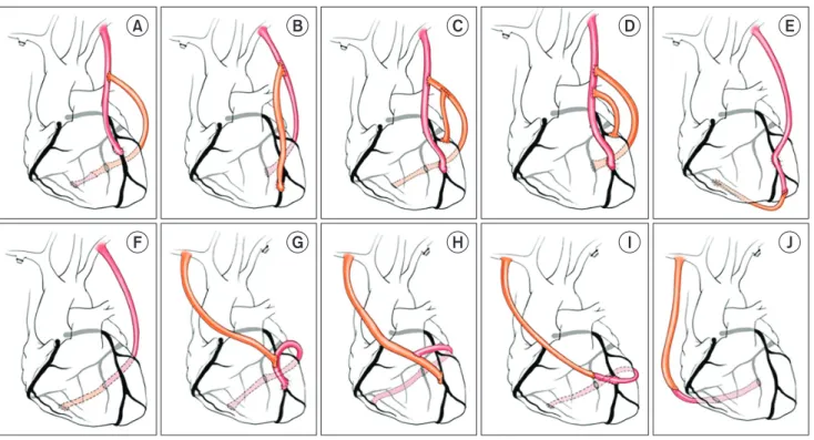
of modifications made. In group 1, the patients required minor alterations of the typical Y graft (Fig. 1A–D). In these patients, 4 different geometric or anastomotic config- urations were identified: (Fig. 1A) a short RITA extended with a remaining segment of the LITA or an additional- ly-harvested saphenous vein graft, (Fig. 1B) a twisted Y configuration used due to size mismatch between the LITA and the LAD, (Fig. 1C) a secondary Y anastomosis made at the proximal or distal end of the RITA, and (Fig. 1D) a double Y or π configuration used for sequential anastomo- sis to non-LAD targets on the RITA.

In group 2, the RITA was anastomosed end-to-end to the LITA to create an I-composite graft (Fig. 1E, F) and was then anastomosed sequentially to the LAD or other territories. Finally, in group 3, the RITA was used as an in- flow graft (Fig. 1G–J). This group also included 3 different geometric or anastomotic configurations: (Fig. 1G, H) a RITA-based I graft, (Fig. 1I) a RITA-based reverse T graft, and (Fig. 1J) a RITA-based Y graft.

Follow-up

All patients enrolled in the study participated in regular outpatient follow-up. The mean length of follow-up was 51.0±42.5 months (range, 3 days to 140 months). Graft pa- tency was evaluated in a total of 116 patients (72.5%) using computed tomography (CT) coronary angiography (CAG) or conventional CAG with a mean interval of 29.9±31.1 months after CABG surgery. The imaging follow-up proto- col at our institution was to perform CT angiography at 9 to 10 months postoperatively and to perform CAG at 5 years postoperatively. We defined graft failure as the total occlusion of the anastomosed graft as revealed on CT an- giography during follow-up.

Statistical analysis

Preoperative demographic and investigative data, opera-

tive variables, 30-day mortality and morbidity, and 5-year

survival were compared among the study groups. Categor-

ical variables were expressed as number and percentage

and were compared using the Fisher exact and Krus-

kal-Wallis tests. Continuous variables were expressed as

mean±standard deviation and compared using the un-

paired t-test. The Kaplan-Meier method was used to ana-

lyze overall survival and major adverse cardiovascular and

cerebrovascular disease (MACCE)-free survival. Multivari-

ate analyses were performed using logistic regression, and

p-values <0.05 were considered to indicate statistical sig-

(3)

https://doi.org/10.5090/kjtcs.2020.53.2.64

KJTCVS

nificance. We used IBM SPSS ver. 25.0 for Windows (IBM Corp., Armonk, NY, USA) for the statistical analysis.

Results

Patient characteristics and early clinical outcomes

Among the 160 patients included in the study, 90 (56.3%), 39 (24.4%) and 31 (19.4%) patients were classified into groups 1, 2, and 3, respectively. The preoperative data are listed in Table 1. The mean age of the patients was 64.9±

10.7 years, and there were no significant demographic and clinical differences between the 3 groups apart from the incidence of chronic renal failure (CRF) and peripheral vascular disease. Group 3 had the highest rates of CRF (48.4%, p<0.001) and left main disease (83.9%). Group 2 had the highest rates of a preoperative history of myocardi- al infarction (28.2%), intra-aortic balloon pump insertion (12.8%), and peripheral vascular disease (31.6%).

Moreover, significant differences were observed in the operative characteristics of the 3 groups, as shown in Table 2. The number of anastomoses in total and in each coro- nary territory differed among groups. Group 1 had a clini-

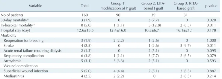
cally significantly greater number of anastomoses (3.8±0.9) than the other groups (2.6±1.0 in group 2 and 3.3±0.9 in group 3) (p<0.001). All 3 deaths (1.9%) that occurred with- in 30 days postoperatively were of noncardiac origin, and all occurred in group 2. In-hospital mortality was also high- est in group 2 at 12.5% (p<0.011); among all patients, the cases of mortality included 2 cases of septic shock and 1 each of cardiac arrest, cardiogenic shock, and pulmonary hemorrhage. Early mortality and the occurrence of stroke differed significantly between groups (Table 3), with group 2 having the highest early mortality rate and group 3 hav- ing the highest rate of stroke.

Long-term survival

The overall 5-year survival rate was 82.6%, and the 5-year survival rates in groups 1, 2, and 3 were 86.7%, 80.1%, and 75.8%, respectively (p=0.076) (Fig. 2). There were no statis- tically significant differences in survival rate among groups. MACCEs were defined as all-cause mortality, stroke, myocardial infarction, and target vessel revascular- ization. The overall 5-year MACCE-free survival rate was 75.9% (p=0.006); group 1 had the highest survival rate to a

Fig. 1. Methods of modifications. Group 1: modification of a typical Y graft (n=90, 56.3%). (A) The short right internal thoracic artery was extended with the remaining LITA or an additional saphenous vein graft (n=65). (B) Twisted Y graft for LITA-left anterior descending artery mismatch (n=12). (C) Secondary Y graft (n=9). (D) Double Y or π graft (n=4). Group 2: LITA-based I composite graft (n=39, 24.4%).

(E) LITA-based I graft to the LAD (n=32). (F) LITA-based I graft to an area other than the LAD (n=7). Group 3: RITA-based composite graft (n=31, 19.4%). (G, H) RITA-based I graft (n=25). (I) RITA-based reverse T or U graft (n=3). (J) RITA-based Y graft (n=3). LITA, left internal thoracic artery; LAD, left anterior descending artery; RITA, right internal thoracic artery.

A B C D E

F G H I J

(4)

Beatrice Chia-Hui Shih, et al. Complex Configuration of Bilateral Internal Thoracic Artery Graft KJTCVS

significant extent at 84.2%, group 2 had a rate of 68.2%,

and group 3 had the lowest rate at 65.7% (Fig. 3). Graft patency

Graft failure was defined as total graft occlusion as shown on coronary CT angiography, according to the Fitz- Table 1. Preoperative characteristics of patients

Characteristic Total Group 1:

modification of Y graft

Group 2: LITA- based I graft

Group 3: RITA-

based graft p-value

No. of patients 160 90 39 31

Age (yr) 64.9±10.7 64.3±11.1 65.5±11.0 65.9±9.1 0.924

Sex (male) 114 (71.3) 64 (71.1) 28 (71.8) 22 (71.0) 0.996

Smoker 83 (51.9) 46 (51.1) 24 (61.5) 13 (41.9) 0.258

Hypertension 122 (76.3) 70 (77.8) 30 (76.9) 22 (71.0) 0.740

Diabetes mellitus 98 (61.3) 57 (63.3) 20 (51.3) 21 (67.7) 0.309

Dyslipidemia 60 (37.5) 38 (42.2) 13 (33.3) 9 (29.0) 0.351

Chronic renal failure 29 (18.1) 9 (10.0) 5 (12.8) 15 (48.4) <0.001

Prior myocardial infarction

Acute 43 (26.9) 27 (30.0) 10 (25.6) 6 (19.4) 0.504

Old 38 (23.8) 23 (25.6) 11 (28.2) 4 (12.9) 0.272

Coronary disease

1-Vessel disease 1 (0.63) 1 (1.11) 0 0

2-Vessel disease 32 (20.3) 10 (11.1) 17 (43.6) 5 (16.1)

3-Vessel disease 125 (79.1) 79 (87.8) 22 (56.4) 26 (83.9)

Left main disease 35 (22.2) 16 (17.8) 7 (17.9) 12 (30.7)

Prior stroke 44 (27.5) 21 (23.3) 12 (30.8) 11 (35.5) 0.371

Chronic obstructive pulmonary disease 14 (8.8) 10 (11.1) 2 (5.4) 2 (6.5) 0.513

Atrial fibrillation 11 (6.9) 5 (5.6) 5 (12.8) 1 (3.2) 0.218

Peripheral vascular disease 29 (18.1) 10 (11.4) 12 (31.6) 7 (22.6) 0.022

Left ventricular ejection fraction ≤40% 42 (26.3) 27 (30.0) 9 (24.3) 6 (19.4) 0.481

Preoperative intra-aortic balloon pump 16 (10.0) 8 (8.9) 5 (12.8) 3 (9.7) 0.790

Values are presented as number, mean±standard deviation, or number (%).

LITA, left internal thoracic artery; RITA, right internal thoracic artery.

Table 2. Operative data

Variable Total Group 1:

modification of Y graft

Group 2: LITA- based I graft

Grouup 3: RITA-

based graft p-value

No. of patients 160 90 39 31

Surgical acuity

Urgent 38 (23.8) 23 (25.6) 7 (17.9) 8 (25.8) 0.619

Emergent 9 (5.6) 4 (4.4) 3 (7.7) 2 (6.5) 0.647

No. of anastomosis

Total 3.4±1.1 3.8±0.9 2.6±1.0 3.3±0.9 <0.001

Left anterior descending artery 1.0±0.2 1.0±0.1 0.9±0.3 0.9±0.2 0.040

Lateral

a)

1.4±0.9 1.7±0.8 0.7±0.8 1.3±0.7 <0.001

Right coronary artery

b)

1.1±0.5 1.1±0.5 1.0±0.5 1.1±0.6 0.597

Isolated CABG 135 (84.4) 82 (91.1) 29 (74.4) 24 (77.4) 0.024

Cardiopulmonary bypass status

Off-pump CABG 95 (59.4) 52 (57.8) 21 (53.8) 22 (71.0) 0.314

On-pump beating CABG 65 (40.6) 38 (42.2) 18 (46.2) 9 (29.0) 0.314

Conventional CABG 33 (20.6) 13 (14.4) 13 (33.3) 7 (22.6) 0.049

Values are presented as number, number (%) or mean±standard deviation.

LITA, left internal thoracic artery; RITA, right internal thoracic artery; CABG, coronary artery bypass grafting.

a)

Diagonal/Ramus/obtuse marginal artery.

b)

Posterolateral branch/posterior descending artery.

(5)

https://doi.org/10.5090/kjtcs.2020.53.2.64

KJTCVS

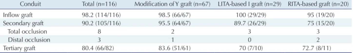
Gibbon grading system [4]. We further categorized graft patency by the anastomosed coronary territory and by the type of conduit. Table 4 demonstrates graft patency for each coronary territory. The total patency rates were 98.7%, 95.3%, and 83.6% for the LAD, left circumflex artery, and right coronary artery territories, respectively. Patency rates for the inflow graft, the secondary graft (anastomosed to the inflow graft), and the tertiary graft (anastomosed to the secondary graft) were 98.2%, 90.2%, and 80.4%, respec- tively at a mean interval of 29.9±31.1 months after CABG surgery (Tables 4, 5).

Discussion

The current literature demonstrates the superiority of BITA over other types of conduits in patients undergoing CABG [5]. These benefits include increased short- and long-term patency, freedom from arteriosclerosis, and a higher survival rate in patients undergoing revasculariza- tion of the left coronary system [4,5]. Statistical adjustment has shown that graft configuration is not an independent predictor of repeat revascularization or mortality [5]. Ad- ditionally, previous studies have revealed that no single BITA graft configuration is superior to the others in terms of mortality or the need for repeat revascularization, apart Table 3. Early clinical outcomes

Variable Total Group 1:

modification of Y graft

Group 2: LITA- based I graft

Group 3: RITA-

based graft p-value

No of patients 160 90 39 31

30-day mortality

a)

3 (1.9) 0 3 (7.7) 0 0.020

In-hospital mortality

b)

8 (5.0) 1 (1.1) 5 (12.8) 2 (6.5) 0.011

Hospital stay (day) 12.6±15.5 12.4±16.0 10.3±6.7 16.1±21.1 0.178

Morbidity

Reoperation for bleeding 3 (1.9) 2 (2.2) 1 (2.6) 0 1.000

Stroke 4 (2.5) 0 1 (2.6) 3 (9.7) 0.011

Acute renal failure requiring dialysis 2 (1.3) 0 2 (5.1) 0 0.095

Respiratory complication 6 (3.8) 1 (1.1) 3 (7.7) 2 (6.5) 0.079

Arrhythmia 5 (3.1) 3 (3.3) 2 (5.1) 0 0.593

Wound complication

Superficial wound infection 5 (5.0) 4 (4.4) 2 (5.1) 2 (6.5) 0.887

Mediastinitis 4 (2.5) 2 (2.2) 0 2 (6.5) 0.214

Values are presented as number, number (%) or mean±standard deviation.

LITA, left internal thoracic artery; RITA, right internal thoracic artery.

a)

Acute respiratory distress syndrome (n=1), cerebral infarction (n=1), and bowel infarction (n=1).

b)

Conditions listed above (n=3), septic shock (n=2), cardiac arrest (n=1), cardiogenic shock (n=1), and pulmonary hemorrhage (n=1).

Fig. 2. Overall survival. (A) All patients. (B) By group. LITA, left internal thoracic artery; RITA, right internal thoracic artery.

1.0

0.8

0.6

0.4

0.2

0.0

144

Cumulative survival rate

Follow-up duration (mo)

0 24 48 72 96 120

Overall survival (total)

5-yr survival: 82.6%

1.0

0.8

0.6

0.4

0.2

0.0

144

Cumulative survival rate

Follow-up duration (mo)

0 24 48 72 96 120

Overall survival (by group)

Modification of Y LITA-based I graft RITA-based graft

86.7%

80.1%

75.8%

5-yr survival p=0.076

A B

(6)

Beatrice Chia-Hui Shih, et al. Complex Configuration of Bilateral Internal Thoracic Artery Graft KJTCVS

from clear evidence supporting the use of the LITA-to- LAD graft. The use of the RITA as a second bypass conduit was a natural technical extension from these data. Howev- er, no consensus currently supports BITA grafting, as not all BITA configurations are equally effective. Our study therefore aimed to investigate whether any particular con- figuration was advantageous in terms of clinical outcomes and to attempt to determine the influence of configuration on graft patency.

Group 1 consisted of patients who underwent a typical LITA in situ Y-composite graft using RITA with additional conduits. In the case of LITA-based in situ conduits, geo-

metric factors such as graft tension or torsion may mitigate any advantage gained by using the RITA as a composite graft. However, the use of composite grafting does allow the left-sided vessels to be bypassed with the BITA, without requiring proximal anastomosis to the aorta, and provides less concern regarding vessel length. A Y-composite graft was constructed when the RITA was too short to reach the left coronary artery territory or when the left coronary ar- tery territory could not be completely revascularized with bilateral in situ ITA grafts. Nonetheless, the modified Y-composite graft allowed for more anastomoses than the single in situ LITA graft. This explains why group 1 had

Fig. 3. MACCE-free survival. (A) All patients. (B) By group. MACCE, major adverse cardiovascular and cerebrovascular event; LITA, left internal thoracic artery; RITA, right internal thoracic artery.

1.0

0.8

0.6

0.4

0.2

0.0

144

Cumulative survival rate

Follow-up duration (mo)

0 24 48 72 96 120

MACCE-free survival (total)

5-yr survival: 75.9%

1.0

0.8

0.6

0.4

0.2

0.0

144

Cumulative survival rate

Follow-up duration (mo)

0 24 48 72 96 120

MACCE-free survival (by group)

Modification of Y LITA-based I graft RITA-based graft

84.2%

68.2%

65.7%

5-yr survival p=0.006

A B

Table 4. Graft patency by coronary territory

Territory Total (n=116) Modification of Y graft (n=67) LITA-based I graft (n=29) RITA-based graft (n=20)

LAD (LAD/D) 98.7 (152/154) 100.0 (96/96) 100.0 (33/33) 92.0 (23/25)

LCX (RI/OM/dLCX) 95.3 (121/127) 98.9 (91/92) 69.2 (9/13) 95.5 (21/22)

RCA (PDA/PLb) 83.6 (97/116) 88.2 (60/68) 79.3 (23/29) 73.7 (14/19)

Values are presented as % (number).

LITA, left internal thoracic artery; RITA, right internal thoracic artery; LAD, left anterior descending artery; D, diagonal; LCX, left circumflex artery;

RI, ramus intermedius artery; OM, obtuse marginal artery; dLCX, distal left circumflex artery; RCA, right coronary artery; PDA, posterior descending artery; PLb, posterolateral branch.

Table 5. Graft patency by type of conduit

Conduit Total (n=116) Modification of Y graft (n=67) LITA-based I graft (n=29) RITA-based graft (n=20)

Inflow graft 98.2 (114/116) 98.5 (66/67) 100 (29/29) 95 (19/20)

Secondary graft 90.2 (105/116) 95.5 (64/67) 89.7 (26/29) 75 (15/20)

Total occlusion 8 2 3 3

Distal occlusion 3 1 0 2

Tertiary graft 80.4 (66/82) 83.6 (51/61) 70 (7/10) 72.7 (8/11)

Values are presented as % (number) or number.

LITA, left internal thoracic artery; RITA, right internal thoracic artery.

(7)

https://doi.org/10.5090/kjtcs.2020.53.2.64

KJTCVS

the highest survival and patency rates; the total number of anastomoses was greater, yielding higher revascularization of the left coronary circulation (Table 2). Our adaptation of RITA as a composite graft to bypass either the right or the left coronary circulation after LITA-to-LAD anastomosis was in congruency with 2 previous large series [6,7], and our results showed that this is likely the most effective by- pass configuration.

Group 2 patients—those who underwent surgery using the LITA-based I-shaped RITA configuration—had the poorest clinical outcomes, with the highest 30-day mortal- ity and in-hospital mortality rates. As mentioned earlier, this could have been due to underlying preoperative condi- tions unfavorable to revascularization as well as to vascular compromise. Whether the I-configuration itself was the paramount contributor to these poor outcomes is still un- determined. According to a previous mechanical study by Fan et al. [8], which assessed the difference in intramural stresses between end-to-side and end-to-end anastomoses, the proliferative influence of increased compliance mis- match on suture-line intimal hyperplasia was greater in end-to-side than in end-to-end anastomoses. In the present study, the configuration used in group 2 was an I-shaped end-to-end anastomosis, first anastomosed to the left coro- nary circulation and then extending to the right coronary circulation. Intramural stress alone therefore cannot be the factor that induced intimal hyperplasia compromising vas- cular integrity; instead, the anastomosis to the distal branches of the RCA with the free RITA in a composite configuration may have resulted in flow competition or limited flow to secondary and tertiary sites. The longer the arterial configuration, the lower the pressure at the distal anastomosis [9], and lower pressure in the distal portion of the Y branch may have compromised the distal anastomo- sis as well. However, the in-hospital and 30-day mortality rates in this group did not result from revascularization.

The mortality cases were mostly of noncardiac origin, meaning that the high mortality rates in group 2 can be at- tributed to that group’s preoperative risk factors and vas- cular compromise due to underlying medical conditions, rather than to the graft configuration. Preoperative in- tra-aortic balloon pump insertion, atrial fibrillation, and peripheral vascular disease likely led to vascular compro- mise and low cardiac function, which resulted in subse- quent in-hospital mortality from causes such as cardiogenic shock and cardiac arrest [10]. Nevertheless, this configura- tion may be assumed in selected patients, since the high mortality rate notwithstanding, overall 5-year survival and MACCE-free survival were higher than in group 3 (Fig. 3).

Lastly, the configuration used in group 3 was a RITA- based composite graft. In this group, the RITA was gener- ally anastomosed to the left coronary circulation first, and the distal end of the LITA was anastomosed to the distal branch of the RCA. As in group 2, anastomosing the distal branch with the free RITA in a composite configuration is associated with a risk of flow competition [8], and due to the lower pressure in the distal portion of the I branch, competitive flow was more frequent at the right coronary bed than at the left. In addition, in accordance with a pre- vious study by Glindeur et al. [11], our study showed that grafting of the intermediate branch or of the distal RCA negatively impacted the prognosis of graft function and thus patency. Group 3 had lower 30-day mortality and in-hospital mortality rates than group 2. However, the pa- tency, overall 5-year survival, and MACCE-free survival rates were lowest in group 3, which could be attributed to ineffective revascularization. The configuration used in this group may cause kinking of the intermediate anasto- moses, especially if the proximal Y anastomoses were per- formed near the pulmonary artery, were inside the pericar- dium, or were compressed by the myocardium [11,12]. Such secondary and tertiary sites had the lowest patency rates.

Although previous studies have shown no difference in long-term survival or freedom from repeat revasculariza- tion between different BITA configurations [12-20], and al- though those studies have also shown that no particular configuration is associated with relatively favorable clinical outcomes, our study revealed that certain aspects of the configuration must be considered. The selection of the ide- al BITA configuration should be based on technical factors associated with the individual patient. The RITA-based composite graft showed the lowest patency rates in the dis- tal RCA territory; thus, in patients in need of revascular- ization of the right coronary system, this configuration must be discouraged. In contrast, when possible, a LITA- based Y-composite graft should be performed, with the LITA-to-LAD graft serving as the gold standard (as in pre- vious studies) because it is expected to yield successful re- vascularization and thus better clinical outcomes. In gen- eral, the technically simplest technique should be selected, as more complex configurations offer no additional benefit to patients. Further studies assessing the mechanical dy- namics of graft configurations are necessary to provide clearer insight into relatively favorable configurations.

This study was limited by its retrospective nature. Graft

patency could be evaluated shortly after surgery in only

about 70% of patients, with most evaluations performed

within 6 months to 2 years postoperatively. The long-term

(8)

Beatrice Chia-Hui Shih, et al. Complex Configuration of Bilateral Internal Thoracic Artery Graft KJTCVS

patency rate could not be uniformly assessed, and the method of patency evaluation was also inconsistent. Post- operative CT angiography and CAG were not previously designated in the postoperative patient groups, and the possibility of the imaging modality either overestimating or underestimating graft patency cannot be excluded. We also could not enroll all patients in imaging follow-up during a specific postoperative period, which may have af- fected the analysis of patency. Unquantifiable factors such as underlying comorbidities, the target vessel, and conduit quality could not be accounted for by risk adjustment. Ad- ditionally, this study did not investigate important clinical endpoints, such as recurrent myocardial infarction and re- peat revascularization, which could reflect graft patency.

In conclusion, recent studies have shown that there is no difference in clinical outcomes between composite graft and in situ graft configurations and that BITA configura- tion does not affect mortality, graft patency, or the need for repeat revascularization. However, our study found that al- though atypical configurations were associated with rela- tively acceptable clinical outcomes and patency rates, the LITA-based Y-composite graft continues to be the gold standard graft configuration. Although the BITA compos- ite configuration can be modified in response to unexpect- ed intraoperative technical difficulties or limitations such as a short length or complex sequential anastomosis, a RI- TA-based composite graft should not be the first resort in patients in need of right coronary revascularization along with LAD revascularization. However, this does not mean that such a configuration must always be discouraged, be- cause existing studies still advocate that no particular graft configuration is better than another, apart from LITA-to- LAD anastomoses. Patients requiring multivessel coronary surgery should be strongly considered for BITA grafting and revascularization, even with atypical configurations, when clinical factors permit.

Conflict of interest

No potential conflict of interest relevant to this article was reported.

Acknowledgments

This work was supported by Grant (Grant no., 02-2013- 062) from the SNUBH research fund. Research associate Hyun Jeong Han (RN) helped us collect and manage the data.

ORCID

Beatrice Chia-Hui Shih: https://orcid.org/0000-0001-9211-0853 Suryeun Chung: https://orcid.org/0000-0002-9619-0640 Hak Ju Kim: https://orcid.org/0000-0003-1384-5240 Hyoung Woo Chang: https://orcid.org/0000-0001-5516-0333 Dong Jung Kim: https://orcid.org/0000-0003-0332-3430 Cheong Lim: https://orcid.org/0000-0003-0913-7014 Kay-Hyun Park: https://orcid.org/0000-0002-1111-9627 Jun Sung Kim: https://orcid.org/0000-0002-3663-5062

References

1. Tabata M, Grab JD, Khalpey Z, et al. Prevalence and variability of internal mammary artery graft use in contemporary multivessel coro- nary artery bypass graft surgery: analysis of the Society of Thoracic Surgeons National Cardiac Database. Circulation 2009;120:935-40.

2. Kappetein AP, Dawkins KD, Mohr FW, et al. Current percutaneous coronary intervention and coronary artery bypass grafting practices for three-vessel and left main coronary artery disease: insights from the SYNTAX run-in phase. Eur J Cardiothorac Surg 2006;29:486-91.

3. Kelleher R, Gimpel D, McCormack DJ, El-Gamel A. Does the use of an in situ or Y-configuration for bilateral internal thoracic arteries in- fluence long-term survival, patency or repeat revascularization in coronary bypass surgery? Interact Cardiovasc Thorac Surg 2019;28:

222-6.

4. FitzGibbon GM, Burton JR, Leach AJ. Coronary bypass graft fate:

angiographic grading of 1400 consecutive grafts early after operation and of 1132 after one year. Circulation 1978;57:1070-74.

5. Taggart DP, D’Amico R, Altman DG. Effect of arterial revascularisa- tion on survival: a systematic review of studies comparing bilateral and single internal mammary arteries. Lancet 2001;358:870-5.

6. Sabik JF 3rd, Stockins A, Nowicki ER, et al. Does location of the second internal thoracic artery graft influence outcome of coronary artery bypass grafting? Circulation 2008;118(14 Suppl):S210-5.

7. Toker ME, Omeroglu SN, Kirali K, Balkanay M, Yakut C. Using the bilateral internal mammary artery in the left or right coronary artery system: 5-year comparison of operation techniques and angiographic results. Heart Surg Forum 2005;8:E462-7.

8. Fan T, Lu Y, Gao Y, et al. Hemodynamics of left internal mammary artery bypass graft: effect of anastomotic geometry, coronary artery stenosis, and postoperative time. J Biomech 2016;49:645-52.

9. Glineur D, Kuschner CE, Grau JB. Bilateral internal thoracic artery graft configuration and coronary artery bypass grafting conduits.

Curr Opin Cardiol 2016;31:625-34.

10. Hori D, Yamaguchi A, Adachi H. Coronary artery bypass surgery in end-stage renal disease patients. Ann Vasc Dis 2017;10:79-87.

11. Glineur D, Hanet C, D’hoore W, et al. Causes of non-functioning

right internal mammary used in a Y-graft configuration: insight from

(9)

https://doi.org/10.5090/kjtcs.2020.53.2.64

KJTCVS

a 6-month systematic angiographic trial. Eur J Cardiothorac Surg 2009;36:129-36.

12. Magruder JT, Young A, Grimm JC, et al. Bilateral internal thoracic artery grafting: does graft configuration affect outcome? J Thorac Cardiovasc Surg 2016;152:120-7.

13. Gaudino M, Taggart D, Suma H, Puskas JD, Crea F, Massetti M. The choice of conduits in coronary artery bypass surgery. J Am Coll Car- diol 2015;66:1729-37.

14. Dar MI, Dar AH, Bilal M, Ahmad M, Haseeb A. Association of in- ternal mammary artery flow with different comorbidities and post- coronary artery bypass graft complications. Cureus 2017;9:e1584.

15. Gansera B, Gunzinger R, Angelis, et al. End of the millennium: end of the single thoracic artery graft?: two thoracic arteries: standard for the next millenium?: early clinical results and analysis of risk factors in 1,487 patients with bilateral internal thoracic artery grafts. Thorac Cardiovasc Surg 2001;49:10-5.

16. Gatti G, Castaldi G, Morosin M, et al. Double versus single source left-sided coronary revascularization using bilateral internal thoracic artery graft alone. Heart Vessels 2018;33:113-25.

17. Di Mauro M, Iaco AL, Allam A, et al. Bilateral internal mammary artery grafting: in situ versus Y-graft: similar 20-year outcome. Eur J Cardiothorac Surg 2016;50:729-34.

18. Lev-Ran O, Matsa M, Ishay Y, Shabtai A, Vodonos A, Sahar G. Ret- roaortic right internal thoracic artery grafting of circumflex artery targets. Asian Cardiovasc Thorac Ann 2015;23:543-51.

19. Bakay C, Onan B, Korkmaz AA, Onan IS, Ozkara A. Sequential in situ left internal thoracic artery grafting to the circumflex and right coronary artery areas. Ann Thorac Surg 2013;95:63-70.

20. Raja SG, Benedetto U, Husain M, et al. Does grafting of the left an-

terior descending artery with the in situ right internal thoracic artery

have an impact on late outcomes in the context of bilateral internal

thoracic artery usage? J Thorac Cardiovasc Surg 2014;148:1275-81.

수치

Fig. 1. Methods of modifications. Group 1: modification of a typical Y graft (n=90, 56.3%)
Table 2. Operative data
Fig. 2. Overall survival. (A) All patients. (B) By group. LITA, left internal thoracic artery; RITA, right internal thoracic artery.
Table 5. Graft patency by type of conduit

참조

관련 문서