Hereditary hemorrhagic telangiectasia (HHT) is an inherited disorder that is char- acterized by abnormal communication between the arteries and veins in the skin, mucosa, and various organs. HHT has been reported to show significant pheno- typic variability and genetic heterogeneity with wide ethnic and geographic varia- tions. Although mutations in the endoglin (ENG) and activin A receptor type II-like 1 (ACVRL1) genes have been known to cause HHT for more than 10 yr, little is known about the clinical features or genetic background of Korean patients with HHT. In addition, mutations in mothers against decapentaplegic homolog 4 (SMAD4) are also seen in patients with the combined syndrome of juvenile polyposis and HHT.
This study examined five Korean patients with the typical manifestations of HHT such as frequent epistaxis and pulmonary arteriovenous malformations. Direct sequencing of the ENG and ACVRL1 genes revealed one known mutation, ENG c.277C>T, in one patient and two novel mutations, ENG c.992-1G>C and ACVRL1 c.81dupT in two patients, respectively. The remaining two patients with negative results were screened for SMAD4 mutations as well as gross deletions of ENG and ACVRL1 using multiple ligation-dependent probe amplification, but none was detected. Despite the small number of patients investigated, we firstly report Kore- an patients with genetically confirmed HHT, and show the genetic and allelic het- erogeneity underlying HHT.
Key Words : Telangiectasia, Hereditary Hemorrhagic; ENG; ACVRL1; SMAD4; Mutation; Korean
INTRODUCTION
Hereditary hemorrhagic telangiectasia (HHT), which is also known as Rendu-Osler-Weber disease, is an autosomal dominantly inherited disorder that is characterized by abnor- mal communications between the arteries and veins (telang- iectasia) in the skin, mucosa, and various organs (1). The preva- lence of this disorder is estimated to be -1:1,300 to -1:
40,000 with some geographical variations (2-9). The clinical features of HHT include spontaneous recurrent epistaxis, mucocutaneous telangiectases (particularly on the tongue, lips, oral cavity, fingers and nose), and arteriovenous malfor- mations (AVM) in the pulmonary, cerebral, hepatic, gas- trointestinal or spinal vessels (10).
HHT is genetically heterogeneous and can be subdivided into HHT-1 and HHT-2 according to mutations in the en- doglin (ENG) gene and the activin A receptor type II-like 1 (ACVRL1) gene, respectively (11, 12). These two subtypes
are clinically indistinguishable and share many phenotypes.
However, there appears to be some differences in the frequen- cy of some their clinical manifestations. Pulmonary AVMs are believed to be more common in patients with HHT-1 than in HHT-2 (7, 13-17). Families with HHT-2 generally tend to show a later onset of the symptoms and a milder phenotype.
The vast majority (-80%) of the ENG mutations in HHT-1 patients lead to premature stop codons and truncated peptides, with no apparent hot focus. On the other hand, more than half (-53%) of the mutations identified in ACVRL1 are mis- sense substitutions, and the majorities of those mutations are located in exons 8, 7, and 3. Some large deletions and inser- tions as well as some splice site mutations in these two genes have also been reported (2, 13, 18). Recently, other loci for HHT, HHT-3, and HHT-4, were identified by linkage anal- ysis (19, 20). Moreover, there are diseases presenting overlap- ping features with HHT such as the Juvenile polyposis/hered- itary hemorrhagic telangiectasia syndrome (JPHT) which is
69
Seung-Tae Lee, Jee-Ah Kim*, Shin-Yi Jang�, Duk-Kyung Kim�, Young Soo Do�, Gee Young Suh�, Jong-Won Kim, and Chang-Seok Ki
Departments of Laboratory Medicine and Genetics, Samsung Medical Center, Sungkyunkwan University School of Medicine, Seoul; Garak High School*, Seoul;
Department of Internal Medicine�, Cardiac and Vascular Center, Samsung Medical Center, Sungkyunkwan University School of Medicine, Seoul;
Department of Radiology�, Samsung Medical Center, Sungkyunkwan University, School of Medicine, Seoul;
Division of Pulmonary and Critical Care Medicine�, Department of Medicine, Samsung Medical Center, Sungkyunkwan University School of Medicine, Seoul, Korea
Address for correspondence Chang-Seok Ki, M.D.
Department of Laboratory Medicine and Genetics, Samsung Medical Center, Sungkyunkwan University School of Medicine, 50 Irwon-dong, Gangnam-gu, Seoul 135-710, Korea
Tel : +82.2-3410-2709, Fax : +82.2-3410-2719 E-mail : [email protected]
*This work was supported by the Samsung Biomedical Research Institute grant, # SBRI C-A6-403-2.
DOI: 10.3346/jkms.2009.24.1.69
Clinical Features and Mutations in the ENG, ACVRL1, and SMAD4 genes in Korean Patients with Hereditary Hemorrhagic Telangiectasia
Received : 30 July 2007 Accepted : 22 May 2008
A clinical investigation and genetic analysis was performed in five Korean patients diagnosed with HHT according to the criteria suggested by Shovlin et al. (10). The frequency of epistaxis was graded according to the category suggested by Bergler et al. (22): grade 1, less than once per week; grade 2, a few times per week; grade 3, more than once per day.
DNA sequence analysis
After obtaining informed consent, the genomic DNA was isolated from the peripheral blood leukocytes using a Wiz- ard Genomic DNA Purification kit according to the manu- facturer’s instructions (Promega, Madison, WI, U.S.A.). DNA sequence analysis of the ENG, ACVRL1, and SMAD4 genes was carried out using a polymerase chain reaction (PCR) with the primers designed by the authors. PCR was initially per- formed using a thermal cycler (model 9600, Applied Biosys- tems, Foster City, CA, U.S.A.), and DNA sequencing was car- ried out using an ABI Prism 3100 Genetic Analyzer with a BigDye Terminator Cycle Sequencing Ready Reaction kit (Applied Biosystems).
tate estimation of fragment sizes. Data analysis was performed using the GeneScan (Applied Biosystems) and GeneMarker (SoftGenetics, State College, PA, U.S.A.) software. Relative peak area values obtained in the patients were compared to those obtained in healthy controls and were expressed as ratios.
Signal ratios of approximately 0.5 were considered patho- logical.
RESULTS Clinical findings
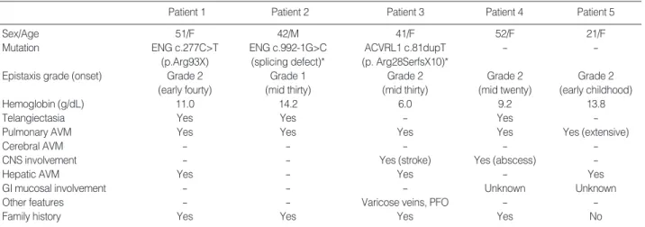
The Clinical features and mutations identified in the five Korean patients were summarized in the Table 1. Patient 1 had recurrent epistaxis and pulmonary AVM along with focal telangiectasia on the fingers (particularly the periungal regi- ons), lips, and oral mucosa (Fig. 1A, B). A history of recur- rent epistaxis was found in her father and 32-yr-old daugh- ter (Fig. 2A). Computed tomography (CT) angiography of the liver showed an enlarged celiac axis and a prominent hep- atic artery with multiple aberrant collateral vessels. Hetero- geneous attenuation of the liver was also noted (Fig. 3A, B).
*Novel mutations.
HHT, hereditary hemorrhagic telangiectasia; AVM, arteriovenous malformation; CNS, central nervous system; GI, gastrointestinal; PFO, patent foramen ovale.
Patient 1 Patient 2 Patient 3 Patient 4 Patient 5
Sex/Age 51/F 42/M 41/F 52/F 21/F
Mutation ENG c.277C>T ENG c.992-1G>C ACVRL1 c.81dupT - -
(p.Arg93X) (splicing defect)* (p. Arg28SerfsX10)*
Epistaxis grade (onset) Grade 2 Grade 1 Grade 2 Grade 2 Grade 2
(early fourty) (mid thirty) (mid thirty) (mid twenty) (early childhood)
Hemoglobin (g/dL) 11.0 14.2 6.0 9.2 13.8
Telangiectasia Yes Yes - Yes -
Pulmonary AVM Yes Yes Yes Yes Yes (extensive)
Cerebral AVM - - - - -
CNS involvement - - Yes (stroke) Yes (abscess) -
Hepatic AVM Yes - Yes - Yes
GI mucosal involvement - - - Unknown Unknown
Other features - - Varicose veins, PFO - -
Family history Yes Yes Yes Yes No
Table 1. Clinical features and mutations identified in five Korean patients with HHT
Patient 2 had pulmonary AVM and telelangiectasia was found on his tongue, along with a history of frequent epistaxis.
Recurrent epistaxis histories were also found in his father, four
sisters, and eight-year-old son. His father was diagnosed with HHT with recurrent massive epistaxis requiring frequent blood transfusions, telangiectasia on the oral mucosa (Fig. 1C), and pulmonary AVM. Pulmonary angiography of patient 2 revealed a large pulmonary AVM with an aneurysmal sac (Fig.
4A). Selective coil embolization of the feeder vessels of the pulmonary AVM was performed and the hemoptysis subsided.
Patient 3 experienced a momentary seizure-like movement and a sudden attack of aphasia. A history of frequent epistaxis and chronic anemia was found. At the age of 17, she had a pulmonary segmentectomy because of a pulmonary AVM.
She had a family history of recurrent epistaxis (the detailed pedigree could not be obtained). Multiphase contrast-enhanced CT of the brain revealed an occlusion of the left intracranial artery at the middle cerebral artery (MCA) bifurcation level with findings of an acute infarct. Abdominal CT revealed a severe tortuous dilatation of the hepatic artery and its intra- hepatic branches with mottled hepatic enhancement (Fig. 3C, D). Chest CT and pulmonary angiography revealed a new small-sized pulmonary AVM in the right middle lobe (Fig.
4B). A wedge resection was performed as a result. The trans- esophageal echocardiogram revealed a patent foramen ovale.
Fig. 1. Clinical features of the HHT patients: (A) telangiectasia on the periungal regions of the fingers of patient 1, (B) telangiectasia on the lips of patient 1, (C) telangiectasia on the oral mucosa of the father of patient 2, (D) bleeding foci in the Kisselbach’s plexus of patient 3;
(E) microtelangia on the tongue of patient 5, and (F) clubbing fingers with one small hemangioma in patient 5.
D E F
B C
A
Fig. 2. Pedigrees of families of (A) patient 1 and (B) patient 2.
I
II
III A
I
II
III
B
Patient 4, a 52-yr-old Korean female, was found to have a mass in the right parietal lobe of the brain. She complained of severe headache with fever, chill, nausea and vomiting. She had neck stiffness and microtelangiectasia on the trunk. She had a history of recurrent epistaxis and chronic anemia. There was a family history of epistaxis in her uncle and daughter (detailed pedigree could not be obtained), and her daughter had died suddenly from a hemangioma one year before the patient visited to our hospital. A brain pre- and post-contrast CT showed a rim-enhancing mass in the right parieto-tem- poral lobe suggesting an abscess with cavity formation. Pul- monary angiography revealed multifocal AVM in both lungs (Fig. 4C). Selective coil embolization of the AVM in the lung was performed, followed by a craniotomy and abscess drainage with the removal of the abscess wall.
Patient 5, a 21-yr-old Korean female, had severe dyspnea and a pulmonary AVM. She had a history of cyanosis, dysp- nea, and clubbing of her fingers at birth, as well as multiple AVM in the lung. She underwent a segmentectomy of both lungs at the age of nine. A visual inspection revealed an ac- neiform eruption on her face, microtelangiectasia on the to-
ngue, and clubbing fingers with one small telangiectasia (Fig.
1E, F). The chest CT and pulmonary angiography revealed extensive peripheral AVM in both lungs (Fig. 4D) and a prob- able arterioportal shunt involving the liver. Selective coil em- bolization of the AVM in the lung was performed twice, and the dyspnea was ameliorated.
Mutation analysis
Direct sequencing of the ENG gene was performed initial- ly for all the five patients with HHT. A nonsense mutation producing a premature stop signal at codon 93 (c.277C>T;
p.Arg93X) in exon 3 of the ENG gene was found in patient 1, which has previously been reported in HHT-1 patients (23). Patient 2 had a base substitution at a consensus splic- ing acceptor site of the 9th intron of the ENG gene (c.992- 1G>C) predicting a splicing defect. This mutation has not been reported previously. We additionally performed direct sequencing of the ACVRL1 gene for three remaining patients and found one patient with mutation; patient 3 had a novel a frameshift mutation as a result of a duplication of one base
Fig. 3. Radiology findings of the HHT patients. (A) and (B) CT angiography of the liver in patient 1 shows an enlarged celiac axis (arrow), a prominent hepatic artery (arrowhead) with multiple aberrant collateral vessels, and heterogeneous attenuation of the liver. (C) and (D) Abdominal CT of patient 3 shows severe tortuous dilatation of the hepatic artery (arrow) and its intrahepatic branches (arrowhead) with mottled hepatic enhancement.
A B
C D
pair in the coding region (exon 2) of the ACVRL1 gene pro- ducing a premature termination codon (c.81dupT; p.Arg28 SerfsX10) (Fig. 5). We could not perform further genetic anal- ysis on the family members of the patients with mutation.
Since patients 4 and 5 did not have any mutations in the ENG and ACVRL1 genes, we additionally screened them for the SMAD4 genes, but the results were negative. Subse- quent MLPA analysis found no gross deletions in the ENG or ACVRL1 genes in these two patients. Because the P093 HHT/PPH1 MLPA kit also covers the 13 exons of the BMPR2 gene, the causative gene for primary pulmonary hyperten- sion which sometimes shares identical pulmonary manifes-
tations with HHT, we could get additional information on this gene. The remaining two patients showed negative results for the BMPR2 gene.
DISCUSSION
Table 1 gives a summary of the clinical features and results of mutation analysis. All the five patients showed the typi- cal manifestations of HHT with variable individual expres- sion of the clinical features. The onset of epistaxis was rather late in these cases (with the exception of patient 5) compared
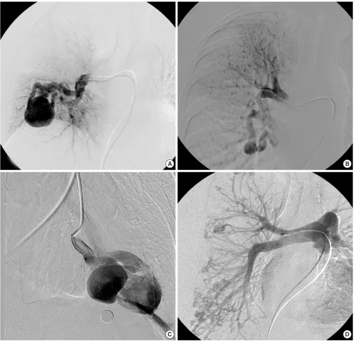
Fig. 4. Pulmonary angiography of the HHT patients. (A) a large pulmonary arteriovenous malformation (AVM) in patient 2; (B) a new small- sized pulmonary AVM in the right middle lobe in patient 3; (C) multifocal AVM in patient 4; (D) extensive peripheral AVM in patient 5.
A B
C D
with normal HHT patients, in whom most manifest recur- rent epistaxis before the age of 20 (15, 24). Pulmonary AVM was observed in all five patients, which is believed to be somewhat unusual considering that only 15-30% of HHT patients have pulmonary AVM (15, 25-27). This might be due to the probable selection bias in this study in those patients with severe clinical manifestations such as pulmonary AVM are referred to our hospital for an evaluation of HHT. How- ever, there remains a possibility that high prevalence of pul- monary AVM might be a unique characteristic in Korean pa- tients with HHT; our review of all the literatures addressing Korean patients with HHT found that 13 out of 22 (-59%) patients had pulmonary AVM (28). Further studies on a large set of patients would be needed to clarify these issues, inves- tigating genetic and environmental factors in Korean popu- lations. Patient 5 had relatively severe clinical manifestations such as infantile cyanosis resulting from a congenital pulmo- nary AVM, which is quite unusual with very few cases (<30) being reported thus far (27, 29). This might be because most cases of congenital AVM are asymptomatic. This suggests that there might be a specific genetic background in this patient.
Molecular analysis of the ENG, ACVRL1, and SMAD4 genes in the five Korean patients with HHT revealed three individuals with mutations in either the ENG or ACVRL1 genes. Endoglin is a membrane protein of the disulphide- linked homodimer that is expressed mainly in the endothe- lial cells of all vessels. This molecule binds the transforming growth factor-β(TGF-β), which is a powerful mediator of vascular remodeling induced by various stimuli such as vas- cular wall stress, and forms a signaling heteromeric complex (26, 30). The vast majority (-80%) of mutations of the ENG gene identified in HHT-1 patients are premature stop codons
tor type II-like 1 encoded by ACVRL1 belongs to the TGF-β superfamily receptor group, which signals through Smad1/5.
Moreover, this molecule is expressed almost exclusively in en- dothelial cells, particularly during angiogenesis (31, 32). Pati- ent 3 had a duplication mutation on exon 2 of the ACVRL1 gene, which is one of mutational hot spot and encodes part of the extracellular domain, predicting a frameshift and very short truncated protein removing the cystein rich (extracel- lular), transmembrane, kinase, and intracellular domain. The truncation mutations in patient 1 and 3 might fit into the haplo-insufficiency model rather than the dominant-negative model, because the mutant proteins lack the transmembrane domain and they would have less chance to form a heterodimer with normal protein.
There is significant phenotypic variability and genetic het- erogeneity in HTT. Sequence analysis of the ENG and AC- VRL1 genes reveals mutations in 60-80% of individuals with HHT (33, 34). Several techniques including quantitative PCR, MLPA and southern blot analysis can identify those deletions undetectable by sequence analysis, which can increase the de- tection rate by up to 10-20% (14, 35). However, mutation analysis of the ENG and ACVRL1 genes can fail to detect mutations in the remaining patients. This suggests the pos- sibility of mutations in the non-coding regions of these genes or mutations in other genes. Recently, linkage analysis in two HHT families identified new HHT loci: HHT-3 in chromo- some 5q31.3-32, and HHT-4 in chromosome 7p14 (19, 20).
It is possible that patients 4 and 5, who tested negative to mutations in the ENG and ACVRL1 genes, might have muta- tions in another unidentified gene. Gallione et al. (21) recent- ly reported that mutations in the SMAD4 tumor suppressor gene might be associated with a combined syndrome of JPHT.
Because some cases with SMAD4 mutations show clinical manifestations of HHT without juvenile polyposis (36), we screened mutations in the SMAD4 gene in patients 4 and 5, and the results were negative.
HHT occurs with a wide ethnic and geographic distribu- tion. The prevalence of HHT has been reported to range from -1:1,300 to -1:40,000 depending on the geographic or eth- nic region (2-9). Moreover, the type and frequency of specif- ic mutations in different localities can vary widely with some regions having a specific founder mutation. For example, a
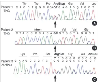
Fig. 5. Sequencing results of three mutation-positive patients with HHT. (A) ENG c.277C>T (p.Arg93X) in patient 1; (B) ENG c.992- 1G>C (splicing defect) in patient 2; (C) ACVRL1 c.81dupT (p. Arg 28SerfsX10) in patient 3.
Patient 3 ACVRL1
G A A G C C G T C T C G G G G C C C G C T
C
study in France and northern Italy revealed higher frequen- cy (-2.7 times) of HHT-2 than HHT-1 (15). The prevalence of HHT in the Korean population appears to be low with only occasional cases being reported thus far. However, the precise incidence, clinical characteristics, and genetic back- ground of HHT in the Korean population are unknown be- cause no epidemiological surveys have been carried out. Al- though only a small number of patients were investigated, this study indicates that there might be a specific clinical dif- ference in Korean HHT patients. This highlights the need for more studies on a large set of patients in this regional population. Furthermore, more studies on the genetic back- ground of Korean patients with HHT as well as the identi- fication of another causative gene is needed. It is expected that such studies will help us better understand this serious but overlooked disorder and develop more appropriate ther- apeutic strategies.
REFERENCES
1. Peery WH. Clinical spectrum of hereditary hemorrhagic telangiec- tasia (Osler-Weber-Rendu disease). Am J Med 1987; 82: 989-97.
2. Abdalla SA, Letarte M. Hereditary haemorrhagic telangiectasia:
current views on genetics and mechanisms of disease. J Med Genet 2006; 43: 97-110.
3. Westermann CJ, Rosina AF, De Vries V, de Coteau PA. The preva- lence and manifestations of hereditary hemorrhagic telangiectasia in the Afro-Caribbean population of the Netherlands Antilles: a family screening. Am J Med Genet A 2003; 116: 324-8.
4. Porteous ME, Burn J, Proctor SJ. Hereditary haemorrhagic telang- iectasia: a clinical analysis. J Med Genet 1992; 29: 527-30.
5. Guttmacher AE, Marchuk DA, White RI Jr. Hereditary hemorrhag- ic telangiectasia. N Engl J Med 1995; 333: 918-24.
6. Dakeishi M, Shioya T, Wada Y, Shindo T, Otaka K, Manabe M, No- zaki J, Inoue S, Koizumi A. Genetic epidemiology of hereditary hem- orrhagic telangiectasia in a local community in the northern part of Japan. Hum Mutat 2002; 19: 140-8.
7. Kjeldsen AD, Vase P, Green A. Hereditary haemorrhagic telangiec- tasia: a population-based study of prevalence and mortality in Dan- ish patients. J Intern Med 1999; 245: 31-9.
8. Bideau A, Plauchu H, Jacquard A, Robert JM, Desjardins B. Genetic aspects of Rendu-Osler disease in Haut-Jura: convergence of me- thodological approaches of historic demography and medical genet- ics. J Genet Hum 1980; 28: 127-47.
9. Plauchu H, de Chadarevian JP, Bideau A, Robert JM. Age-related clinical profile of hereditary hemorrhagic telangiectasia in an epidemi- ologically recruited population. Am J Med Genet 1989; 32: 291-7.
10. Shovlin CL, Guttmacher AE, Buscarini E, Faughnan ME, Hyland RH, Westermann CJ, Kjeldsen AD, Plauchu H. Diagnostic criteria for hereditary hemorrhagic telangiectasia (Rendu-Osler-Weber syn- drome). Am J Med Genet 2000; 91: 66-7.
11. McAllister KA, Grogg KM, Johnson DW, Gallione CJ, Baldwin MA, Jackson CE, Helmbold EA, Markel DS, McKinnon WC, Murrell J,
McCormick MK, Pericak-Vance MA, Heutink P, Oostra BA, Hait- jema T, Westerman CJ, Porteous ME, Guttmacher AE, Letarte M, Marchuk DA. Endoglin, a TGF-beta binding protein of endothelial cells, is the gene for hereditary haemorrhagic telangiectasia type 1.
Nat Genet 1994; 8: 345-51.
12. Johnson DW, Berg JN, Gallione CJ, McAllister KA, Warner JP, Helm- bold EA, Markel DS, Jackson CE, Porteous ME, Marchuk DA. A sec- ond locus for hereditary hemorrhagic telangiectasia maps to chro- mosome 12. Genome Res 1995; 5: 21-8.
13. Bayrak-Toydemir P, McDonald J, Markewitz B, Lewin S, Miller F, Chou LS, Gedge F, Tang W, Coon H, Mao R. Genotype-phenotype correlation in hereditary hemorrhagic telangiectasia: mutations and manifestations. Am J Med Genet A 2006; 140: 463-70.
14. Bossler AD, Richards J, George C, Godmilow L, Ganguly A. Novel mutations in ENG and ACVRL1 identified in a series of 200 individ- uals undergoing clinical genetic testing for hereditary hemorrhagic telangiectasia (HHT): correlation of genotype with phenotype. Hum Mutat 2006; 27: 667-75.
15. Lesca G, Olivieri C, Burnichon N, Pagella F, Carette MF, Gilbert- Dussardier B, Goizet C, Roume J, Rabilloud M, Saurin JC, Cottin V, Honnorat J, Coulet F, Giraud S, Calender A, Danesino C, Buscarini E, Plauchu H. Genotype-phenotype correlations in hereditary hem- orrhagic telangiectasia: data from the French-Italian HHT network.
Genet Med 2007; 9: 14-22.
16. Letteboer TG, Mager JJ, Snijder RJ, Koeleman BP, Lindhout D, Ploos van Amstel JK, Westermann CJ. Genotype-phenotype relationship in hereditary haemorrhagic telangiectasia. J Med Genet 2006; 43:
371-7.
17. Berg J, Porteous M, Reinhardt D, Gallione C, Holloway S, Umasun- thar T, Lux A, McKinnon W, Marchuk D, Guttmacher A. Hereditary haemorrhagic telangiectasia: a questionnaire based study to delin- eate the different phenotypes caused by endoglin and ALK1 mutations.
J Med Genet 2003; 40: 585-90.
18. Pece N, Vera S, Cymerman U, White RI Jr, Wrana JL, Letarte M.
Mutant endoglin in hereditary hemorrhagic telangiectasia type 1 is transiently expressed intracellularly and is not a dominant negative.
J Clin Invest 1997; 100: 2568-79.
19. Cole SG, Begbie ME, Wallace GM, Shovlin CL. A new locus for hered- itary haemorrhagic telangiectasia (HHT3) maps to chromosome 5. J Med Genet 2005; 42: 577-82.
20. Bayrak-Toydemir P, McDonald J, Akarsu N, Toydemir RM, Calderon F, Tuncali T, Tang W, Miller F, Mao R. A fourth locus for hereditary hemorrhagic telangiectasia maps to chromosome 7. Am J Med Genet A 2006; 140: 2155-62.
21. Gallione CJ, Repetto GM, Legius E, Rustgi AK, Schelley SL, Tej- par S, Mitchell G, Drouin E, Westermann CJ, Marchuk DA. A com- bined syndrome of juvenile polyposis and hereditary haemorrhagic telangiectasia associated with mutations in MADH4 (SMAD4). Lancet 2004; 363: 852-9.
22. Bergler W, Sadick H, Gotte K, Riedel F, Hormann K. Topical estro- gens combined with argon plasma coagulation in the management of epistaxis in hereditary hemorrhagic telangiectasia. Ann Otol Rhi- nol Laryngol 2002; 111 (3 Pt 1): 222-8.
23. Cymerman U, Vera S, Pece-Barbara N, Bourdeau A, White RI Jr,
M, Plauchu H, Cordier JF. Pulmonary arteriovenous malformations in hereditary hemorrhagic telangiectasia: a series of 126 patients.
Medicine (Baltimore) 2007; 86: 1-17.
28. Korean Association of Medical Journal Editors. KoreaMed. Avail- able at http://www.koreamed.org
29. Butter A, Emran M, Al-Jazaeri A, Bouron-Dal Soglio D, Bouchard S. Pulmonary arteriovenous malformation mimicking congenital cys- tic adenomatoid malformation in a newborn. J Pediatr Surg 2006;
41: e9-11.
30. Llorca O, Trujillo A, Blanco FJ, Bernabeu C. Structural model of human endoglin, a transmembrane receptor responsible for heredi-
Hum Mutat 2004; 23: 289-99.
34. Schulte C, Geisthoff U, Lux A, Kupka S, Zenner HP, Blin N, Pfister M. High frequency of ENG and ALK1/ACVRL1 mutations in Ger- man HHT patients. Hum Mutat 2005; 25: 595.
35. Cymerman U, Vera S, Karabegovic A, Abdalla S, Letarte M. Char- acterization of 17 novel endoglin mutations associated with heredi- tary hemorrhagic telangiectasia. Hum Mutat 2003; 21: 482-92.
36. Gallione CJ, Richards JA, Letteboer TG, Rushlow D, Prigoda NL, Leedom TP, Ganguly A, Castells A, Ploos van Amstel JK, Wester- mann CJ, Pyeritz RE, Marchuk DA. SMAD4 mutations found in uns- elected HHT patients. J Med Genet 2006; 43: 793-7.