Preoperative Serum Thyroglobulin as a Useful Predictive Marker to Differentiate Follicular Thyroid Cancer from Benign Nodules in Indeterminate Nodules
Indeterminate cytology results increase the number of repetitive procedure and
unnecessary surgery. This study was designed to find useful and simple predictive tools to differentiate malignant thyroid nodules from indeterminate nodules. We retrospectively enrolled 164 patients who had undergone thyroid surgery as a result of indeterminate cytology in the National Cancer Center. We reviewed patients’ age at diagnosis, sex, preoperative biochemical markers such as thyroglobulin (Tg), anti-Tg antibody, free T4 and TSH level, and sonographical and pathological findings, which were subjected to statistical analysis. We found several clinical and sonographical predictive factors that showed significant differences. Young age, male, preoperative high Tg level, and hypoechoic nodule on sonography all increased cancer probability significantly in multivariate analysis.
With a cut-off value of 187.5 ng/mL Tg, sensitivity and specificity were 54.8% and 90.1%, respectively (AUC 0.748, P < 0.001). In the case of nodule size > 1.7 cm, elevated serum Tg predicts the risk of malignancy; especially Tg > 70 ng/mL (odds ratio 3.245, 95%
confidence interval 1.115-9.450, P = 0.038). Preoperative Tg levels had very high specificity in predicting thyroid cancer in case of suspicious follicular neoplasm. Therefore, Tg levels may be a useful marker for differentiating thyroid cancer from benign thyroid nodules in the cytological diagnosis of indeterminate nodules.
Key Words: Thyroid Nodule; Fine Needle Aspiration; Thyroglobulin Eun Kyung Lee*, Ki-Wook Chung*,
Hye Sook Min, Tae Sung Kim, Tae Hyun Kim, Jun Sun Ryu, Yoo Seok Jung, Seok Ki Kim, and You Jin Lee
Division of Endocrinology and Metabolism, Department of Internal Medicine, Center for Thyroid Cancer, National Cancer Center, Goyang, Korea
*Eun Kyung Lee and Ki-Wook Chung contributed equally to this work.
Received: 28 June 2011 Accepted: 2 July 2012 Address for Correspondence:
You Jin Lee, MD
Division of Endocrinology and Metabolism, Department of Internal Medicine, Center for Thyroid Cancer, National Cancer Center, 323 Ilsan-ro, Ilsandong-gu, Goyang 410-769, Korea Tel: +82.31-920-1664, Fax: +82.31-920-2798 E-mail: [email protected]
This study was supported by the National Cancer Center Research Grant No. 0910033-1.
http://dx.doi.org/10.3346/jkms.2012.27.9.1014 • J Korean Med Sci 2012; 27: 1014-1018
INTRODUCTION
Thyroid nodules are very common (1), and most do not impact life expectancy of the affected person (2). However, along with increased detection rates of non-palpable thyroid nodules, the incidence rate of thyroid cancer has been raised (3, 4) and the absolute number of indeterminate thyroid nodule cytology re- sults has also markedly increased. Although fine-needle aspira- tion cytology (FNAC) is a primary method for detecting malig- nant nodules, 10%-25% of thyroid nodules are categorized as indeterminate nodules (5). Classifications for indeterminate nodules are defined as “atypia of undetermined significance (AUS) or follicular lesion of undetermined significance (FLUS)”
and “follicular neoplasm (FN) or suspicious for a follicular neo- plasm (SFN)” under the Bethesda classification systems (6), and the risk of malignancy of the FN/SFN category nodules is approximately 15%-30%. These renewed classifications also cannot resolve the difficulties clinicians face in differentiating malignant from benign nodules.
Numerous studies were performed in order to differentiate benign and malignant thyroid nodules using clinical factors other than cytology. Unfortunately, there are currently no use- ful differentiating markers except for some well-known risk fac- tors such as positive family history, tumor size, and age (7-10).
Therefore, we tried to elucidate the role of preoperative se- rum thyroglobulin (Tg) as a novel marker that could distinguish between benign and malignant thyroid nodules, especially in the rigorously selected FN/SFN category.
MATERIALS AND METHODS Materials
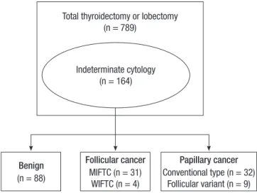
We reviewed FNAC results from 789 patients who underwent thyroid surgery at the National Cancer Center from May 2002 to July 2009 and selected 164 cases compatible with indetermi- nate cytology results according to the new Bethesda system category (Fig. 1) (6). One pathologist re-examined the patient cytology and pathology slides under the criteria as followed; 1)
FN/SFN: follicular cells are arranged predominantly in micro- follicular or trabecular arrangements whether nuclear atypia may or may not be present, 2) AUS/FLUS : neither convincingly benign nor sufficiently atypical to place into a different category (11). In our analysis we selected 164 cases showing cytology re- sults of FN/SFN category and also reviewed patients’ clinical, biochemical, sonographical and pathological findings. We ex- cluded the cases whose cytological results have been reported as another category (benign or malignancy) after repeated as- pirations.
Measurement of biochemical and ultrasonographical parameters
The serum TSH levels were measured by an immunoradiomet- ric assay (IRMA) using a commercial kit (Immunotech, Mar- seille, France). Tg and anti-Tg antibody (TgAb) levels were mea- sured using IRMA and a commercial radioimmunoassay kit, respectively (Brahms Aktiengesellschaft, Hennigsdorf, Germa- ny). The reference ranges for TSH, Tg and TgAb were 0.17-4.05 mU/L, 0-40 ng/mL and 0-100 U/mL, respectively.
We obtained patients’ sex, age at diagnosis and TSH, Tg, and TgAb levels from medical records. Reviewing ultrasonography, cytology slide, and pathology specimens, we also obtained tu- mor size, characteristics of tumor such as margin, component, echogenicity, hypoechoic rim, vascularity, calcification and presence of Hashimoto’s thyroiditis and final pathological diag- nosis. Hashimoto’s thyroiditis was defined as diffuse thyroid disease measured by ultrasonography and/or positive TgAb or microsomal antibody (12, 13).
Statistical analysis
We grouped patients into the benign or cancer groups (papil- lary thyroid carcinoma [PTC] or follicular thyroid carcinoma [FTC]) according to the final diagnosis. Continuous variables are expressed as means ± standard deviation (SD), whereas categorical variables are presented as absolute values and per- centages. Differences between continuous variables were ana- lyzed by unpaired Student’s t-test or ANOVA, and differences between categorical variables were analyzed using the chi- square test. Multivariate and logistic regression analysis was employed to identify independent predictors of malignancy.
An ROC curve was plotted to determine the Tg cut-off levels for differentiating cancerous from benign nodules. A P value of
< 0.05 was considered statistically significant, and all analyses were performed using Stata statistical software, version 9 (Stata Corporation, College Station, TX, USA).
Ethics statement
This study was approved by the institutional review board of National Cancer Center (IRB No. NCCNCS-09-225) and in- formed consent was waived.
RESULTS
Comparison of biochemical and sonographical findings between benign and malignant groups
First, we compared basal clinical findings between benign and malignant groups that had shown indeterminate nodule cytol- ogy results. The benign group and malignant group comprised Fig. 1. Patients enrolled in our study. Indeterminate cytology included all of “atypical
of undetermined significance or follicular lesion of undetermined significance” and
“follicular neoplasm or suspicious for a follicular neoplasm.” MIFTC, minimally inva- sive follicular thyroid carcinoma; WIFTC, widely invasive follicular thyroid carcinoma.
Total thyroidectomy or lobectomy (n = 789)
Benign (n = 88)
Follicular cancer MIFTC (n = 31)
WIFTC (n = 4)
Papillary cancer Conventional type (n = 32)
Follicular variant (n = 9) Indeterminate cytology
(n = 164)
Table 1. Comparison of biochemical findings between benign and malignant group
Parameters Benign MIFTC WIFTC Follicular variant PTC PTC
Total, No. (%) 88 (53.7) 31 (18.9) 4 (2.4) 9 (5.5) 32 (19.5)
Female, No. (%) 77 (87.5) 22 (71.0) 4 (100) 8 (88.9) 27 (84.4)
Age at diagnosis (yr) 48 ± 11 50 ± 13 50 ± 17 45 ± 10 46 ± 12
Tumor size (cm) 2.4 ± 1.2 2.9 ± 1.9* 4.3 ± 1.3* 1.2 ± 0.7* 0.8 ± 0.1*
TSH (mU/L) 1.6 ± 1.2 1.2 ± 1.1 1.5 ± 0.7 2.4 ± 1.5 1.6 ± 1.3
Tg (ng/mL, median [range]) 15.4 [1-1499] 188.0* [2.3-7940.0] 2078.5* [31.7-6860.0] 9.3 [2.4-398.0) 8.8 [0.3-181.0]
TgAb (U/mL, median) < 20 < 20 49.1 < 20 < 20
Hashimoto, N (%) 17 (19.3) 3 (9.7) 0 (0.0) 3 (33.3) 3 (9.4)
*P value < 0.05 when compared with benign group. Tg, thyroglobulin; TgAb, anti-thyroglobulin antibody; MIFTC, minimally invasive follicular thyroid carcinoma; WIFTC, widely invasive follicular thyroid carcinoma; PTC, papillary thyroid carcinoma.
88 (53.7%) and 76 (46.3%) of the total patients, respectively. Ma- lignant groups were composed of 32 conventional PTCs, 9 fol- licular variant PTC (FVPTC), 31 minimally invasive FTC (MIFTC), and 4 widely invasive FTC (WIFTC). Benign groups included 53 follicular adenoma, 32 nodular hyperplasia, 2 hy- perplastic nodules, and 1 Hashimoto’s thyroiditis.
Table 1 shows the results of comparison between groups. Pa- tients’ sex, age at diagnosis and TSH levels did not differ be- tween groups. The size of tumors was larger in FTCs and small- er in PTCs than in benign nodules (benign vs MIFTC vs WIFTC, 2.4 ± 1.2 vs 2.9 ± 1.1 vs 4.3 ± 1.3 cm, P = 0.001; benign vs PTC, 2.4 ± 1.2 vs 0.8 ± 0.1 cm, P < 0.001, benign vs FVPTC, 2.4 ± 1.2 vs 1.2 ± 0.7 cm, P = 0.025). Besides tumor size, preoperative Tg levels were higher in the FTC group than in the benign group:
benign vs MIFTC vs WIFTC, median (range), 15.4 (1-1,499) vs
188.0 (2.3-7,940) vs 2,078.5 ng/mL (31.7-6,860), P < 0.001.
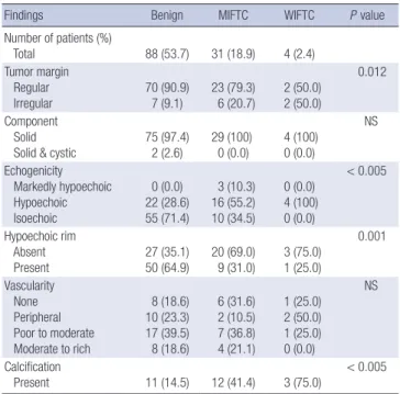
When we compared sonographical findings between groups, we found that irregular margin, low echogenicity, absence of hypoechoic rim and presence of calcification were detected more commonly in malignant thyroid nodules (Table 2).
Multivariate analysis to find predictive factors of malignancy
We also performed multivariate analysis to find predictive fac- tors of thyroid malignancy. As shown in Table 3, younger age, being male, higher Tg levels, hypoechoic nodules and the pres- ence of calcification were independent and significant risk fac- tors for FTC.
We performed ROC curve analyses to find cut-off levels of age at diagnosis, tumor size, and Tg to differentiate the benign and malignant groups. Age at diagnosis, using a cut-off value of 52.5 yr, had a sensitivity of 48.5% and a specificity of 68.2% for detecting the malignant group with an area under the curve (AUC) of 0.524 (95% confidence interval [CI] 0.404-0.645, P = 0.674), which failed to show significant differences between groups (data not shown). Mean tumor size, using a cut-off of 1.7 cm, had a sensitivity of 85.7% and a specificity of 32.3% with an AUC of 0.663 and P = 0.005 (95% CI 0.562-0.764) (data not shown). Preoperative Tg levels (AUC 0.748, 95% CI 0.634-0.861, P < 0.001) showed a sensitivity of 48.5% and a high specificity of 91.5% with cut-off value of 187.5 ng/mL (Fig. 2).
The risk of malignancy was increased in nodules more than 1.7 cm in size; especially in the case of Tg > 70 ng/mL, the odds ratio [OR] was 3.245 (95% CI 1.115-9.450, P = 0.038), and the sensitivity and specificity was 67.7% and 60.7%, respectively.
Without consideration of nodule size, the nodule with preoper- ative Tg > 100 ng/mL showed increased risk (OR 2.913, 95% CI 1.134-7.483, P = 0.029).
Table 2. Comparison of ultrasonographical findings between benign and malignant group
Findings Benign MIFTC WIFTC P value
Number of patients (%)
Total 88 (53.7) 31 (18.9) 4 (2.4)
Tumor margin Regular
Irregular 70 (90.9)
7 (9.1) 23 (79.3)
6 (20.7) 2 (50.0) 2 (50.0)
0.012
Component Solid
Solid & cystic 75 (97.4)
2 (2.6) 29 (100)
0 (0.0) 4 (100) 0 (0.0)
NS
Echogenicity Markedly hypoechoic Hypoechoic Isoechoic
0 (0.0) 22 (28.6) 55 (71.4)
3 (10.3) 16 (55.2) 10 (34.5)
0 (0.0) 4 (100) 0 (0.0)
< 0.005
Hypoechoic rim Absent Present
27 (35.1) 50 (64.9)
20 (69.0) 9 (31.0)
3 (75.0) 1 (25.0)
0.001
Vascularity None Peripheral Poor to moderate Moderate to rich
8 (18.6) 10 (23.3) 17 (39.5) 8 (18.6)
6 (31.6) 2 (10.5) 7 (36.8) 4 (21.1)
1 (25.0) 2 (50.0) 1 (25.0) 0 (0.0)
NS
Calcification
Present 11 (14.5) 12 (41.4) 3 (75.0) < 0.005
Table 3. Multivariate analysis for risk of follicular thyroid carcinoma
Variables Odds ratio 95% Confidence
interval P value
Age 0.917 0.850-0.990 0.026
Male 8.036 1.230-52.499 0.030
Nodule size 1.116 0.698-1.783 NS
TSH (mU/L) 0.804 0.410-1.575 NS
Tg (ng/mL) 1.987 1.248-3.165 0.004
TgAb (U/mL) 0.768 0.232-2.540 NS
Hashimoto’s thyroiditis 4.643 0.300-71.831 NS
Irregular margin 0.422 0.039-4.556 NS
Calcification 6.413 1.097-37.712 0.040
Hypoechogenicity 5.662 1.126-28.466 0.035
Hypoechoic rim 0.190 0.035-1.024 NS
NS, not significant. Fig. 2. ROC curve of Tg levels to predict malignancy in indeterminate nodules.
Sensitivity
1-specificity
0.0 0.2 0.4 0.6 0.8 1.0
1.0 0.8 0.6 0.4 0.2 0.0
DISCUSSION
We compared the clinical, biochemical, and ultrasonographi- cal findings between benign follicular adenomas and carcino- mas. We found that preoperative Tg cut-off levels of 187.5 ng/
mL might predict increased risk of malignancy and discrimi- nate between benign and malignant nodules (AUC 0.748, 95%
CI 0.634-0.861, P < 0.001).
Thyroid nodules are very common and have recently begun to be detected more frequently (1, 14). Ultrasonography-guided fine needle aspiration (FNA) is a widely used, cost effective, and accurate diagnostic tool for thyroid cancer. However, over 20%
of patients undergoing FNA of a thyroid nodule have indeter- minate cytology results and usually require thyroid operation for the purpose of definite diagnosis. Pathologic diagnosis re- sults in benign nodules for most patients, and if the patients are diagnosed with thyroid cancer, they should undergo comple- tion thyroidectomy (15).
The malignancy rate of indeterminate cytology was assumed to be 20%-30% (6, 14, 16), however, Granados-Garcia et al. (17) also reported high rate of malignancy (40%, 30/75). Recently, Rossi et al. (18) also reported 34.9% of malignancy risk in inde- terminate cytology even though it had conducted as a prospec- tive study. Similarly, percentage of malignancy was relatively high (46.3%, 46/164) in our study. Considering that we exclud- ed patients who did not undergo surgery, high malignancy rate may be thought to lie in acceptable range.
Lots of studies were performed to classify indeterminate nodules into benign and malignant groups. Young or old age, male gender, nodule size, family history of thyroid cancer (19), and history of benign nodule (20) are known to be associated with a high risk of malignant thyroid nodule. However, until now, there have been no studies showing any definite differen- tiating risk factor. In addition, risk factors of FTC are still not well understood except low iodine intake (21). Only a few re- cent studies such as studies of Kim et al. (9) and Sippel et al. (8) suggested tumor size and age as risk factors of Hurthle cell thy- roid carcinoma.
We explored rigorously several clinical, biochemical, and so- nographical findings; young age and male gender were inde- pendent risk factors for predicting high risk of FTC similar to previous findings (Table 3, OR, [95% CI], P value; old vs young age, 0.917 [0.850-0.990], P = 0.026; male vs female, 8.036 [1.230- 52.499], P = 0.030) (15, 16). Interestingly, preoperative Tg levels were independent risk factors of FTC (OR 1.987, 95% CI 1.248- 3.165, P = 0.004, Table 3). Nodule size, however, was not differ- ent between the benign and cancer groups after multivariate analysis (OR 1.116, 95% CI 0.698-1.783, P = 0.683) in our study.
These insignificant differences may come from characteristics of retrospective study itself; higher preference to surgery in pa- tients with larger nodules, which was the limitation of our
study.
Serum Tg levels can be elevated in most proliferative thyroid diseases and are known to be an insensitive and nonspecific test for thyroid cancer. Routine measurement of serum Tg levels for initial evaluation of thyroid nodules is not recommended (7). Our result was also compatible with previous reports in PTC or FVPTC groups; that is, for most prevalent thyroid cancer cases, preoperative Tg levels did not differ from those of benign nodules (Table 1, benign vs FVPTC vs PTC, 15.4 [1-1499] vs 9.3 [2.4-398] vs 8.8 ng/mL [0.3-181]). Recently, there were two re- ports which lead opposite conclusions about the efficacy of measurement of preoperative serum Tg; Sands et al. (22) re- ported that a combination of indeterminate cytology and pre- operative Tg ≥ 75 ng/mL increased diagnostic efficacy com- pared to indeterminate cytology alone and suggested that ele- vated preoperative Tg levels may be predictive of well-differen- tiated thyroid cancer (benign vs cancer, Tg levels; 223 vs 53 ng/
mL, P = 0.007). However, Suh et al. (23) concluded that Tg has poor accuracy for predicting malignancy in follicular or Hurthle cell neoplasms. These studies were conducted in smaller popu- lation than that of our study, and the experimental approach was different from ours.
In our study, preoperative Tg levels were significantly higher in the FTC group, and according to the severity of follicular can- cer, Tg levels increased steeply (Table 2, benign vs MIFTC vs WIFTC, Tg levels, [95% CI], P value; 15.4 [1-1,499] vs 188.0 [2.3- 7,940] vs 2,078.5 ng/mL [31.7-6,860], P < 0.001). ROC curves also showed good sensitivity and specificity for the cut-off value 187.5 ng/mL (AUC 0.748, 95% CI 0.634-0.861, P < 0.001, sensi- tivity of 48.5%, and specificity of 91.5%).
Taken together, the above results indicate that preoperative Tg levels may be one of the most useful single predictors for dis- tinguishing benign nodules from FTCs.
In summary, preoperative Tg levels had very high specificity in predicting FTC in the case of indeterminate nodules. There- fore, in practice, preoperative Tg measurement may be a very useful single tool for differentiating FTCs from benign thyroid nodules in the case of indeterminate cytology results.
REFERENCES
1. Ezzat S, Sarti DA, Cain DR, Braunstein GD. Thyroid incidentalomas.
Prevalence by palpation and ultrasonography. Arch Intern Med 1994;
154: 1838-40.
2. Rago T, Chiovato L, Aghini-Lombardi F, Grasso L, Pinchera A, Vitti P.
Non-palpable thyroid nodules in a borderline iodine-sufficient area: de- tection by ultrasonography and follow-up. J Endocrinol Invest 2001; 24:
770-6.
3. Jung KW, Park S, Kong HJ, Won YJ, Boo YK, Shin HR, Park EC, Lee JS.
Cancer statistics in Korea: incidence, mortality and survival in 2006- 2007. J Korean Med Sci 2010; 25: 1113-21.
4. Jung KW, Won YJ, Park S, Kong HJ, Sung J, Shin HR, Park EC, Lee JS.
Cancer statistics in Korea: incidence, mortality and survival in 2005. J Korean Med Sci 2009; 24: 995-1003.
5. Rosen JE, Stone MD. Contemporary diagnostic approach to the thyroid nodule. J Surg Oncol 2006; 94: 649-61.
6. Cibas ES, Ali SZ. The Bethesda System for reporting thyroid cytopatholo- gy. Thyroid 2009; 19: 1159-65.
7. Cooper DS, Doherty GM, Haugen BR, Kloos RT, Lee SL, Mandel SJ, Mazzaferri EL, McIver B, Pacini F, Schlumberger M, et al. Revised American Thyroid Association management guidelines for patients with thyroid nodules and differentiated thyroid cancer. Thyroid 2009; 19:
1167-214.
8. Sippel RS, Elaraj DM, Khanafshar E, Zarnegar R, Kebebew E, Duh QY, Clark OH. Tumor size predicts malignant potential in Hurthle cell neo- plasms of the thyroid. World J Surg 2008; 32: 702-7.
9. Kim TH, Lim JA, Ahn HY, Lee EK, Min HS, Kim WK, Choi YH, Park YJ, Park DJ, Kim KH, et al. Tumor size and age predict the risk of malignan- cy in Hurthle cell neoplasm of the thyroid and can therefore guide the ex- tent of initial thyroid surgery. Thyroid 2010; 20: 1229-34.
10. Ron E, Kleinerman RA, Boice JD Jr, LiVolsi VA, Flannery JT, Fraumeni JF Jr. A population-based case-control study of thyroid cancer. J Natl Cancer Inst 1987; 79: 1-12.
11. Bongiovanni M, Cibas ES, Faquin WC. The role of thyroid fine needle as- piration cytology and the Bethesda system for reporting thyroid cytopa- thology. Diagn Histopathol 2011; 17: 95-105.
12. Ciccone MM, De Pergola G, Porcelli MT, Scicchitano P, Caldarola P, Ia- coviello M, Pietro G, Giorgino F, Favale S. Increased carotid IMT in over- weight and obese women affected by Hashimoto’s thyroiditis: an adipos- ity and autoimmune linkage? BMC Cardiovasc Disord 2010; 10: 22.
13. Schmidt M, Voell M, Rahlff I, Dietlein M, Kobe C, Faust M, Schicha H.
Long-term follow-up of antithyroid peroxidase antibodies in patients with chronic autoimmune thyroiditis (Hashimoto’s thyroiditis) treated with levothyroxine. Thyroid 2008; 18: 755-60.
14. Gharib H, Goellner JR. Fine-needle aspiration biopsy of the thyroid: an
appraisal. Ann Intern Med 1993; 118: 282-9.
15. Baloch ZW, Fleisher S, LiVolsi VA, Gupta PK. Diagnosis of “follicular neoplasm”: a gray zone in thyroid fine-needle aspiration cytology. Diagn Cytopathol 2002; 26: 41-4.
16. Carpi A, Nicolini A, Gross MD, Fig LM, Shapiro B, Fanti S, Rampin L, Polico C, Rubello D. Controversies in diagnostic approaches to the inde- terminate follicular thyroid nodule. Biomed Pharmacother 2005; 59:
517-20.
17. Granados-Garcia M, Cortes-Flores AO, del Carmen Gonzalez-Ramirez I, Cano-Valdez AM, Flores-Hernandez L, Aguilar-Ponce JL. Follicular neoplasms of the thyroid: importance of clinical and cytological correla- tion. Cir Cir 2010; 78: 473-8.
18. Rossi M, Buratto M, Bruni S, Filieri C, Tagliati F, Trasforini G, Rossi R, Beccati MD, Degli Uberti EC, Zatelli MC. Role of ultrasonographic/clin- ical profile, cytology, and BRAF V600E mutation evaluation in thyroid nodule screening for malignancy: a prospective study. J Clin Endocrinol Metab 2012; 97: 2354-61.
19. Hemminki K, Eng C, Chen B. Familial risks for nonmedullary thyroid cancer. J Clin Endocrinol Metab 2005; 90: 5747-53.
20. Franceschi S, Preston-Martin S, Dal Maso L, Negri E, La Vecchia C, Mack WJ, McTiernan A, Kolonel L, Mark SD, Mabuchi K, et al. A pooled analysis of case-control studies of thyroid cancer. IV. Benign thyroid dis- eases. Cancer Causes Control 1999; 10: 583-95.
21. Nagataki S, Nystrom E. Epidemiology and primary prevention of thyroid cancer. Thyroid 2002; 12: 889-96.
22. Sands N, Karls S, Rivera J, Tamilia M, Hier MP, Black MJ, Gologan O, Payne RJ. Preoperative serum thyroglobulin as an adjunct to fine-needle aspiration in predicting well-differentiated thyroid cancer. J Otolaryngol Head Neck Surg 2010; 39: 669-73.
23. Suh I, Vriens MR, Guerrero MA, Griffin A, Shen WT, Duh QY, Clark OH, Kebebew E. Serum thyroglobulin is a poor diagnostic biomarker of ma- lignancy in follicular and H¨urthle-cell neoplasms of the thyroid. Am J Surg 2010; 200: 41-6.