Perinatology Vol. 29, No. 4, December, 2018 https://doi.org/10.14734/PN.2018.29.4.185
Case report
Perinatology
pISSN 2508-4887•eISSN 2508-4895
Chan Young Lee, MD1, Na Mi Lee, MD, PhD1, Dae Yong Yi, MD, PhD1, Sin Weon Yun, MD, PhD1, Soo Ahn Chae, MD, PhD1, In Seok Lim, MD, PhD1, Gwang Jun Kim, MD, PhD2 Departments of 1Pediatrics,
2Obstetrics and Gynecology, Chung-Ang University College of Medicine, Seoul, Korea
Jarcho-Levin syndrome is a congenital disorder characterized by several vertebral and costal ano- malies. Other abnormalities have also been described, including neural tube defects, Arnold-Chiari malformation, renal/urinary tract abnormalities, hydrocephalus, hydroureteronephrosis, and meningo - myelocele. We describe a spondylocostal dysplasia form of Jarcho-Levin syndrome that was prena tally diagnosed at 11 weeks of gestation and surviving. Although the patient had sporadic-type Jarcho- Levin syndrome, with normal karyotype and no family history of disease, the assessment of inheri tance patterns and genetic counseling for the parents was important to inform them about the po tential risks.
Key Words: Jarcho-Levin syndrome, Prenatal diagnosis, Scoliosis, Ultrasonography
Introduction
Jarcho-Levin syndrome was first described in 1938, and is a rare genetic disorder.1 It is characterized by short-trunk skeletal dysplasia with vertebral and rib anomalies, leading to respiratory insufficiency, and has an incidence of 1 per 40,000 births. Approximately 400 cases have been described in the worldwide literature, five of which are from Korea.2-6 None of the cases reported in Korea were diagnosed prenatally, and four cases with prenatally diagnosed Jarcho-Levin syndrome were aborted.7-10 We report a prenatally diagnosed and surviving patient with Jarcho-Levin syndrome.
Case
A 26-year-old woman (gravida 0, para 0) was referred for evaluation. She had a three- quarter pack-year smoking history, but stopped at about 12 weeks of gestation. She had moderate obesity (body mass index, 33.79 kg/m2). The maternal and paternal medical and family histories were unremarkable. Prenatal ultrasound at 11 weeks of gestation showed incomplete development of one side of a vertebral body, resulting in a wedge shape around the T11 area, and thickened nuchal translucency (3.2 mm). Targeted sonographic examination at 14 weeks of gestation demonstrated displacement and rotation of the heart into the right side of the chest, with a thoracic hemivertebra, short crown-rump length, and suspected absence of the left kidney (Fig. 1).
A female infant was born at 37+2 weeks of gestation by cesarean section (birth weight, 3,164 g). The Apgar scores were 7 and 9 at 1 and 5 minutes, respectively. The infant had a short neck, accessory nipples on the left side, and grossly abnormal left rib cage (Fig. 1). The abdomen was protuberant, and there was a single umbilical artery. At about Received: 29 June 2018
Revised: 29 August 2018 Accepted: 7 September 2018 Correspondence to Na Mi Lee, MD, PhD
Department of Pediatrics, Chung- Ang University College of Medicine, 102 Heukseok-ro, Dongjak-gu, Seoul 06973, Korea
Tel: +82-2-6299-3181 Fax: +82-2-6264-2167 E-mail: [email protected] Copyright© 2018 by The Korean Society of Perinatology
This is an Open Access article distributed under the terms of the Creative Com- mons Attribution Non-Commercial License (http://creativecommons.org/
license/by-nc/4.0/), which permits unrestricted non-commercial use, distribution, and reproduction in any medium, provided that the original work is properly cited.
Prenatally Diagnosed and Surviving Pati
ent with JarchoLevin Syndrome: Case
Report with Literature Review
Lee CY, et al. Jarcho-Levin syndrome
186
https://doi.org/10.14734/PN.2018.29.4.185www.e-kjp.org
Perinatology
2 hours after birth, the infant was intubated and began to re- ceive ventilator care owing to respiratory difficulty due to rib cage abnormality. On day 3, she briefly tolerated extubation and required supplemental oxygen; however, she required re- intubation for tachypnea, dyspnea, and desaturation on day 8.
She was extubated after administration of antibiotics for 7 days.
Chest and abdominal radiographs revealed multiple vertebral deformities with hemivertebrae, scoliosis in the thoracolumbar spine, and absence of third to seventh ribs on the left side. The heart was displaced farther right owing to the vertebral and rib defects. Dynamic 3-dimensional computed tomography (CT) of the spine revealed left T1, T2, and T10 hemivertebrae and a fusion anomaly in T6 and T7, with lateral wedging (Fig. 2).
Echocardiography showed no anomalies except for isolated dextrocardia owing to the thoracic deformity. On abdominal and pelvic CT, the left kidney was not visible in the left renal fossa and an ectopic kidney was not visible in the pelvis; thus, the contralateral right kidney had compensatory hypertrophy.
Cranial ultrasound was normal. Chromosomal studies with GTG-banding showed normal results (46,XX). No mutation was detected in the coding region of the MESP2 gene.
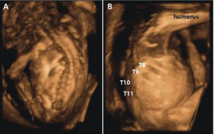
After extubation, the infant could take food well, showed no desaturation, and was discharged at 27 days of age (weight, 3,130 g). However, she was readmitted at 5 months of age because of severe cough, fever, and respiratory difficulty. She was treated for pneumonia for 16 days. The duration of treat- Fig. 1. Three-dimensional posterior image of the affected fetus at 14 weeks of
gestation. (A) Kyphoscoliosis of the spine. (B) Absence of the T10 and T11 ribs in the left lateral thoracic cage.
A B C
Fig. 2. Gross and computed tomography (CT) images of the infant. (A) The infant has a short neck, accessory nipples, and abnormal rib cage on the left side. (B) Three-dimensional spinal CT shows left T1, T2, and T10 hemivertebrae and a fusion anomaly in T6 and T7, with lateral wedging and scoliosis of the thoracic spine with convexity to the right. (C) Chest anteroposterior image shows dextrocardia and abnormal rib cage on the left side.
2018 December;29(4):185-188
www.e-kjp.org
https://doi.org/10.14734/PN.2018.29.4.185187
Perinatology
ment was longer because of the rib cage abnormality, but she was discharged without respiratory problems. She has severe scoliosis; however, neurologic examination at 4 years of age showed normal results. She is being followed by pediatric, orthopedic, and thoracic surgeons.
Discussion
Jarcho-Levin syndrome is a rare genetic disorder. Muta tions in the delta-like 3 (DLL3) gene on chromosome 19, and the LFNG, HES7, and MESP2 genes, which are important com- ponents of the notch signaling pathway, are responsible for the development of spondylocostal dysostosis in the Jarcho-Levin syndrome.11,12 Our case was sporadic, without a family history or genetic abnormality (no mutation of the MESP2 gene, with other gene tests unavailable in Korea). The mother had been smoking in the first trimester, which might have affected the development of vertebrae and ribs in the infant. The associa- tion between Jarcho-Levin syndrome and maternal smoking has not yet been reported. However, a significant positive as- sociation between maternal smoking and congenital malforma- tions has been reported in various studies. For example, ma- ternal smoking has been reported to increase the risk of mu- sculoskeletal defects in an infant by 16%.13
Jarcho-Levin syndrome reportedly presents with various cli- nical signs, with most cases being postnatally diagnosed. How- ever, prenatally diagnosed Jarcho-Levin syndrome is rare, especially in the absence of a family history. All cases of Jar- cho-Levin syndrome diagnosed during the first trimester have involved termination of pregnancy.7-10 Most cases showed in- creased fetal nuchal translucency thickness,7,10 although a case with normal thickness was also reported.8
Solomon et al.14 classified Jarcho-Levin syndrome into 2 subtypes based on survival rate, inheritance pattern, and ex- tent of skeletal anomalies, which helps to establish the progno- sis and understand this disorder. First, spondylothoracic dyso- stosis (STD), an autosomal recessive disorder associated with MESP2 gene mutation, is characterized by posterior symme- tric fusion of the ribs and defects of vertebral develop ment, resulting in a crab-like or fan-like appearance.15 There are no intrinsic rib defects. In STD, death may occur because of pul-
monary insufficiency or pneumonia.16 Second, spondylocostal dysplasia (SCD), an autosomal dominant and recessive disor- der, shows intrinsic rib anomalies such as bifurcation and broadening.14 Patients with SCD are known to have mutations in the DLL3 gene on chromosome 19. However, the karyotypes of almost all such patients are typically normal. Biochemical assays and chromosome banding studies are not available for diagnostic confirmation, and evaluation of distinctive clinical and radiological features is preferable.
The anomalies associated with Jarcho-Levin syndrome are complex congenital heart disease, and urogenital and anal anomalies. Patients with musculoskeletal anomalies are more likely to have genitourinary anomalies, because both the mus- culoskeletal and genitourinary systems have a mesodermal origin. Rai et al.17 reported that 26.7% of patients with congenital vertebral abnormalities also have genitourinary ab normalities, the most frequent being unilateral renal agenesis. The two reported patients in Korea had renal anomalies (fused kidneys and an intrathoracic kidney).4,6 In particular, rib dysplasia and hemivertebra on the left side might have been associated with additional defects of the left side in our case, involving both mesodermal origin, with renal agenesis, and ectodermal origin, with accessory nipples.
We report an SCD form of Jarcho-Levin syndrome that was prenatally diagnosed. Although the Jarcho-Levin syndrome in our case was sporadic, with normal karyotype and no family history, the assessment of the inheritance patterns and sug- gestion of genetic counseling to the parents was necessary to inform them about the potential risks. Since Jarcho-Levin syn- drome is associated with a high risk of respiratory difficulty, we recommend that an affected infant should be transferred to a hospital with a neonatal intensive care unit if prenatal ultrasonography reveals rib and vertebral defects.
Conflict of Interest
No potential conflict of interest relevant to this article was reported.
Lee CY, et al. Jarcho-Levin syndrome
188
https://doi.org/10.14734/PN.2018.29.4.185www.e-kjp.org
Perinatology
Acknowledgments
This case was reviewed and approved by the Institutional Review Board of Chung-Ang University (IRB No. 1707-015- 16086).
References
1) Jarcho S. Hereditary malformation of the vertebral bodies. Bull Johns Hopkins Hosp 1938;62:216-26.
2) Park Y, Gong G, Choe G, Yu E, Kim KS, Lee I. Jarcho-levin syndrome--a report of an autopsy case with cytogenetic analysis. J Korean Med Sci 1993;8:471-5.
3) Lee WJ, Lee BK, Cho YS, Park MH, Hoon RJ, Oh KY, et al. Prenatal dia- gnosis of spondylothoracic dysplasia (Jarcho-levin syndrome) by ultra- sound. Korean J Obstet Gynecol 2002;45:2075-80.
4) Byun SY, Sung MH, Choi JM, Kim TH, Hwang KG, Jung JA. A case of Jarcho-levin syndrome with intrathoracic kidney. Korean J Pediatr 2004;
47:1225-7.
5) Park WH, Choi SO, Lee HJ. Jarcho-levin syndrome associated with im- perforate anus and thoracoabdominal wall hernia. J Korean Surg Soc 2007;73:188-90.
6) Kim JY, Hwang SJ, Lee SM, Oh JW, Yum MK, Kim CR. A case of Jarcho- levin syndrome with fusion of both kidneys in a newborn infant. J Korean Soc Neonatol 2008;15:84-8.
7) Kauffmann E, Roman H, Barau G, Dumas H, Laffitte A, Fourmaintraux A, et al. Case report: a prenatal case of Jarcho-levin syndrome diagnosed during the first trimester of pregnancy. Prenat Diagn 2003;23:163-5.
8) Dane C, Yayla M, Dane B. Prenatal diagnosis of Jarcho-levin syndrome in the first trimester. Gynecol Obstet Invest 2007;63:200-2.
9) Eliyahu S, Weiner E, Lahav D, Shalev E. Early sonographic diagnosis of Jarcho-levin syndrome: a prospective screening program in one family.
Ultrasound Obstet Gynecol 1997;9:314-8.
10) Hull AD, James G, Pretorius DH. Detection of Jarcho-levin syndrome at 12 weeks' gestation by nuchal translucency screening and three- dimensional ultrasound. Prenat Diagn 2001;21:390-4.
11) Kansal R, Mahore A, Kukreja S. Jarcho-levin syndrome with diastemato- myelia: a case report and review of literature. J Pediatr Neurosci 2011;
6:141-3.
12) Kurup PM, Tanigasalam V, Bhat BV. Spondylocostal dysostosis (Jarcho levin syndrome). Indian J Pediatr 2018;85:486.
13) Hackshaw A, Rodeck C, Boniface S. Maternal smoking in pregnancy and birth defects: a systematic review based on 173 687 malformed cases and 11.7 million controls. Hum Reprod Update 2011;17:589-604.
14) Solomon L, Jimenez RB, Reiner L. Spondylothoracic dysostosis: report of two cases and review of the literature. Arch Pathol Lab Med 1978;
102:201-5.
15) Berdon WE, Lampl BS, Cornier AS, Ramirez N, Turnpenny PD, Vitale MG, et al. Clinical and radiological distinction between spondylothoracic dysostosis (Lavy-Moseley syndrome) and spondylocostal dysostosis (Jarcho-levin syndrome). Pediatr Radiol 2011;41:384-8.
16) Cornier AS, Ramírez N, Arroyo S, Acevedo J, García L, Carlo S, et al.
Pheno type characterization and natural history of spondylothoracic dysplasia syndrome: a series of 27 new cases. Am J Med Genet A 2004;
128A:120-6.
17) Rai AS, Taylor TK, Smith GH, Cumming RG, Plunkett-Cole M. Congenital abnormalities of the urogenital tract in association with congenital vertebral malformations. J Bone Joint Sur Br 2002;84:891-5.