INTRODUCTION
After introduction of helical computed tomography (CT) in the late 1980s, imaging of the aorta has become an accepted and widely used procedure for the evaluation of patients with aortic dissection, stenosis, or aneurysm formation (1, 2). Aortic aneu- rysm is a common, potentially lethal, but treatable disease, par- ticularly if detected before dissection or rupture. Recently, the incidence of thoracic aortic aneurysms has been estimated to be increasing and there are around 10.4 cases per 100000 person- years (3). According to the American College of Cardiology Foundation/American Heart Association guidelines, for pa- tients with isolated aortic arch aneurysms between 3.5-4.4 cm in
diameter, it is reasonable to reimage using computed tomo- graphic imaging or magnetic resonance imaging at 12-month intervals to detect enlargement of the aneurysm. And for pa- tients with degenerative or traumatic aneurysms of the descend- ing thoracic aorta exceeding 5.5 cm, saccular aneurysms, or postoperative pseudoaneurysms, endovascular stent grafting should be strongly considered when feasible (4). Accurate assess- ment of aortic size is a key component in the detection of aneu- rysms and in guiding therapeutic decisions. CT has evolved to be the mainstay of evaluation owing to its accuracy and reproduc- ibility, as well as its speed, simplicity, and true 3-dimensional ca- pabilities. In spite of the pivotal role of CT in aortic evaluation, only limited measurements of the aorta have been published (5-
J Korean Soc Radiol 2013;69(2):105-112 http://dx.doi.org/10.3348/jksr.2013.69.2.105
Received April 8, 2013; Accepted May 3, 2013 Corresponding author: Whal Lee, MD
Department of Radiology, Seoul National University College of Medicine, 101 Daehak-ro, Jongno-gu, Seoul 110-744, Korea.
Tel. 82-2-2072-2584 Fax. 82-2-743-6385 E-mail: [email protected]
This is an Open Access article distributed under the terms of the Creative Commons Attribution Non-Commercial License (http://creativecommons.org/licenses/by-nc/3.0) which permits unrestricted non-commercial use, distri- bution, and reproduction in any medium, provided the original work is properly cited.
Purpose: To determine normal reference values for aortic diameters in asymptom- atic Korean adults.
Materials and Methods: Three hundred adults without signs or symptoms of car- diovascular diseases were enrolled in this study. Aortic diameters were measured at nine predetermined levels on CT images. Aortic diameter measurements were ad- justed for body surface area. Analysis of data was performed with regard to age, sex, weight, height and hypertension.
Results: Aortic diameters were 2.99 ± 0.57 cm at the ascending aorta, 2.54 ± 0.35 cm at the transverse aortic arch, 2.36 ± 0.35 cm at the proximal descending thorac- ic aorta (DTA), 2.23 ± 0.37 cm at the mid DTA, 2.17 ± 0.38 cm at the distal DTA, 2.16
± 0.37 cm at the thoracoabdominal junction, 2.10 ± 0.35 cm at the level of the celi- ac axis, 1.94 ± 0.36 cm at the suprarenal aorta, 1.58 ± 0.24 cm at the aortic bifurca- tion. Men had slightly larger diameters than women (p < 0.05). All diameters in- creased with age and hypertension, with statistical significance (p < 0.01). And all aortic diameters increased with height (p < 0.05) except at the level of the aortic arch (p = 0.056), and increased with weight (p < 0.05) except at the level of the su- prarenal aorta (p = 0.067).
Conclusion: Male sex, higher weight and height, age and hypertension are associ- ated with larger aortic diameters in asymptomatic Korean adults.
Index terms Aorta
Normal Reference Computed Tomography
Measurement of the Aortic Diameter in the Asymptomatic Korean Population: Assessment with Multidetector CT
나선형 전산화단층촬영에서 측정한 무증상 한국 성인의 정상 대동맥 직경
Sang Hwan Lee, MD, Whal Lee, MD, Hyuck Jae Choi, MD, Dae Jin Kim, MD, Eun-Ah Park, MD, Jin Wook Chung, MD, Jae Hyung Park, MD
Department of Radiology, Seoul National University College of Medicine and Institute of Radiation Medicine, SNUMRC (Seoul National University Medical Research Center), Clinical Research Institute, Seoul National University Hospital, Seoul, Korea
proved this study (R-0603-188-170).
Image Acquisition
Single slice CT examination was performed using Somatom Plus-4 scanner (Siemens Medical System, Erlangen, Germany).
All patients fasted for 8 hours or longer prior to the examina- tion. The postcontrast scan was started 60-seconds after starting the intravenous injection of contrast medium that contained 300 mg I/mL iopromide (Ultravist 370®, Bayer Healthcare, Ber- lin, Germany) in a total volume of 120 mL; this was given via the antecubital vein of the upper extremity at a rate of 3 mL/sec.
Measurement parameters included 10 mm/sec table speed, 5 mm thickness, 120 kVp, and 220 mA. The scan levels ranged from the trifurcation level of the aortic arch to the proximal por- tion of the aortic bifurcation. Helical acquisitions were obtained with one or two breath-holds. Aorta-focused reconstruction with 16 × 16 cm field of view was achieved every 5 mm with the 180° linear interpolation algorithm.
Measurements
The aortic diameters were measured at the following nine ana- tomic levels of the aorta: 1) ascending at the middle level of the right main pulmonary artery, 2) transverse aortic arch, 3) proxi- mal descending thoracic aorta (DTA) at the middle level of the left main pulmonary artery, 4) mid DTA at the level of the mitral valve, 5) distal DTA at the top of the diaphragmatic level, 6) tho- racoabdominal junction, 7) celiac axis, 8) suprarenal aorta just above the orifices of the renal arteries, and 9) aortic bifurcation (Fig. 1). The measurement of the maximal external aortic diame- ter was made on the picture archiving and communication sys- tem (Marotec, Seoul, Korea). On transverse images, the shortest 14). To distinguish the normal from the enlarged aorta, it is nec-
essary to standardize the values of “normal” aortic dimensions.
But, to our knowledge, no publication up until now has reported on these aortic measurements in a population of Korean adults.
The purposes of this study were to establish reference values of the aorta obtained by helical CT in asymptomatic Korean adults and to analyze the relationship between these values and sex, weight, height, age and hypertension.
MATERIALS AND METHODS
Patients
Aortic diameters were measured prospectively in 300 Korean adults who were scheduled to undergo a CT for a variety of non-vascular clinical problems. The subjects agreed to undergo an extension of their portal phase scan range to cover the entire aorta for participation in this study. The reasons for CT exami- nation of the patients included malignant neoplasm (n = 197), benign neoplasm (n = 28), infectious disease (n = 25), inflam- matory disease (n = 24), routine check-up (n = 21), and autoim- mune disease (n = 5). Patients were excluded if they had the fol- lowing: signs or symptoms of cardiovascular disease, paraaortic disease or obvious aortic disease, such as aneurysm, thrombus or dissection. And we excluded patients with obvious athero- sclerotic plaque on CT in the patient group. In the total patient group, risk factors of atherosclerosis such as smoking and diabe- tes mellitus were 9.7% and 9%, respectively. The total of 300 pa- tients consisted of 6 age groups [age groups 21-30, 31-40, 41-50, 51-60, 61-70, and 71-80, each group with 25 males and 25 fe- males (Table 1)]. Informed consent was obtained from each subject and the Institutional Review Board of our institute ap- Table 1. Demographic Data
Female Male Total
No. 150 150 300
Age (y) (mean ± SD) 50.9 ± 16.6 50.8 ± 16.9 50.6 ± 16.7
Weight (kg) (mean ± SD) 55.4 ± 9.0 66.5 ± 10.8 60.9 ± 11.4
Height (cm) (mean ± SD) 163.8 ± 9.1 172.3 ± 2.5 168.1 ± 6.0
Adjusted body surface area (m2) (mean ± SD) 1.51 ± 0.1 1.76 ± 0.1 1.63 ± 0.2
Cholesterol (mg/dL) (mean ± SD) 177.9 ± 35.1 171.0 ± 36.8 174.5 ± 36.0
Blood pressure (mm Hg) (mean) 131.2/84.0 127.1/83.1 129.2/83.6
Hypertension (number of patients) (%) 62 (41.3) 53 (35.3) 115 (38.3)
Diabetes mellitus (number of patients) (%) 14 (9.3) 13 (8.6) 27 (9)
Smoker (number of patients) (%) 3 (2) 26 (17.3) 29 (9.7)
Note.-SD = standard deviation
Statistical Analysis
Measurements were stored in a database and exported to a statistical software package (SPSS, ver 17; SPSS Inc., Chicago, IL, USA) for analysis. A normal distribution of diameters was as- sumed. Adjusted body surface area (BSA) was evaluated for ad- justing the aortic diameter. Analysis was performed to test for influence of the following factors on the aortic diameter: sex, age, weight, height, and hypertension. Variables that showed an influence were analyzed in detail using simple linear regression for age, analysis of covariance (ANCOVA) adjusted by weight, height, age and hypertension with regard to the influence of sex, diameter of the aorta at a predetermined level was measured to
avoid overestimation from the non-perpendicular ovoid cross- section of the aorta. The aortic diameter was measured from the outer edge of the wall to the outer edge of the opposite wall, per- pendicular to the axis of rotation of the aorta. Wherever possible, magnified images were used in order to monitor the scanning and to reduce operator errors. All images were measured by an experienced radiologist (DJK). The interobserver reliability was evaluated using the intraclass correlation coefficient (ICC) in 30 patients by two experienced radiologists (DJK and LSH). We list- ed the measured aortic diameter (Fig. 2).
A B
Fig. 1. Diagram of aorta with the levels at which the diameters were measured.
A. The curved multi-planar reconstruction image shows: 1) ascending at the mid level of the right main pulmonary artery; 2) transverse aortic arch; 3) proximal descending thoracic aorta (DTA) at the mid level of the left main pulmonary artery; 4) mid DTA at the level of the mitral valve; 5) distal DTA at the top of the diaphragmatic level.
B. The maximum intensity projection image shows: 6) thoracoabdominal junction; 7) celiac axis; 8) suprarenal aorta just above the orifices of the renal arteries; 9) aortic bifurcation.
Fig. 2. Mean aortic diameters of both genders at various levels by helical CT in 300 adults.
Note.-AA = ascending aorta, Ao = aorta, TA = thoracic aorta 0
1 2 3
Diameter (cm)
Asc Ao AA Prox. TA Mid. TA Dista. TA Subdiaph. Ao Celiac trunk Suprarenal Bifurcation
Level
Female Male 1
2
3
4
5
6 7 8
9
0.35 cm at the transverse aortic arch, 2.36 ± 0.35 cm at the proxi- mal DTA, 2.23 ± 0.37 cm at the mid DTA, 2.17 ± 0.38 cm at the distal DTA, 2.16 ± 0.37 cm at the thoracoabdominal junction, 2.10 ± 0.35 cm at the level of the celiac axis, 1.94 ± 0.36 cm at the suprarenal aorta, and 1.58 ± 0.24 cm at the aortic bifurcation (Ta- ble 2). The mean aortic diameters of both genders at various lev- els are shown (Fig. 3). A good interobserver agreement of the av- erage measures (ICC, 0.957; 95% confidence interval, 0.930 to 0.977) was present in 30 patients.
ANCOVA adjusted by age, sex, weight and height with regard to the influence of hypertension, and partial correlation coefficient adjusted by age, sex and hypertension with regard to the influ- ence of weight and height.
RESULTS
Aortic diameters (mean ± standard deviation) had the follow- ing measurements: 2.99 ± 0.57 cm at the ascending aorta, 2.54 ±
Table 2. Measured Aortic Diameters and Body Surface Area-Adjusted Aortic Diameters of Nine Different Levels on Helical CT in 300 Adults
Measured Aortic Diameters Female (n = 150) Male (n = 150) Total (n = 300) p Value
Ascending aorta 2.92 ± 0.56 3.06 ± 0.58 2.99 ± 0.57 0.036
Transverse aortic arch 2.49 ± 0.35 2.59 ± 0.36 2.54 ± 0.35 0.015
Proximal thoracic aorta 2.27 ± 0.31 2.46 ± 0.36 2.36 ± 0.35 0.000
Middle thoracic aorta 2.12 ± 0.35 2.34 ± 0.36 2.23 ± 0.37 0.000
Distal thoracic aorta 2.07 ± 0.35 2.27 ± 0.38 2.17 ± 0.38 0.000
Thoracoabdominal junction 2.05 ± 0.35 2.28 ± 0.36 2.16 ± 0.37 0.000
Celiac trunk 1.98 ± 0.31 2.21 ± 0.36 2.10 ± 0.35 0.000
Suprarenal 1.84 ± 0.39 2.04 ± 0.31 1.94 ± 0.36 0.000
Bifurcation 1.47 ± 0.22 1.68 ± 0.22 1.58 ± 0.24 0.000
Body Surface Area-Adjusted Aortic Diameters Female (n = 150) Male (n = 150) Total (n = 300) p Value
Ascending aorta 1.95 ± 0.41 1.74 ± 0.37 1.84 ± 0.41 0.000
Transverse aortic arch 1.66 ± 0.27 1.47 ± 0.23 1.57 ± 0.27 0.000
Proximal thoracic aorta 1.51 ± 0.23 1.40 ± 0.23 1.46 ± 0.23 0.000
Middle thoracic aorta 1.41 ± 0.24 1.33 ± 0.24 1.37 ± 0.24 0.008
Distal thoracic aorta 1.38 ± 0.25 1.29 ± 0.25 1.34 ± 0.25 0.003
Thoracoabdominal junction 1.37 ± 0.26 1.30 ± 0.24 1.33 ± 0.25 0.015
Celiac trunk 1.32 ± 0.22 1.26 ± 0.23 1.29 ± 0.23 0.017
Suprarenal 1.23 ± 0.28 1.16 ± 0.20 1.19 ± 0.24 0.015
Bifurcation 0.98 ± 0.14 0.95 ± 0.13 0.97 ± 0.14 0.124
Note.-Measurements are expressed as mean ± SD in centimeters. Body surface area-adjusted measurements are expressed as mean ± SD in cm/m2. SD = standard deviation
Fig. 3. Mean aortic diameters at the ascending aorta (A), the distal thoracic aorta (B) and aortic bifurcation (C) derived from helical CT mea- surements in 300 adults 21-80 years old.
0.0 0.0 0.0
0.5 0.5 0.5
1.0 1.0 1.0
1.5 1.5 1.5
2.0 2.0 2.0
2.5 2.5 2.5
3.0 3.0 3.0
3.5 3.5 3.5
4.0 4.0 4.0
Ascending aorta (cm) Distal thoracic aorta (cm) Aortic bifurcation (cm)
20s 30s 40s 50s 60s 70s 20s 30s 40s 50s 60s 70s 20s 30s 40s 50s 60s 70s
Age (years) Age (years) Age (years)
Gender Female Male
Gender Female Male
Gender Female Male
A B C
an important independent determinant of cardiovascular mor- bidity and mortality (21-23). The aorta is subject to constant pulsatile stress, so that the elastic components of the aortic me- dia fragment and eventually break down to be partially replaced by mostly fibrotic nonelastic tissue (24). These histological pro- cesses lead to stiffening of the aortic wall and increased mean aortic blood pressure, and finally to transverse dilation of the aorta. And we found that women’s aortic diameters were mean- ingfully bigger than men’s in terms of the ascending aorta and aortic arch level, while the opposite was true from the proximal descending thoracic aorta to the aortic bifurcation, when adjust- ed by weight, height, age and hypertension with regard to the influence of sex. Previous studies have shown that there is no difference between men and women in cerebral blood flow (25, 26), while others have suggested a higher cerebral blood flow in women (27-30). We have not yet found the reason why woman’s ascending aorta and aortic arch have larger diameters while men’s aortas shows a larger diameter at the level of the proximal descending thoracic aorta to below, and so further study is re- quired to investigate this.
The influence of weight, and height on aortic dimensions in adults was apparent in this study. Previous studies have shown that weight and height have a significant bearing on the aortic diameter (15, 16, 19). We speculate that increased peripheral vascular resistance is related to both weight gain and increase in aortic diameter. Blood pressure is well recognized for its effect on the aortic diameter. Previous studies have shown that hyper- tension has a significant bearing on the aortic diameter (19, 20).
According to a German study by Hager et al. (10), the mean aortic diameters were as follows: at the ascending aorta, 3.09 cm in Germans, and 2.99 cm in Korean; at the transverse arch, 2.77 Aortic diameters and BSA adjusted aortic diameters decreased
continuously from the ascending aorta to the bifurcation level.
Men had slightly larger aortic diameters than women (p < 0.05).
Women had slightly larger BSA-adjusted aortic diameters than men (p < 0.05), but the difference was not statistically significant at the level of the aortic bifurcation (p = 0.124). Women’s aortic diameters were bigger than men’s in terms of the ascending aorta and aortic arch level, while the opposite was true for the aorta be- tween the proximal descending thoracic aorta and the aortic bi- furcation (p < 0.01), when adjusted by age, hypertension, height and weight. All aortic diameters increased with height (p < 0.05), except at the level of the aortic arch (p = 0.056), and all aortic di- ameters increased with weight (p < 0.05), except at the level of the suprarenal aorta (p = 0.067).
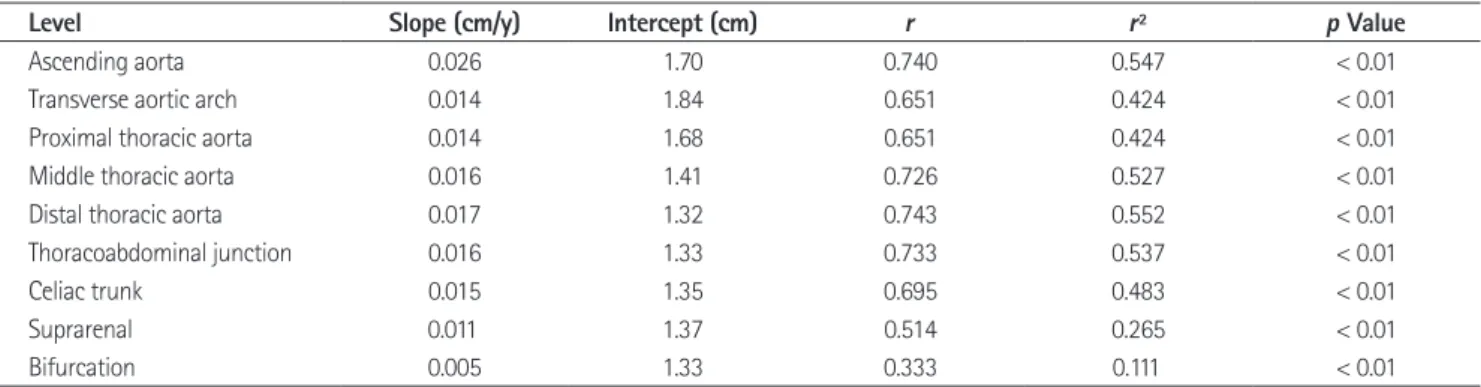
All diameters increased with hypertension when adjusted by sex, age, height, and weight (p < 0.01). Age as an influence was also analyzed in detail by simple linear regression analysis (Table 3). There was a significant increase of aortic diameter at all levels throughout adult life (p < 0.01).
DISCUSSION
In this study, we showed that aortic diameters in adults vary with sex, weight, height, age, and hypertension. This study match- es with the study of Hager et al. (10), which showed that the aor- tic diameter increased about 1 mm per decade during adult- hood.
Previous studies have shown that age and gender have a sig- nificant bearing on the aortic diameter (7, 15-18). Dixon et al.
(9) concluded that aortic dilatation is part of the natural aging process. Age-related arterial function change is considered to be
Table 3. Simple Linear Regression Analysis of the Influence of Age on Aortic Diameter at Nine Different Levels
Level Slope (cm/y) Intercept (cm) r r2 p Value
Ascending aorta 0.026 1.70 0.740 0.547 < 0.01
Transverse aortic arch 0.014 1.84 0.651 0.424 < 0.01
Proximal thoracic aorta 0.014 1.68 0.651 0.424 < 0.01
Middle thoracic aorta 0.016 1.41 0.726 0.527 < 0.01
Distal thoracic aorta 0.017 1.32 0.743 0.552 < 0.01
Thoracoabdominal junction 0.016 1.33 0.733 0.537 < 0.01
Celiac trunk 0.015 1.35 0.695 0.483 < 0.01
Suprarenal 0.011 1.37 0.514 0.265 < 0.01
Bifurcation 0.005 1.33 0.333 0.111 < 0.01
Note.-The slope describes increasing diameters with age.
Lutterbey G, et al. Aortic dissection: a comparative study of diagnosis with spiral CT, multiplanar transesophageal echocardiography, and MR imaging. Radiology 1996;199:
347-352
3. Clouse WD, Hallett JW Jr, Schaff HV, Gayari MM, Ilstrup DM, Melton LJ 3rd. Improved prognosis of thoracic aortic aneurysms: a population-based study. JAMA 1998;280:
1926-1929
4. Hiratzka LF, Bakris GL, Beckman JA, Bersin RM, Carr VF, Casey DE Jr, et al. 2010 ACCF/AHA/AATS/ACR/ASA/SCA/
SCAI/SIR/STS/SVM guidelines for the diagnosis and man- agement of patients with Thoracic Aortic Disease: a report of the American College of Cardiology Foundation/Ameri- can Heart Association Task Force on Practice Guidelines, American Association for Thoracic Surgery, American Col- lege of Radiology, American Stroke Association, Society of Cardiovascular Anesthesiologists, Society for Cardiovascular Angiography and Interventions, Society of Interventional Radiology, Society of Thoracic Surgeons, and Society for Vascular Medicine. Circulation 2010;121:e266-e369
5. Lu TL, Huber CH, Rizzo E, Dehmeshki J, von Segesser LK, Qanadli SD. Ascending aorta measurements as assessed by ECG-gated multi-detector computed tomography: a pilot study to establish normative values for transcatheter therapies. Eur Radiol 2009;19:664-669
6. Guthaner DF, Wexler L, Harell G. CT demonstration of car- diac structures. AJR Am J Roentgenol 1979;133:75-81 7. Aronberg DJ, Glazer HS, Madsen K, Sagel SS. Normal tho-
racic aortic diameters by computed tomography. J Com- put Assist Tomogr 1984;8:247-250
8. Horejs D, Gilbert PM, Burstein S, Vogelzang RL. Normal aortoiliac diameters by CT. J Comput Assist Tomogr 1988;
12:602-603
9. Dixon AK, Lawrence JP, Mitchell JR. Age-related changes in the abdominal aorta shown by computed tomography.
Clin Radiol 1984;35:33-37
10. Hager A, Kaemmerer H, Rapp-Bernhardt U, Blücher S, Rapp K, Bernhardt TM, et al. Diameters of the thoracic aorta throughout life as measured with helical computed tomog- raphy. J Thorac Cardiovasc Surg 2002;123:1060-1066 11. Wolak A, Gransar H, Thomson LE, Friedman JD, Hachamo-
vitch R, Gutstein A, et al. Aortic size assessment by non- cm in Germans, and 2.54 cm in Koreans; and at the proximal
DTA, 2.47 cm in Germans, and 2.36 cm in Korean. The median age was 50.2 years in Germans, and 50.6 years in Koreans; mean height was 172.4 cm in Germans, and 168.1 cm in Koreans; and mean weight was 73.1 kg in Germans and 60.9 kg in Koreans.
The German people have a larger aortic diameter than Koreans.
Considering that the two groups have almost the same median age, weight and height play an important role in explaining the aortic diameter differences.
The limitation of our study is the use of data from non-gated helical CT scans. In order to establish more solid normative ta- bles, electrocardiography (ECG)-gated multidetector CT (MDCT) measurements are needed. ECG-gated MDCT pro- vides high resolution images in near isotropic conditions (31).
The major difference in the diameter was at the level of the as- cending aorta. In two studies with non-gated CT in adult pa- tients, the diameter of the aortic sinus measured between 29.8 and 36.2 mm, and the diameter of the ascending aorta mea- sured between 30.9 and 35.1 mm (7, 10). A Dutch group used gated CT to measure the distance between the aortic valve and the right brachiocephalic artery in 14 patients (32). Their results ranged from 72 to 99 mm. This group also showed that motion and stress forces in the ascending aorta are higher than in the abdominal aorta, with a maximum difference of diameter of up to 27.5% during the cardiac cycle.
Nevertheless, considering that we examined a relatively larger population of up to 300 patients, we expect the mean of the mea- sured values to effectively reflect the true mean value, minimiz- ing possible errors arising from non-gated CT.
And this study is the first step in determining normal refer- ence values for the aorta diameter of Korean adults.
This study reemphasizes that aortic dilatation is a part of the natural aging process. The CT measurement of the diameter of the normal aorta for differing genders and age may prove useful when assessing the abnormal state in a variety of disease pro- cesses.
REFERENCES
1. Trerotola SO. Can helical CT replace aortography in tho- racic trauma. Radiology 1995;197:13-15
2. Sommer T, Fehske W, Holzknecht N, Smekal AV, Keller E,
applications. Eur Heart J 2006;27:2588-2605
22. Mitchell GF, Hwang SJ, Vasan RS, Larson MG, Pencina MJ, Hamburg NM, et al. Arterial stiffness and cardiovascular events: the Framingham Heart Study. Circulation 2010;
121:505-511
23. Vlachopoulos C, Aznaouridis K, Stefanadis C. Prediction of cardiovascular events and all-cause mortality with arterial stiffness: a systematic review and meta-analysis. J Am Coll Cardiol 2010;55:1318-1327
24. O’Rourke MF, Hashimoto J. Mechanical factors in arterial aging: a clinical perspective. J Am Coll Cardiol 2007;50:1-13 25. ILSI Risk Science Institute Working Group. Physiological Parameter Values for PBPK Models. Washington D.C.: ILSI, 1994:40-66
26. Moon DH, Lee HK, Song HC, Lee J, Bom HS, Sohn HK, et al.
Change of cerebral blood flow distribution and vascular reserve according to age in Koreans measured by Tc-99m HMPAO brain SPECT. Korean J Nucl Med 1999;33:247-261 27. Cosgrove KP, Mazure CM, Staley JK. Evolving knowledge
of sex differences in brain structure, function, and chem- istry. Biol Psychiatry 2007;62:847-855
28. Hatazawa J, Iida H, Shimosegawa E, Sato T, Murakami M, Miura Y. Regional cerebral blood flow measurement with iodine-123-IMP autoradiography: normal values, reproduc- ibility and sensitivity to hypoperfusion. J Nucl Med 1997;38:
1102-1108
29. Gur RE, Gur RC. Gender differences in regional cerebral blood flow. Schizophr Bull 1990;16:247-254
30. Esposito G, Van Horn JD, Weinberger DR, Berman KF. Gen- der differences in cerebral blood flow as a function of cognitive state with PET. J Nucl Med 1996;37:559-564 31. Horiguchi J, Kiguchi M, Fujioka C, Shen Y, Arie R, Sunasaka
K, et al. Radiation dose, image quality, stenosis measure- ment, and CT densitometry using ECG-triggered coronary 64-MDCT angiography: a phantom study. AJR Am J Roent- genol 2008;190:315-320
32. van Prehn J, Vincken KL, Muhs BE, Barwegen GK, Bartels LW, Prokop M, et al. Toward endografting of the ascend- ing aorta: insight into dynamics using dynamic cine-CTA.
J Endovasc Ther 2007;14:551-560 contrast cardiac computed tomography: normal limits by
age, gender, and body surface area. JACC Cardiovasc Im- aging 2008;1:200-209
12. Mao SS, Ahmadi N, Shah B, Beckmann D, Chen A, Ngo L, et al. Normal thoracic aorta diameter on cardiac comput- ed tomography in healthy asymptomatic adults: impact of age and gender. Acad Radiol 2008;15:827-834
13. Lin FY, Devereux RB, Roman MJ, Meng J, Jow VM, Jacobs A, et al. Assessment of the thoracic aorta by multidetector computed tomography: age- and sex-specific reference values in adults without evident cardiovascular disease. J Cardiovasc Comput Tomogr 2008;2:298-308
14. Euathrongchit J, Deesuwan P, Kuanprasert S, Woragitpoo- pol S. Normal thoracic aortic diameter in Thai people by multidetector computed tomography. J Med Assoc Thai 2009;92:236-242
15. Vasan RS, Larson MG, Levy D. Determinants of echocardio- graphic aortic root size. The Framingham Heart Study. Cir- culation 1995;91:734-740
16. Roman MJ, Devereux RB, Kramer-Fox R, O’Loughlin J.
Two-dimensional echocardiographic aortic root dimen- sions in normal children and adults. Am J Cardiol 1989;64:
507-512
17. Cohen GI, White M, Sochowski RA, Klein AL, Bridge PD, Stewart WJ, et al. Reference values for normal adult trans- esophageal echocardiographic measurements. J Am Soc Echocardiogr 1995;8:221-230
18. Reed CM, Richey PA, Pulliam DA, Somes GW, Alpert BS.
Aortic dimensions in tall men and women. Am J Cardiol 1993;71:608-610
19. Kim M, Roman MJ, Cavallini MC, Schwartz JE, Pickering TG, Devereux RB. Effect of hypertension on aortic root size and prevalence of aortic regurgitation. Hypertension 1996;28:
47-52
20. Jakrapanichakul D, Chirakarnjanakorn S. Comparison of aortic diameter in normal subjects and patients with sys- temic hypertension. J Med Assoc Thai 2011;94 Suppl 1:S51- S56
21. Laurent S, Cockcroft J, Van Bortel L, Boutouyrie P, Gi- annattasio C, Hayoz D, et al. Expert consensus document on arterial stiffness: methodological issues and clinical
나선형 전산화단층촬영에서 측정한 무증상 한국 성인의 정상 대동맥 직경
이상환 · 이 활 · 최혁재 · 김대진 · 박은아 · 정진욱 · 박재형
목적: 무증상 한국 남녀 성인 대동맥 직경의 정상 참조치를 측정하고자 하였다.
대상과 방법: 심혈관 질환의 징후나 증상이 없는 남녀 성인 300명이 연구 대상에 포함되었다. 이들의 나선형 전산화단층 촬영 사진에서 사전에 정해진 9개 레벨에서 대동맥 직경을 측정하였고, 또한 체표면적으로 보정한 대동맥 직경도 평가하 였다. 그리고 대동맥 직경 데이터와 연령, 성별, 몸무게, 키, 고혈압과의 관계에 대해 분석하였다.
결과: 남녀 성인의 대동맥 평균 직경은 각 부위에서 다음과 같았다. 상행대동맥(2.99 ± 0.57 cm), 대동맥궁(2.54 ± 0.35 cm), 근위부 흉부대동맥(2.36 ± 0.35 cm), 중간부 흉부대동맥(2.23 ± 0.37 cm), 원위부 흉부대동맥(2.17 ± 0.38 cm), 가슴배연결부(2.16 ± 0.37 cm), 복강축(2.10 ± 0.35 cm), 콩팥위대동맥(1.94 ± 0.36 cm), 대동맥분기 (1.58 ± 0.24 cm). 대동맥 직경은 전체 부위에서 남성의 평균 직경이 여성에 비해 더 컸으며, 연령과 혈압이 증가함에 따 라 통계적으로 유의하게 증가하였다. 대동맥궁 부위를 제외하고 키가 클수록 각 부위에서 대동맥 직경이 통계적으로 유 의하게 증가하였다. 또한 콩팥위대동맥 부위를 제외하고 몸무게가 무거울수록 각 부위에서 대동맥 직경이 통계적으로 유 의하게 증가하였다.
결론: 한국 성인의 대동맥 직경은 남성, 고혈압, 그리고 연령, 몸무게, 키가 증가할수록 통계적으로 유의하게 크다.
서울대학교 의과대학 영상의학과, 서울대학교 의학연구원 방사선의학연구소, 서울대학교병원 임상의학연구원