Effects of propofol-induced autophagy against oxidative stress in human osteoblasts
Eun-Jung Kim
1, In-Seok Choi
1, Ji-Young Yoon
1, Bong-Soo Park
2, Ji-Uk Yoon
3, Cheul-Hong Kim
11Department of Dental Anesthesia and Pain Medicine, School of Dentistry, Pusan National University, Dental Research Institute,
2Department of Oral Anatomy, School of Dentistry, Pusan National University, 3Department of Anesthesia and Pain Medicine, School of Medicine, Pusan National University, Gyeongnam, Korea
Background: Oxidative stress occurs during the aging process and other conditions such as bone fracture, bone diseases, and osteoporosis, but the role of oxidative stress in bone remodeling is unknown. Propofol exerts antioxidant effects, but the mechanisms of propofol preconditioning on oxidative stress have not been fully explained. Therefore, the aim of this study was to evaluate the protective effects of propofol against H2O2–induced oxidative stress on a human fetal osteoblast (hFOB) cell line via activation of autophagy.
Methods: Cells were randomly divided into the following groups: control cells were incubated in normoxia (5% CO2, 21% O2, and 74% N2) without propofol. Hydrogen peroxide (H2O2) group cells were exposed to H2O2 (200 μM) for 2 h, propofol preconditioning (PPC)/H2O2 group cells were pretreated with propofol then exposed to H2O2, 3-methyladenine (3-MA)/PPC/H2O2 cells were pretreated with 3-MA (1 mM) and propofol, then were exposed to H2O2. Cell viability and apoptosis were evaluated. Osteoblast maturation was determined by assaying bone nodular mineralization. Expression levels of bone related proteins were determined by western blot.
Results: Cell viability and bone nodular mineralization were decreased significantly by H2O2, and this effect was rescued by propofol preconditioning. Propofol preconditioning effectively decreased H2O2-induced hFOB cell apoptosis. However, pretreatment with 3-MA inhibited the protective effect of propofol. In western blot analysis, propofol preconditioning increased protein levels of collagen type I, BMP-2, osterix, and TGF-β1.
Conclusions: This study suggests that propofol preconditioning has a protective effect on H2O2-induced hFOB cell death, which is mediated by autophagy activation.
Key Words: Propofol, Oxidative Stress, Osteoblasts, Autophagy
Copyrightⓒ 2016 Journal of Dental
Anesthesia and Pain Medicine Received: 2016. March. 9.•Revised: 2016. March. 16.•Accepted: 2016. March. 17.
Corresponding Author: Cheul-Hong Kim, Department of Dental Anesthesia and Pain Medicine, Pusan National University Dental Hospital, Geumo-ro 20, Yangsan, Gyeongnam, 626-787, Korea
Tel: +82-55-360-5370 Fax: +82-55-360-5369 E-mail: [email protected]
INTRODUCTION
Human bone is a highly active organ that maintains its homeostasis through an exquisite balance between bone formation by osteoblasts and bone resorption by osteoclasts. The energetic balance between these two cell types results in bone remodeling [1-4]. Imbalance of free radical production and redox mechanisms can result in oxidative stress, which damages cellular components and their functions. Oxidative stress is responsible for the
aging process and many diseases such as bone fracture, bone diseases, and osteoporosis [5]. The role of free radicals and oxidative stress in bone remodeling is unknown [6].
There is a clear difference between reactive oxygen species (ROS) required for basic cellular mechanisms and excessive ROS production that might cause oxidative stress and contribute to the pathogenesis of major diseases such as diabetes, neurodegeneration, and cancer [7].
Among the various ROS, hydrogen peroxide (H2O2) is perhaps the most ubiquitous of these species, which is
overt cell death [13-15].
Propofol (2,6-diisopropylphenol) is an intravenous sedative-hypnotic agent used for general anesthesia and sedation of patients in intensive care unit [16,17]. Pro- pofol has a similar structure to the endogenous antioxi- dant vitamin E and exhibits antioxidant activities [18,19].
In other studies, administration of anesthetic agents was found to result in pharmacologic preconditioning against oxidative injury [20,21].
Autophagy is a conserved cellular catabolic process that can engulf cytoplasmic components and degrade them through the lysosomal pathway, thus, they can then be recycled. Generally, autophagy is thought to be induced under stress conditions [22-24]. However, whether the induction of autophagy contributes to cell survival or cell death remains elusive. Oxidative injury is one of the most common causes of bone remodeling inhibition. Therefore, a sustainable drug is needed to identify better, safer anabolic agents with low cytotoxicity that act by either increasing osteoblast proliferation or differentiation to enhance bone formation [25]. Several lines of evidence have shown that anesthesia drugs may have a beneficial effect on bone loss and fracture outcomes [26]. On the basis of this, it was hypothesized that the antioxidant properties of propofol may protect human fetal osteoblast (hFOB) cells against oxidative injury caused by H2O2. Therefore, the aim of this study was to evaluate the protective effects of propofol- mediated autophagy activation on oxidative stress induced by H2O2 treatment in the hFOB cell line.
USA). Antibodies against collagen type I, bone mor- phogenic protein (BMP)-2, osterix, and transforming growth factor-beta (TGF-β1) were purchased from Abcam (Cambridge, UK). The GAPDH, mouse anti- rabbit IgG, and rabbit anti-mouse IgG antibodies were purchased from Santa Cruz Biotechnology (Santa Cruz, CA, USA). All other chemicals and reagents were pur- chased from Sigma unless otherwise specified.
2. Cell culture
Human fetal osteoblast cell line (hFOB 1.19) was purchased from ATCC (Rockville, MD, USA). Cells were cultured in Dulbecco’s Modified Eagle Medium: Nutrient Mixture F-12 (DMEM/F-12) containing 4 mM L-glutamine, 1.5 g/L sodium bicarbonate, 4.5 g/L glucose, and 1.0 mM sodium pyruvate supplemented with 10% fetal bovine serum (FBS) and 1% penicillin–streptomycin (GIBCO- BRL, Rockville, MD, USA). Cells were incubated at 37°C in a humidified 5% CO2 –95% air incubator.
3. Propofol treatment
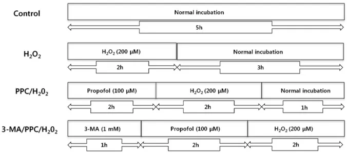
The propofol stock (56 mM) was kept at room tem- perature until use and diluted to the proper concentration with DMEM/F-12 when needed. Prior to propofol treat- ment, cells were grown to about 75% confluence and then exposed to propofol at various concentrations (0, 3, 30, 100, or 300 μM) for 2 h. The cells were divided into the following groups: control cells were incubated at 37°C in a humidified atmosphere with 5% CO2 and without any additional treatment. H2O2 cells were exposed to 200 μM H2O2 for 2 h. PPC/H2O2 cells were pretreated with 100 μM propofol for 2 h and then exposed to 200 μM H2O2 for 2 h. 3-MA/PPC/H2O2 cells were
Fig. 1. The in vitro experimental protocols. Control = normal incubation, H2O2 = normal incubation after H2O2 treatment, PPC/H2O2 = propofol preconditioning (PPC) before H2O2 treatment, 3-MA/PPC/H2O2 = 3-methyladenine treatment followed by propofol treatment and H2O2 treatment.
pretreated with 1 mM 3-MA and 100 μM propofol for 1 h and 2 h, respectively. After treatment of 3-MA and propofol, cells were exposed to 200 μM H2O2 for 2 h.
The experimental paradigm as just described is visualized in Fig. 1.
4. Cell viability assay
The cell viability of hFOB cells was determined using an MTT assay. Cells were cultured in 96-well plates (4
× 103 cells/well). The cells were then treated with the indicated concentrations of drug. At the end of the treatment, 100 μL of MTT solution (500 mg/mL) was added to each well. The cells were incubated for 4 h at 37°C. The formazan crystals were then solubilized in DMSO (200 μL/well) by constant shaking for 15 min.
The cell viability was measured by an ELISA reader (Tecan, Männedorf, Switzerland) by determining the wells’ absorbance at 620 nm.
5. Fluorescence microscopy
Cells were harvested and cytocentrifuged onto a clean glass slide. Cells were stained in 1 μg/mL Hoechst 33342 for 15 min at 37°C in the dark and subsequently washed in phosphate buffered saline (PBS). The slides were mounted with glycerol. The samples were observed and photographed under an epifluorescence microscope (Carl
Zeiss, Goettingen, Germany). Cells were grown on coverslips and treated with propofol. After 24 h, cells were stained with 0.05 mM monodansylcadaverine (MDC), a selective fluorescent marker for autophagic vacuoles, at 37°C for 1 h. The cellular fluorescence changes were observed using a fluorescence microscope (Axioskop, Carl Zeiss, Germany). For further detection of the acidic cellular compartment, we used acridine orange (AO), which emits bright red fluorescence in acidic vesicles but fluorescence green in the cytoplasm and nucleus. Cells were stained with 1 μg/ml AO for 15 min and washed with PBS. Acidic vesicular organelles (AVOs) formation was obtained under a confocal microscope LSM 700 (Carl Zeiss, Germany).
6. Flow cytometer analysis
Quantification of apoptotic cells was determined by Annexin V-FITC/PI staining. Adherent cells were collec- ted by trypsinization and centrifugation, and then were resuspended in 500 μL 1X binding buffer. Then they were stained with 5 μL Annexin V-FITC and propidium iodide (PI) (50 μg/mL) and were incubated at room temperature for 5 min in the dark. The cells were analyzed with a CYTOMICS FC500 flow cytometer system (Beckman Coulter, CA, USA). The results were shown as quadrant dot plots with intact cells (Annexin V-/PI-), early apopto-
(A) (B)
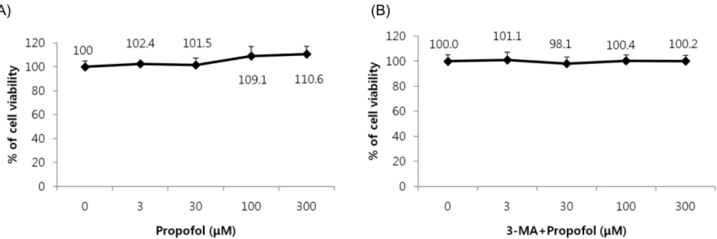
Fig. 2. Effect of propofol and 3-MA on cell viability. (A) The viability of propofol-treated hFOB cells. Propofol did not elicit any significant toxic effect on hFOB cells. (B) 3-MA did not elicit any significant toxic effect on hFOB cells.
hyde (PFA) and were stained with 2% Alizarin Red solution according to the manufacturer’s protocol. For quantification, absorbance of the released Alizarin Red was measured at 550 nm with a microplate reader.
8. Western blot analysis
Cells (1.5×10⁶) were washed twice in ice-cold PBS, resuspended in 200 μL ice-cold solubilizing buffer (300 mM NaCl, 50 mM Tris-HCl (pH 7.6), 0.5% Triton X-100, 2 mM PMSF, 2 μL/mL aprotinin, and 2 μL/mL leupep- tin),and incubated at 4°C for 1 h. The lysates were centrifuged at 13,200 rpm for 30 min at 4°C. Protein concentrations of cell lysates were determined using a Bradford protein assay (Bio-Rad, Richmond, CA, USA), and 20 μg protein were loaded per lane and resolved by 10% SDS/PAGE. Gels were transferred to polyvinyli- dene fluoride (PVDF) membranes (Millipore, Billerica, MA, USA). After blocking, the membranes were reacted with the appropriate primary antibodies. Immunostaining with secondary antibodies was detected using Super-
RESULTS
1. Effect of propofol treatment on cell proliferation The effect of propofol on hFOB cells was investigated over a wide concentration range. We pretreated hFOB cells with various doses of propofol, exposed the cells to oxidative stress, and then measured cell viability by an MTT assay. The viability of propofol-treated hFOB cells increased in a dose-dependent manner. Propofol and 3-MA alone did not show any significant toxic effects on hFOB cells (Fig. 2A, 2B). Incubation with 200 μM H2O2 markedly decreased cell viability, which was significantly attenuated when treated with propofol, suggesting that propofol abated H2O2-induced oxidative stress in osteoblasts (Fig. 3).
Fig. 6. Fluorescence microscopy (400X) analysis of autophagosomes in hFOB cells after oxidative stress. Images were taken after staining with monodansylcadaverine (MDC). Blue fluorescence indicates autophagic vacuoles and autophagosomes can be seen as punctate areas of fluorescence.
Fig. 3. Effect of H2O2, PPC, and 3-MA on cell viability. Cell viability was determined by 3-(4,5-dimethylthiazol-2-yl)-2,5-diphenylterazolium bromide (MTT) assay. *P < 0.05 as compared with control group.
Fig. 5. Flow cytometry analysis of apoptosis and necrosis with Annexin- V-FITC and propidium iodide staining.
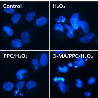
Fig. 4. Morphological changes after H2O2, PPC, and/or 3-MA treatment in hFOB cells. Fluorescence microscopy images are shown and were analyzed for presence of apoptotic bodies.
2. Propofol protected against H2O2-induced apoptosis in hFOB cells
The effect of propofol on apoptosis was examined by Hoechst 33342 staining of hFOB cells exposed to control, H2O2, PPC/H2O2, and 3-MA/PPC/H2O2. Cells were viewed under a fluorescence microscope (400X magnifi- cation) (Fig. 4). The majority of hFOB cells in the control and PPC/H2O2 groups showed normal morphology with round regular nuclei. In contrast, apoptotic bodies were seen in the H2O2 group and the 3-MA/PPC/H2O2 group
cells. Pretreatment with propofol effectively reduced hFOB cell apoptosis according to the restored mor- phology. Annexin-V FITC/PI staining quantitatively con- firmed the antiapoptotic effects of propofol (Fig. 5).
Fig. 9. Western blot analysis. Expression of collagen type I, bone morphogenic protein (BMP)-2, osterix, and transforming growth factor-beta (TGF-β)1 in the experimental groups indicated. Propofol preconditioning was found to increase the expression of collagen type I, BMP-2, osterix, and TGF-β1.
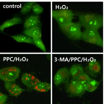
Fig. 7. Fluorescence microscopy (400X) analysis of autophagosomes in hFOB cells after oxidative stress. Images were taken after staining with acridine orange. Green indicates where the dye has stained the nucleus and red indicates where the cell is starting to 'digest' parts of itself in small capsules called autophagosomes.
Fig. 8. Photomicrographs of cultures stained with Alizarin Red after 28 days of culture. Red indicates positive staining for Ca2+ in the matrix.
Compared with control group, the portion of Annexin- V(+)/PI(−) cells in H2O2 group increased from 3.4% to 10.3% (P < 0.05). However, pretreatment with propofol 2 h prior to PPC/H2O2 significantly attenuated the percentage of Annexin-V(+)/PI(−) cells to 1.2% (P <
0.05), demonstrating the anti-apoptotic effect of propofol.
3. Effect of propofol treatment on autophagy activation Prominent accumulation of autophagy-specific MDC staining was observed around the nuclei in the PPC/H2O2
group (Fig. 6). Similarly, after AO staining, red fluores- cent spots appeared in PPC/H2O2 group hFOB cells, while the control and 3-MA groups showed mainly green
cytoplasmic fluorescence (Fig. 7).
4. Effect of propofol preconditioning on bone nodular mineralization
Abundant bone nodular mineralization occurred in the control group, and propofol preconditioning had no influence on this. Cells in the PPC/H2O2 group showed higher bone nodular mineralization compared to the control group and bone nodular mineralization increased gradually with increasing propofol preconditioning (Fig. 8).
5. Effect of Propofol preconditioning on collagen type I, BMP-2, osterix, and TGF-β1
Propofol preconditioning was found to increase the expression of bone-related proteins such as collagen type I, BMP-2, osterix, and TGF-β1 (Fig. 9). These results suggest that pretreatment with propofol induces the expression of collagen type I, BMP-2, osterix, and TGF-β 1 in osteoblasts under H2O2 injury.
DISCUSSION
We demonstrated that propofol preconditioning in- creased the proliferation, differentiation, and maturation of osteoblasts during oxidative injury in our study. The healing process is very important to recover from frac- tures, and delayed healing after bone implants can cause premature loosening or failure. In our cell viability analysis, there is a distinction between our study and a previous study, which reported an inhibitory effect of non-steroidal anti-inflammatory drugs on fracture healing [27].
In this study, we demonstrated that propofol precondi- tioning has positive effects on the expression of collagen type I, BMP-2, osterix, and TGF-β1. BMP-2 and TGF- β1 play a crucial role in inducing osteoblast differentia- tion and bone formation at the preosteoblast stage [28,29].
These proteins play an important role in the differentia- tion of osteoblast progenitor cells, with significant up- regulation observed in both matrix synthesis and minerali- zation [30-32]. Collagen type I plays a crucial role in cell adhesion, proliferation, and differentiation of osteoblasts [33]. Osterix is a transcription factor that regulates important osteoblast genes such as collagen type I, osteocalcin, and osteopontin [34].
The results of our study reveal a new direction of research on the mechanisms of propofol-mediated cyto- protection in oxidative injury. Because of the antioxida- tive effect of propofol, propofol preconditioning enhan- ced autophagic activity in hFOB cells under oxidative injury. Mechanisms underlying these effects of propofol may be largely attributed to the deactivation of mito- chondrial stress pathways.
Chen et al. [35] suggested that ROS play a key role in the regulation of autophagy. In this study, propofol preconditioning not only decreased H2O2-induced oxida- tive stress, but also activated autophagy. Many studies have focused on oxidative injury in cells, but agents that could effectively protect against oxidative injury remain unidentified. The relationship between apoptosis and
autophagy is complex. Autophagy represents a stress adaptation for inhibition of cell death, however in other cellular settings, autophagy represents an alternative pathway to autophagic cell death [36,37].
This study shows that propofol pretreatment increases the osteoblast proliferation rate. No functional studies were performed to investigate the effects of propofol on the bone healing process. Therefore, although the find- ings of this study are limited to an in vitro interpretation, we suggest that propofol may have a beneficial effect in the recovery from bone stress injury.
Acknowledgments: This work was supported by a 2-Year Research Grant from Pusan National University.
REFERENCES
1. Teitelbaum SL. Bone resorption by osteoclasts. Science 2000; 289: 1504-8.
2. Boyle WJ, Simonet WS, Lacey DL. Osteoclast differentia- tion and activation. Nature 2003; 423: 337-42.
3. Yamaguchi A, Komori T, Suda T. Regulation of osteoblast differentiation mediated by bone morphogenetic proteins, hedgehogs, and Cbfa1. Endocr Rev 2000; 21: 393-411.
4. Wozney JM, Rosen V, Celeste AJ, Mitsock LM, Whitters MJ, Kriz RW, et al. Novel regulators of bone formation:
Molecular clones and activities. Science 1988; 242: 1528-34.
5. Xu ZS, Wang XY, Xiao DM, Hu LF, Lu M, Wu ZY, et al. Hydrogen sulfide protects MC3T3-E1 osteoblastic cells against H2O2-induced oxidative damage-implications for the treatment of osteoporosis. Free Radic Biol Med 2011; 50: 1314-23.
6. Lean JM, Jagger CJ, Kirstein B, Fuller K, Chambers TJ.
Hydrogen peroxide is essential for estrogen-deficiency bone loss and osteoclast formation. Endocrinology 2005;
146: 728-35.
7. Panieri E, Gogvadze V, Norberg E, Venkatesh R, Orrenius S, Zhivotovsky B. Reactive oxygen species generated in different compartments induce cell death, survival, or
A key messenger that modulates protein phosphorylation through cysteine oxidation. Sci STKE 2000; 2000: pe1.
11. Finkel T. Signal transduction by reactive oxygen species.
J Cell Biol 2011; 194: 7-15.
12. Zhu Y, Park SH, Ozden O, Kim HS, Jiang H, Vassilopoulos A, et al. Exploring the electrostatic repulsion model in the role of Sirt3 in directing MnSOD acetylation status and enzymatic activity. Free Radic Biol Med 2012; 53:
828-33.
13. Eno CO, Zhao G, Venkatanarayan A, Wang B, Flores ER, Li C. Noxa couples lysosomal membrane permeabili- zation and apoptosis during oxidative stress. Free Radic Biol Med 2013; 65: 26-37.
14. Kim YN, Jung HY, Eum WS, Kim DW, Shin MJ, Ahn EH, et al. Neuroprotective effects of PEP-1-carbonyl reductase 1 against oxidative-stress-induced ischemic neuronal cell damage. Free Radic Biol Med 2014; 69:
181-96.
15. Ito H, Watanabe Y, Isshiki A, Uchino H. Neuroprotective properties of propofol and midazolam, but not pento- barbital, on neuronal damage induced by forebrain ischemia, based on the GABAA receptors. Acta Anaesthesiol Scand 1999; 43: 153-62.
16. Yano T, Nakayama R, Ushijima K. Intracerebroventricular propofol is neuroprotective against transient global ischemia in rats: Extracellular glutamate level is not a major determinant. Brain Res 2000; 883: 69-76.
17. Bayona NA, Gelb AW, Jiang Z, Wilson JX, Urquhart BL, Cechetto DF. Propofol neuroprotection in cerebral ischemia and its effects on low-molecular-weight antioxidants and skilled motor tasks. Anesthesiology 2004;
100: 1151-9.
18. Wu GJ, Chen WF, Hung HC, Jean YH, Sung CS,
Pharmacol Ther 2001; 89: 123-37.
21. Tanaka K, Ludwig LM, Kersten JR, Pagel PS, Warltier DC. Mechanisms of cardioprotection by volatile anesthetics. Anesthesiology 2004; 100: 707-21.
22. Luciani A, Villella VR, Esposito S, Brunetti-Pierri N, Medina D, Settembre C, et al. Defective CFTR induces aggresome formation and lung inflammation in cystic fibrosis through ROS-mediated autophagy inhibition. Nat Cell Biol 2010; 12: 863-75.
23. Mizushima N. The pleiotropic role of autophagy: From protein metabolism to bactericide. Cell Death Differ 2005;
12 Suppl 2: 1535-41.
24. Chen W, Sun Y, Liu K, Sun X. Autophagy: A double-edged sword for neuronal survival after cerebral ischemia. Neural Regen Res 2014; 9: 1210-6.
25. Schelonka EP, Usher A. Ipriflavone and osteoporosis.
JAMA 2001; 286: 1836-7.
26. Myers G, Prince RL, Kerr DA, Devine A, Woodman RJ, Lewis JR, et al. Tea and flavonoid intake predict osteoporotic fracture risk in elderly australian women: A prospective study. Am J Clin Nutr 2015; 102: 958-65.
27. Gerstenfeld LC, Thiede M, Seibert K, Mielke C, Phippard D, Svagr B, et al. Differential inhibition of fracture healing by non-selective and cyclooxygenase-2 selective non-steroidal anti-inflammatory drugs. J Orthop Res 2003; 21: 670-5.
28. Li X, Cao X. BMP signaling and skeletogenesis. Ann N Y Acad Sci 2006; 1068: 26-40.
29. Noda M, Camilliere JJ. In vivo stimulation of bone formation by transforming growth factor-beta. Endocrinology 1989;
124: 2991-4.
30. Pavlin D, Zadro R, Gluhak-Heinrich J. Temporal pattern of stimulation of osteoblast-associated genes during mechanically-induced osteogenesis in vivo: Early responses
of osteocalcin and type I collagen. Connect Tissue Res 2001; 42: 135-48.
31. Ryoo HM, Hoffmann HM, Beumer T, Frenkel B, Towler DA, Stein GS, et al. Stage-specific expression of dlx-5 during osteoblast differentiation: Involvement in regulation of osteocalcin gene expression. Mol Endocrinol 1997; 11:
1681-94.
32. Jagodzinski M, Drescher M, Zeichen J, Hankemeier S, Krettek C, Bosch U, et al. Effects of cyclic longitudinal mechanical strain and dexamethasone on osteogenic differentiation of human bone marrow stromal cells. Eur Cell Mater 2004; 7: 35,41; discussion 41.
33. Ducy P, Desbois C, Boyce B, Pinero G, Story B, Dunstan
C, et al. Increased bone formation in osteocalcin-deficient mice. Nature 1996; 382: 448-52.
34. Chen Y, Gibson SB. Is mitochondrial generation of reactive oxygen species a trigger for autophagy? Autophagy 2008;
4: 246-8.
35. Baehrecke EH. Autophagy: Dual roles in life and death?
Nat Rev Mol Cell Biol 2005; 6: 505-10.
36. Levine B, Yuan J. Autophagy in cell death: An innocent convict? J Clin Invest 2005; 115: 2679-88.
37. Kondo Y, Kanzawa T, Sawaya R, Kondo S. The role of autophagy in cancer development and response to therapy.
Nat Rev Cancer 2005; 5: 726-34.