Usefulness of Plaque Magnetic Resonance Imaging in Identifying High-Risk Carotid Plaques Irrespective of the Degree of Stenosis
Jinseong Lee, Jinsang Kil, Dae-Won Kim, Sung-Don Kang
Institute of Wonkwang Medical Science, Department of Neurosurgery, Wonkwang University School of Medicine, Iksan, Korea
Objective : Measurement of the degree of stenosis is not enough to de- cide on the treatment strategy for patients with carotid stenosis. Plaque morphology examination is needed for such a decision-making. Thus, we evaluated the usefulness of plaque magnetic resonance imaging (MRI) to decide on the modality of treatment for patients with carotid athero- sclerotic plaques.
Materials and Methods : Fifteen patients presenting with carotid stenosis between 2014 and 2016 were included. They underwent angiography for measurement of the degree of stenosis. Carotid plaques were visualized using MRI.
Results : There were six (40%) stable and nine (60%) unstable plaques.
Seven symptomatic patients (77.7%) had unstable lesions and two symp- tomatic patients (33.3%) had stable lesions (p = 0.096). There were six (40%) intraplaque hemorrhage (IPH) cases. There were six symptomatic patients (100%) in the IPH group and three symptomatic patients (33.3%) in the non-IPH group (p = 0.013). The mean stenosis degree was 58.9% in the IPH group and 70.4% in the non-IPH group (p = 0.094). Symptoms oc- curred irrespective of the degree of the stenosis in the IPH groups. In the IPH group, the recurrent ischemic cerebrovascular event rate was 33.3%.
Particularly, the recurrent ischemic cerebrovascular event rate was 66.7%
in the IPH group with mild stenosis treated with medications.
Conclusion : IPH in plaque MRI is significantly associated with ischemic symptoms and has a high risk for subsequent ischemic cerebrovascular events irrespective of the degree of stenosis. Plaque MRI is a useful tool in predicting symptomatic risks for carotid stenosis irrespective of the de- gree of such stenosis.
J Cerebrovasc Endovasc Neurosurg.
2017 December;19(4):291-300 Received : 6 December 2017 Revised : 20 December 2017 Accepted : 31 December 2017 Correspondence to Sung-Don Kang Department of Neurosurgery, Wonkwang University Hospital, Wonkwang University School of Medicine, 895 Muwang-ro, Iksan 54538, Korea
Tel : 82-63-859-1463 Fax : 82-63-852-2606 E-mail : kangsd@wku.ac.kr
ORCID : http://orcid.org/0000-0002-3965-1674 This paper was supported by Wonkwang University in 2016.
This is an Open Access article distributed under the terms of the Creative Commons Attribution Non- Commercial License (http://creativecommons.org/li- censes/by-nc/3.0) which permits unrestricted non- commercial use, distribution, and reproduction in any medium, provided the original work is properly cited.
Keywords Atherosclerotic plaque, Magnetic resonance imaging, Hemorrhage, Carotid stenosis
INTRODUCTION
Ischemic stroke and transient ischemic attack (TIA) are commonly caused by carotid atherosclerosis.
Measurement of carotid artery stenosis is a standard method for evaluating the stroke risk. However, Ambrose et al.2) suggested that myocardial infarction (MI) frequently occurs from non-severe coronary ar-
TOF T1 T2 T1-enhanced
Fibrous tissue Iso/Hypo Iso Hyper +
Lipid-rich Iso Iso/Hyper Hypo -
Calcification Hypo Hypo Hypo -
Hemorrhage Hyper Hyper Variable -
MRI = magnetic resonance imaging; TOF = time-of-flight Table 1. MRI findings of the plaque components
tery lesion on coronary angiogram. Thus, they did not estimate MI risk with simple coronary artery stenosis.2) About 90% of conservatively treated patients with car- otid artery stenosis did not have a cerebrovascular event for 5 years.13)19) It showed that the evaluation of carotid artery stenosis is not enough to determine on the treatment strategy of patients with carotid athero- sclerotic plaques. Histopathological studies of plaque morphology identified specific parameters indicating plaque rupture. Carotid plaques characterized by a thinned fibrous cap with a lipid-rich necrotic core or by intraplaque hemorrhage (IPH) indicate unstable, rupture-prone lesions with a high risk of spontaneous thromboembolic events.3)5)8)10)12)18)
High-resolution magnetic resonance imaging (MRI) is suitable for evaluation of plaque morphology, be- cause it is noninvasive and has excellent capabilities of discriminating tissue characteristics compared with other imaging modalities.11)16)17)20) Therefore, we eval- uated the usefulness of plaque MRI to decide on the modality of treatment for patients with carotid athero- sclerotic plaques.
MATERIALS AND METHODS
Study population
Between April 2014 and January 2016, we retro- spectively evaluated 15 patients who were treated for carotid stenosis at our stroke center. All patients un- derwent carotid artery MRI examination and carotid artery angiography.
Plaque MRI
The patients underwent MRI performed on a 3.0T
Achieva Tx (Phillips, Amsterdam, The Netherlands).
Carotid plaques were visualized using MRI. T1, T2, time-of-flight (TOF), T1-enhanced, simultaneous non-con- trast angiography and intraplaque hemorrhage (SNAP), and magnetization-prepared rapid gradient-echo (MPRAGE) studies were performed. The MRI findings of each plaque component were as follows: the lipid-rich ne- crotic core showed a hyposignal on the T2-weighted images; calcification showed a hyposignal on the TOF, T1-weighted, and T2-weighted images; hemorrhage showed a hypersignal on the TOF and T1-weighted images; and fibrous tissues showed enhancements on the T1-enhanced image (Table 1). Each lesion was classified using the modified American Heart Association (AHA) classification for MRI.4) Type I-II has near nor- mal wall thickness with no calcification. Type III has diffuse intimal thickening or small eccentric plaques with no calcification. Type IV-V involves a plaque with a lipid or necrotic core surrounded by fibrous tissues with possible calcifications. Type VI involves a complex plaque with a possible surface defect, hemor- rhage, or thrombus. Type VII involves a calcified plaque.
Type VIII involves a fibrotic plaque without a lipid core and with possible small calcifications. Among them, Types IV-V and VI were classified as unstable plaques, while the others were classified as stable plaques. Further, we classified them according to the presence of IPH.
RESULTS
The mean patient age was 70.2 ± 8.2 (range, 53-80) years. The male-to-female sex ratio was 10:5. Nine pa-
Sex Age Modified AHA Stability IPH
F 66 III Stable X
M 75 IV-V Unstable X
F 53 IV-V Unstable X
M 75 VI Unstable O
F 77 VI Unstable O
M 66 VII Stable X
M 74 VII Stable X
M 69 IV-V Unstable X
M 67 VI Unstable O
M 80 VI Unstable O
M 80 VI Unstable O
F 54 III Stable X
F 69 VII Stable X
M 75 VI Unstable O
M 73 VII Stable X
MRI = magnetic resonance imaging; AHA = American Heart Association; IPH = intraplaque hemorrhage; M = male; F = female Table 2. Patient data (carotid plaque status via MRI)
Stable Unstable p-value
N (%) 6 (40.0) 9 (60.0)
Symptomatic 2 (33.3) 7 (77.7) 0.096
Age range, mean (years) 53-73 (64.7 ± 9.1) 66-80 (73.9 ± 5.3) 0.088
Male sex 3 (50.0) 7 (77.7) 0.280
Hypertension 5 (83.3) 7 (77.7) 0.799
Current or former smoker 2 (33.3) 1 (11.1) 0.309
Hypercholesterolemia 3 (50.0) 4 (44.4) 0.838
Coronary heart disease 3 (50.0) 4 (44.4) 0.838
Stenosis (%) 72.8 ± 5.7 61.1 ± 22.8 0.247
Values are presented as mean ± standard deviation or number (%) unless otherwise indicated Table 3. Patient data (grouped according to stability)
tients were symptomatic (60%). There were Four (26.7%) cases of mild stenosis (< 50%), two (13.3%) of moder- ate stenosis (50-69%), and nine (60%) of severe steno- sis (> 70%). Diabetes mellitus (DM) was diagnosed in five patients (33.3%). Hypertension was diagnosed in 12 patients (80%). Hyperlipidemia was diagnosed in seven patients (46.7%). Coronary heart disease was di- agnosed in seven patients (46.7%). One patient was lost to follow-up. One patient died shortly due to pneumonia. The mean follow-up duration was 19.7 months (range, 5-33 months). During this period, two patients (13.3%) developed an ipsilateral ischemic cer-
ebrovascular event. Three patients (20%) presented with bilateral stenosis. Nine patients (60%) underwent carotid angioplasty and stenting (CAS). One patient (6.7%) underwent carotid endarterectomy (CEA). Five patients (33.3%) underwent medical treatment.
Carotid plaque MRI lesion types
The lesion types of the carotid plaques were as fol- lows: two lesions were classified under type III (13.3%), three lesions under type IV-V (20%), six le- sions under type VI (40%), and four lesions under type VII (26.7%). There were six (40%) stable (types
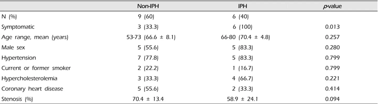
Non-IPH IPH p-value
N (%) 9 (60) 6 (40)
Symptomatic 3 (33.3) 6 (100) 0.013
Age range, mean (years) 53-73 (66.6 ± 8.1) 66-80 (70.4 ± 4.8) 0.257
Male sex 5 (55.6) 5 (83.3) 0.280
Hypertension 7 (77.8) 5 (83.3) 0.799
Current or former smoker 2 (22.2) 1 (16.7) 0.799
Hypercholesterolemia 3 (33.3) 4 (66.7) 0.221
Coronary heart disease 5 (55.6) 2 (33.3) 0.414
Stenosis (%) 70.4 ± 13.4 58.9 ± 24.1 0.094
Values are presented as mean ± standard deviation or number (%) unless otherwise indicated.
IPH, intraplaque hemorrhage
Table 4. Patient data (grouped according to the presence of IPH)
III and VII) and nine (60%) unstable (types IV-V and VI) plaques. There were six (40%) IPH cases (Table 2).
In the stable lesion types, there were one (16.7%) mild stenosis, two (33.3%) moderate stenosis, and three (50%) severe stenosis. The mean stenosis degree was 72.8%. In the unstable lesion types, there were three (33.3%) mild stenosis, no moderate stenosis, and six (66.7%) severe stenosis. The mean stenosis degree was 61.1%. There were seven symptomatic patients (77.7%) with unstable lesions and two symptomatic patients (33.3%) with stable lesions (p = 0.096) (Table 3).
All patients with IPH (n = 6) were symptomatic (100%). There were three symptomatic patients (33.3%) in the non-IPH group (p = 0.013). The mean stenosis degree was 58.9% in the IPH group and 70.4% in the non-IPH group (p = 0.094) (Table 4). In the non-IPH group, the symptomatic patients had severe stenosis.
However, the symptoms occurred irrespective of the degree of stenosis in the IPH group.
In the stable lesion types, the patients with mild and moderate stenosis (n = 3, 50%) were treated with medications and those with severe stenosis (n = 3, 50%) with CAS. In the unstable lesion types, three pa- tients with mild stenosis (33.3%) were treated with medications; however, two patients were treated for ischemic cerebrovascular events during the follow-up period (66.7%). Further, the patients with severe stenosis (n = 6, 66.7%) were treated with CAS. In the stable le- sion types, none of the patients (0%) presented with
ischemic cerebrovascular events during the follow-up period. In the unstable lesion types, two patients (22.2%) had recurrent ischemic cerebrovascular events during the follow-up period. The plaque MRI of these patients revealed IPH. In the IPH group, the recurrent ischemic cerebrovascular event rate was 33.3%. Particularly, the recurrent ischemic cerebrovascular event rate was 66.7% in the IPH group with mild stenosis treated with medications.
Illustrative case 1
A 67-year-old man was admitted due to mild motor weakness of the right upper extremity (grade 4+) and mild dysarthria. He had hypertension and hyperlipidemia.
He had a history of cerebral infarction twice, 4 years and 4 months earlier. A month ago, he was treated with medications for a mild left cervical internal car- otid artery (ICA) stenosis (Fig. 1A). MRI on admission showed multiple cerebral infarctions at the left cere- bral hemisphere (Fig. 1B). The degree of carotid steno- sis was not changed (14.7%), however, small ulcer- ation was demonstrated on cerebral angiogram (Fig.
1C). Plaque MRIs showed IPH (Fig. 2). On T1 en- hanced image, focal disruption of fibrous cap was demonstrated (Fig. 2D, E). He was treated conservatively.
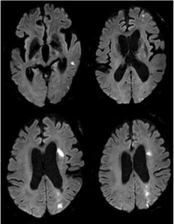
After 8 days, he had a recurrent cerebral infarction (Fig. 3). His motor weakness was aggravated at the right upper extremity (grade 4) and lower extremity (grade 4+). He underwent CEA. In the operative find-
A B C
Fig. 1. (A) CT angiography shows mild stenosis of left ICA before cerebral infarction. (B) MRI showing multiple cerebral infarctions at the left cerebral hemisphere. (C) Left CCA angiogram shows no significant change of stenosis degree except small ulceration (black arrow) after recurrent cerebral infarction. CT = computed tomography; ICA = internal carotid artery; MRI = magnetic resonance imag- ing; CCA = common carotid artery.
A B C
D E F G
Fig. 2. Plaque MRIs show IPH. IPH (pink arrow) shows high-signal intensity on T1-weighted (A), TOF (B), SNAP (F), MPRAGE (G) images. On T2-weighted image (C), IPH (arrow) shows iso-signal intensity. On T1 enhanced image (D, E), focal disruption of fibrous cap (white arrow) is demonstrated. Focal disruption of fibrous cap in (E) is corresponding to plaque ulcer shown in DSA. MRI = magnetic resonance imaging; IPH = intraplaque hemorrhage; TOF = time-of-flight; SNAP = simultaneous non-contrast angiography and intraplaque hemorrhage; MPRAGE = magnetization-prepared rapid gradient-echo; DSA = digital subtraction angiography.
ings, the carotid plaque had an IPH and ulcer as shown in the MRI (Fig. 4).
Illustrative case 2
A 75-year-old man was admitted due to dizziness.
He had no DM, hypertension, and hyperlipidemia. He was treated for coronary heart disease 20 years prior.
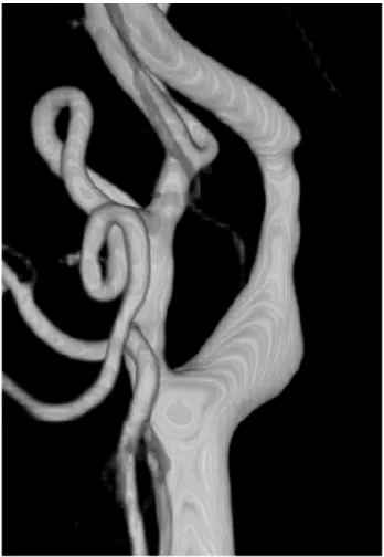
He had no motor weakness. The degree of carotid stenosis was mild (35%) (Fig. 5). Plaque MRIs showed
Fig. 3. MRI shows a new cerebral infarction at border zone of left hemisphere. MRI = magnetic resonance imaging.
A B
Fig. 4. Operative findings. (A, B) Carotid plaque has IPH and ulcer as shown in the MRI. IPH = intraplaque hemorrhage; MRI = mag- netic resonance image.
IPH in the right ICA and chronic complete occlusion
in the left ICA (Fig. 6). He was treated with medications.
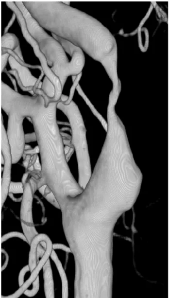
After 27 months, he had a recurrent ischemic cere- brovascular event, amaurosis fugax. Cervical angio- gram showed progression of carotid stenosis to 80%
(Fig. 7). He underwent CAS. Right ICA angiogram showed more dilatation of ICA after CAS (Fig. 8).
DISCUSSION
High-resolution MRI is ideal for the evaluation of the plaque status. Cai et al. modified the AHA classi- fication of atherosclerotic lesions by MRI.4) Using this modified classification, plaques with a lipid-rich ne- crotic core surrounded by fibrous tissues with possi- ble calcification can be classified as lesion types IV-V and complex plaques with a possible surface defect, hemorrhage, or thrombus as lesion type VI.6)21) Cai et al. reported that high sensitivity and specificity were achieved from type III to type VII lesions.4) The sensi- tivity and specificity of MRI in categorizing each le- sion type were as follows: type I-II, 67% and 100%;
type III, 81% and 98%; type IV-V, 84% and 90%; type VI, 82% and 91%; type VII, 80% and 94%; and type VIII, 56% and 100%, respectively.4)
Fig. 5. Right CCA 3D angiogram shows mild stenosis of right ICA. CCA = common carotid artery; ICA = internal carotid artery.
A B C
D E F
Fig. 6. Plaque MRIs show IPH. IPH (pink arrow) shows high-signal intensity on T1-weighted (A), TOF (B), SNAP (E), MPRAGE (F) images. On T2-weighted image (C), IPH shows high-signal intensity (white arrow). On T1 enhanced images (D), fibrous cap is intact (yellow arrowhead). MRI = magnetic resonance image; IPH = intraplaque hemorrhage; TOF = time-of-flight; SNAP = simultaneous non-contrast angiography and intraplaque hemorrhage; MPRAGE = magnetization-prepared rapid gradient-echo.
Lipid-rich necrotic core and IPH demonstrated in atherosclerotic plaques are strongly associated with increased risks of ischemic cerebrovascular events as well as plaque progression. IPH has also been re- garded to be a potentially important factor in treat- ment planning. Altaf et al.1) found MRI-defined vul- nerable plaque features, such as IPH, to be related to recurrent cerebral events when evaluating sympto- matic patients with mild to moderate carotid artery stenosis. The presence of IPH significantly predicted recurrence of various ischemic event (e.g., stroke, TIA, and amaurosis fugax) with a hazard ratio (HR) of 9.8 (95% confidence interval [CI] 1.3-75.1); all other pa- tient-related factors, including age (HR = 1.0), hyper- tension (HR = 1.8), ischemic heart disease (HR = 0.4), DM (HR = 1.4), smoking (HR = 1.3), and degree of stenosis (HR = 1.5; 95% CI 0.5-4.9) had lower HRs.1) Esposito-Bauer et al.7) also found that MRI-defined vulnerable plaques are at a high risk for future cere- brovascular events in asymptomatic patients. Only pa- tients harboring with high-risk lesion types IV-V and VI occurred an ipsilateral cerebrovascular event; none of the patients presenting with stable lesion types III, VII, and VIII (11.7% vs. 0%) during the follow-up de-
Fig. 7. Right CCA 3D angiogram showing severe focal stenosis of the right proximal cervical ICA. CCA = common carotid ar- tery; ICA = internal carotid artery.
Fig. 8. Right CCA angiogram shows dilatation of ICA after CAS. CCA = common carotid artery; ICA = internal carotid artery;
CAS = carotid angioplasty and stenting.
veloped such an event. The carotid plaques of four (44.4%) of these nine patients who suffered an ische- mic event during the follow-up were classified as le- sion type IV-V; five (55.6%) of these patients pre- sented with lesion type VI. The event-free survival rate was higher among the patients with the MRI-de- fined stable lesion types (III, VII, and VIII) than among patients with the MRI-defined high-risk lesion types (IV-V and VI) (58-month event-free probability 100% vs. 67.8%; log rank test p < 0.0001).7) Our findings are consistent with such results. In our study, only
the patients with unstable lesions (22.2% vs. 0%) pre- sented with ischemic cerebrovascular events (ischemic stroke) during the follow-up period.
Rothwell et al. reported that the benefits of CEA de- pend upon patient-specific factors other than the de- gree of stenosis in symptomatic moderate stenosis groups.14) Plaque characters, such as the integrity of the fibrous cap and the size of the lipid core, are im- portant for contributing to the stability of the plaque
and are found in both symptomatic and asympto- matic carotid arteries.9)15)
In our study, the patients with mild stenosis suf- fered recurrent cerebral infarction during the fol- low-up period. They had IPH on MRI. However, the symptomatic patients in the non-IPH group had se- vere stenosis. We think that if IPH is present, CAS or CEA should be considered irrespective of the degree of stenosis.
CONCLUSION
Plaque MRI is a useful tool in predicting sympto- matic risks for carotid stenosis irrespective of the de- gree of such stenosis. IPH in plaque MRI is sig- nificantly associated with ischemic symptoms and has a high risk for subsequent ischemic cerebrovascular events irrespective of the degree of stenosis. If IPH is demonstrated in plaque MRI, considering proactive treatment may be reasonable to prevent future ische- mic event.
Disclosure
The authors report no conflict of interest concerning the materials or methods used in this study or the findings specified in this paper.
REFERENCES
1. Altaf N, Daniels L, Morgan PS, Auer D, MacSweeney ST, Moody AR, et al. Detection of intraplaque hemorrhage by magnetic resonance imaging in symptomatic patients with mild to moderate carotid stenosis predicts recurrent neurological events. J Vasc Surg. 2008 Feb;47(2):337-42.
2. Ambrose JA, Tannenbaum MA, Alexopoulos D, Hjemdahl- Monsen CE, Leavy J, Weiss M, et al. Angiographic pro- gression of coronary artery disease and the development of myocardial infarction. J Am Coll Cardiol. 1988 Jul;12(1):
56-62.
3. Bassiouny HS, Sakaguchi Y, Mikucki SA, McKinsey JF, Piano G, Gewertz BL, et al. Juxtalumenal location of pla- que necrosis and neoformation in symptomatic carotid stenosis. J Vasc Surg. 1997 Oct;26(4):585-94.
4. Cai JM, Hatsukami TS, Ferguson MS, Small R, Polissar NL, Yuan C. Classification of human carotid atherosclerotic lesions with in vivo multicontrast magnetic resonance imaging. Circulation. 2002 Sep;106(11):1368-73.
5. Carr S, Farb A, Pearce WH, Virmani R, Yao JS.
Atherosclerotic plaque rupture in symptomatic carotid artery stenosis. J Vasc Surg. 1996 May;23(5):755-65; dis- cussion 765-6.
6. Chu B, Kampschulte A, Ferguson MS, Kerwin WS, Yarnykh VL, O'Brien KD, et al. Hemorrhage in the atherosclerotic carotid plaque: a high-resolution MRI study. Stroke.
2004 May;35(5):1079-84.
7. Esposito-Bauer L, Saam T, Ghodrati I, Pelisek J, Heider P, Bauer M, et al. MRI plaque imaging detects carotid plaques with a high risk for future cerebrovascular events in asymptomatic patients. PLoS One. 2013 Jul 24;8(7):e67927.
8. Falk E. Why do plaques rupture? Circulation. 1992 Dec;86 (6 Suppl):III30-42.
9. Golledge J, Greenhalgh RM, Davies AH. The sympto- matic carotid plaque. Stroke. 2000 Mar;31(3):774-81.
10. Lusby RJ, Ferrell LD, Ehrenfeld W, Stoney R, Wylie EJ.
Carotid plaque hemorrhage: its role in production of cerebral ischemia. Arch Surg. 1982 Nov;117(11):1479-88.
11. Maynor CH, Charles HC, Herfkens RJ, Suddarth SA, Johnson GA. Chemical shift imaging of atherosclerosis at 7.0 Tesla. Invest Radiol. 1989 Jan;24(1):52-60.
12. Mofidi R, Crotty TB, McCarthy P, Sheehan SJ, Mehigan D, Keaveny TV. Association between plaque instability, angiogenesis and symptomatic carotid occlusive disease.
Br J Surg. 2001 Jul;88(7):945-50.
13. Robless P, Baxter A, Byrd S, Emson M, Halliday A.
Prevalence of asymptomatic CT infarcts in the ongoing Asymptomatic Carotid Surgery Trial (ACST). Int Angiol.
1998 Sep;17(3):194-200.
14. Rothwell PM, Eliasziw M, Gutnikov SA, Fox AJ, Taylor DW, Mayberg MR, et al. Analysis of pooled data from the randomised controlled trials of endarterectomy for symp- tomatic carotid stenosis. Lancet. 2003 Jan 11;361(9352):107-16.
15. Takaya N, Yuan C, Chu B, Saam T, Underhill H, Cai J, et al. Association between carotid plaque characteristics and subsequent ischemic cerebrovascular events: a pro- spective assessment with MRI--initial results. Stroke.
2006 Mar;37(3):818-23.
16. Toussaint JF, LaMuraglia GM, Southern JF, Fuster V, Kantor HL. Magnetic resonance images lipid, fibrous, calcified, hem- orrhagic, and thrombotic components of human athero- sclerosis in vivo. Circulation. 1996 Sep;94(5):932-8.
17. Toussaint JF, Southern JF, Fuster V, Kantor HL.
T2-weighted contrast for NMR characterization of hu- man atherosclerosis. Arterioscler Thromb Vasc Biol. 1995 Oct;15(10):1533-42.
18. Virmani R, Kolodgie FD, Burke AP, Finn AV, Gold HK, Tulenko TN, et al. Atherosclerotic plaque progression and vulnerability to rupture: angiogenesis as a source of intraplaque hemorrhage. Arterioscler Thromb Vasc Biol.
2005 Oct;25(10):2054-61.
19. Walker MD, Marler JR, Goldstein M, Grady PA, Toole JF, Baker WH, et al. Endarterectomy for asymptomatic carotid artery stenosis. JAMA. 1995 May;273(18):1421-8.
20. Yuan C, Hatsukami TS, Beach KW, Hayes CE, Nelson JA, Ferguson MS, et al. In vivo MR evaluation of athe- rosclerosis in human carotid artery with use of phased-ar- ray coils. J Vasc Interv Radiol. 1996 Jan-Feb;7(1):46-8.
21. Yuan C, Mitsumori LM, Ferguson MS, Polissar NL, Echelard D, Ortiz G, et al. In vivo accuracy of multispectral magnetic resonance imaging for identifying lipid-rich ne-
crotic cores and intraplaque hemorrhage in advanced hu- man carotid plaques. Circulation. 2001 Oct;104(17):2051-6.