INTRODUCTION
Uterine papillary serous carcinoma (UPSC) and uterine clear cell carcinoma (UCCC) represent aggressive histologic
subtypes of endometrial cancer that account for greater than 50% of relapses and deaths from endometrial cancer [1-4].
Multi-modality therapy is often recommended, but given the relative rarity of these histologies compared to the more common endometrioid adenocarcinoma, there is a lack of prospective evidence to guide management. The role of lymph node-directed radiation therapy in particular is not well defined, as UPSC is thought to spread predominantly transperitoneally, and UCCC is thought to spread as much lymphatically as hematogenously [1-4]. Controversy over the use of lymph node-directed radiation therapy stems in part from the fact that the risk factors for lymphatic spread are not
The incidence of pelvic and para-aortic lymph node metastasis in uterine papillary serous and clear cell carcinoma according to the SEER registry
Malcolm D. Mattes1, Jennifer C. Lee2, Daniel J. Metzger2, Hani Ashamalla2, Evangelia Katsoulakis2
1Department of Radiation Oncology, West Virginia University, Mograntown, WV; 2Department of Radiation Oncology, New York Methodist Hospital, Brooklyn, NY, USA
Received May 28, 2014, Revised Sep 11, 2014, Accepted Sep 11, 2014 Data was presented in abstract form at the ACRO 2014 Annual Meeting (Orlando, FL, USA).
Correspondence to Malcolm D. Mattes
Department of Radiation Oncology, West Virginia University, PO Box 9234, Health Sciences Center, Morgantown, WV 26506, USA. E-mail: malcolm.
Objective: In this study we utilized the Surveillance, Epidemiology and End-Results (SEER) registry to identify risk factors for lymphatic spread and determine the incidence of pelvic and para-aortic lymph node metastases in patients with uterine papillary serous carcinoma (UPSC) and uterine clear cell carcinoma (UCCC) who underwent complete surgical staging and lymph node dissection.
Methods: Nine hundred seventy-two eligible patients diagnosed between 1998 to 2009 with International Federation of Gynecology and Obstetrics (FIGO) 1988 stage IA-IVA UPSC (n=685) or UCCC (n=287) were identified for analysis. Binomial logistic regression was used to determine risk factors for lymph node metastasis, with the incidence of pelvic and para-aortic lymph node metastases reported for each FIGO primary tumor stage. The Cox proportional hazards regression model was used to determine factors associated with overall survival.
Results: FIGO primary tumor stage was the only independent risk factor for lymph node metastasis (p<0.01). The incidence of pelvis-only and para-aortic lymph node involvement according to the FIGO primary tumor stage were as follows: IA (2.3%/3.8%), IB (7.5%/5.2%), IC (22.5%/16.9%), IIA (20.8%/13.2%), IIB (25.7%/14.9%), and III/IV (25.7%/24.3%). Prognostic factors for overall survival included lymph node involvement (hazard ratio [HR], 1.42; 95% confidence interval [CI], 1.09 to 1.85; p<0.01), patient age >60 years (HR, 1.70; 95% CI, 1.21 to 2.41; p<0.01), and advanced FIGO primary tumor stage (p<0.01). Tumor grade, histologic subtype, and patient race did not predict for either lymph node metastasis or overall survival.
Conclusion: There is a high incidence of both pelvic and para-aortic lymph node metastases for FIGO stages IC and above uterine papillary serous and clear cell carcinomas, suggesting a potential role for lymph node-directed therapy for these patients.
Keywords: Adenocarcinoma Clear Cell; Lymphatic Metastasis; Registries; Risk Factors; Pelvis
pISSN 2005-0380·eISSN 2005-0399
well defined. For instance, known risk factors for lymphatic spread in endometrioid adenocarcinoma like primary tumor grade, depth of invasion, and lymphovascular space invasion have not consistently been correlated with lymphatic spread for UPSC and UCCC in several small, single-institution series [5-18].
In order to determine which patients are most likely to derive the greatest benefit from lymph node-directed radia- tion therapy or full lymphadenectomy, the primary objective of this study was to use the Surveillance, Epidemiology and End-Results (SEER) registry to identify risk factors for lymphatic spread in patients with UPSC or UCCC, and describe the inci- dence and anatomic distribution of lymph node metastases according to the risk factors identified. We also report on prognostic factors for overall survival in this cohort of patients.
MATERIALS AND METHODS
The SEER registry of the National Cancer Institute is a com- prehensive source of population-based information for all newly diagnosed cancer patients residing in SEER participat- ing areas, including approximately 26% of the United States population in seventeen regions over the years of this study.
All data are provided such that individual patient identification may not be readily accessed, and no effort to acquire such information was carried out for the purposes of this study.
Eligibility criteria for this study included patients of age 30 to 99 diagnosed between 1998 and 2009 with papillary serous cystadenocarcinoma (SEER code: 8460/3) or clear cell adenocarcinoma (SEER code: 8310/3), who underwent either total abdominal hysterectomy/bilateral salpingoophorectomy (TAH/BSO) or modified radical hysterectomy/BSO, with at least 11 lymph nodes dissected during surgery. Of note, although the definition of an adequate lymphadenectomy is debatable for endometrial adenocarcinoma and is not well studied for UPSC and UCCC, we chose a cutoff of 11 lymph nodes in this study because this has been associated with a survival benefit in endometrial cancer [19,20]. The data retrieved from the SEER registry included the following patient demographics and disease characteristics: patient age at diagnosis and race, type of surgery performed, number of lymph nodes dissected, tumor grade, tumor histology, International Federation of Gynecology and Obstetrics (FIGO) 1988 primary tumor stage [21], and regional lymph node involvement. It should be noted that the FIGO nodal stage is coded independently from the primary tumor stage, allowing for collection of informa- tion on extent of primary tumor invasion independent from information on lymph node involvement. Notable pathologic information that is not readily accessible in the SEER registry
includes the presence of lymphovascular space invasion and location of dissected lymph nodes (i.e., extent of dissection).
As such, we were unable to determine which patients under- went only pelvic lymphadenectomy versus combined pelvic and para-aortic lymphadenectomy. Patients were excluded if they had metastatic disease or positive peritoneal washings at diagnosis, since these would have upstaged the patient according to the FIGO system, and as such no information on the primary tumor depth of invasion would have been available. Other excluded patients included those with incom- pletely coded information pertaining to the primary tumor stage, grade, or site of regional lymph node involvement.
Descriptive statistics were used in reporting the patient demographics, disease characteristics, and the likelihood of pelvic and para-aortic lymph node involvement for the entire cohort of patients. Binomial logistic regression was used to determine which factors (age, race, grade, histologic subtype, FIGO primary tumor stage, and number of lymph nodes examined beyond 11 nodes) were independently associated with lymph node metastasis. Based on this analysis, the inci- dence of pelvis-only and para-aortic lymph node involvement
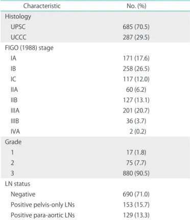
Table 1. Tumor characteristics (n=972)
Characteristic No. (%)
Histology
UPSC 685 (70.5)
UCCC 287 (29.5)
FIGO (1988) stage
IA 171 (17.6)
IB 258 (26.5)
IC 117 (12.0)
IIA 60 (6.2)
IIB 127 (13.1)
IIIA 201 (20.7)
IIIB 36 (3.7)
IVA 2 (0.2)
Grade
1 17 (1.8)
2 75 (7.7)
3 880 (90.5)
LN status
Negative 690 (71.0)
Positive pelvis-only LNs 153 (15.7) Positive para-aortic LNs 129 (13.3)
FIGO, International Federation of Gynecology and Obstetrics; LN, lymph node; UCCC, uterine clear cell carcinoma; UPSC, uterine papillary serous carcinoma.
was described according to FIGO primary tumor stage for all high grade carcinomas. Finally, univariate and multivariate Cox proportional hazards regression models were applied to assess factors associated with overall survival, and the Kaplan- Meier method/log-rank test were used to compare survival for subgroups of patients. Statistical analysis was performed using SPSS ver. 20.0 (IBM Co., Armonk, NY, USA). A p-value less than 0.05 was considered statistically significant.
RESULTS
A total of 972 eligible patients with stage IA-IVA UPSC and
UCCC were identified and included in the subsequent analy- sis. In the overall study group, the median age of diagnosis was 67 years (range, 30 to 99 years) and the median number of lymph nodes dissected was 20 (range, 11 to 81) . Seventy- eight percent of patients were white, whereas 14% were black and 8% were of unknown/other race. Additional tumor characteristics are shown in Table 1. A total of 282 patients (29.0%) had positive lymph nodes, of which 153 (54.3%) had pelvis-only lymph node involvement, 129 (45.7%) had para- aortic lymph node involvement.
According to binomial logistic regression analysis, only the FIGO primary tumor stage was an independent risk factor for lymph node metastasis (p<0.01). Additional factors tested which were not correlated with lymph node metastasis included patient age (p=0.58), patient race (p=0.30), tumor grade (p=0.38), tumor histologic subtype (p=0.51), and number of lymph nodes dissected beyond 11 nodes (p=0.75).
The incidence of pelvis-only and para-aortic lymph node involvement for all high grade carcinomas according to each FIGO primary tumor stage is shown in Table 2, and although histology (UPSC vs. UCCC) was not found to independently predict for lymph node metastasis, the histology-specific inci- dence of pelvis-only and para-aortic lymph node involvement according to each FIGO primary tumor stage is also shown in Tables 3, 4.
Univariate analysis demonstrated that older patient age (p<0.01), more advanced FIGO primary tumor stage (p<0.01), Table 2. Incidence of lymph node metastases for high grade UPSC
and UCCC combined, according to stage
Stage Pelvis-only (%) Para-aortic (%) Total (%)
IA (n=150) 2.3 3.8 6.1
IB (n=231) 7.5 5.2 12.7
IC (n=113) 22.5 16.9 39.4
IIA (n=53) 20.8 13.2 34.0
IIB (n=115) 25.7 14.9 40.6
III/IV (n=218) 25.7 24.3 50.0
UCCC, uterine clear cell carcinoma; UPSC, uterine papillary serous car- cinoma.
Table 3. Incidence of lymph node metastases for high grade UPSC, according to stage
Stage Pelvis-only (%) Para-aortic (%) Total (%)
IA (n=94) 1.3 2.6 3.9
IB (n=163) 8.4 4.2 12.6
IC (n=81) 18.0 21.3 39.3
IIA (n=42) 19.0 16.7 35.7
IIB (n=71) 28.6 15.9 44.5
III/IV (n=171) 26.7 20.6 47.3
UPSC, uterine papillary serous carcinoma.
Table 4. Incidence of lymph node metastases for high grade UCCC, according to stage
Stage Pelvis-only (%) Para-aortic (%) Total (%)
IA (n=56) 3.7 5.6 9.3
IB (n=68) 5.5 7.3 12.8
IC (n=32) 32.1 7.1 39.2
IIA (n=11) 27.3 0.0 27.3
IIB (n=44) 21.1 13.2 34.3
III/IV (n=47) 22.2 37.8 60.0
UCCC, uterine clear cell carcinoma.
Table 5. Multivariate Cox's proportional hazards model for overall survival by time periods since diagnosis
Factor Hazard ratio (95% CI) p-value
Age (yr) at diagnosis ≤60
>60
Reference
1.70 (1.21–2.41) <0.01 FIGO stage
IA/B IC/IIA IIB III/IVA
Reference 1.42 (1.00–2.05) 1.59 (1.08–2.35) 1.84 (1.34–2.53)
0.05 0.02
<0.01 Lymph node status
Negative Positive
Reference
1.42 (1.09–1.85) <0.01 Histology
UPSC UCCC
Reference
0.88 (0.67–1.16) 0.36 CI, confidence interval; FIGO, International Federation of Gynecology and Obstetrics; UCCC, uterine clear cell carcinoma; UPSC, uterine papillary serous carcinoma.
and the presence of positive lymph nodes (p<0.01) were associated with lower overall survival, whereas there was a trend towards lower overall survival for UPSC as compared to UCCC (p=0.09). These factors were included in a multivariate analysis, with hazard ratios and 95% confidence intervals shown in Table 5. Older age at diagnosis, more advanced FIGO primary tumor stage, and presence of positive lymph nodes all independently predicted for lower overall survival.
The Kaplan-Meier curves for patients stratified according to lymph node status and histology are shown in Fig. 1. Patients with positive lymph nodes had a lower 5-year overall survival than patients with negative lymph nodes (50% vs. 67%, respectively; p<0.01), and patients with UPSC trended towards having a lower 5-year overall survival than those with UCCC (60% vs. 67%, respectively; p=0.09).
DISCUSSION
In this study, we have shown that FIGO primary tumor stage is an important independent predictor of lymphatic spread for UPSC and UCCC, while the presence of lymph node metastasis, patient age at diagnosis, and FIGO primary tumor stage are all prognostic of overall survival. We have described the incidence and anatomical distribution of lymph node metastases for these aggressive histologies according to each FIGO primary tumor stage, finding that there is a high incidence of pelvic and para-aortic lymph node metastases for FIGO (1988) stages IC and above (39% to 50%), and a lower likelihood of lymph node involvement for stages IA/IB (<15%). While decisions on the use of lymph node-directed radiation therapy will inevitably also rely upon other factors
like the presence or absence of lymphovascular invasion and the pathologic findings from complete surgical staging, our results suggest that the FIGO primary tumor stage should also be strongly considered in determining which patients are most likely to benefit from treatment.
In several small (generally 50 patients or fewer) single- institution series, a wide range of 0% to 36% has been reported for the incidence of lymph node metastasis in UPSC and UCCC confined to the endometrial lining with no myo- metrial invasion [5-12]. This has led some to speculate that the depth of invasion is not a meaningful predictive factor for extrauterine disease for these aggressive histologies. However, this wide range likely stems from a variety of factors, including the small numbers of patients in each study, the inclusion of tumors with varying percentages of the aggressive histologic subtype in the specimen, and inclusion of patients with other elements of extrauterine disease (e.g., peritoneal deposits).
Our data suggests that among surgically staged patients that underwent an adequate lymph node dissection with negative washings and no other evidence of extrauterine metastases, the rates of lymphatic spread for tumors confined to endome- trium or inner half of the myometrium is relatively low. Other studies also support our findings that with increasing invasion beyond the endometrium, there is increasing prevalence of lymph node metastasis and other extra-uterine disease [5,17].
Another interesting finding from our study is that the incidence of para-aortic lymph node involvement was compa- rable to the incidence of pelvis-only lymph node involvement for each FIGO stage, which may have implications for the lymph node volume irradiated if radiation therapy is to be given. With the advent of more advanced technologies like CT-based three dimensional treatment planning and intensity Fig. 1. Kaplan-Meier curves for patients with uterine papillary serous and clear cell carcinoma: (A) negative (solid yellow) and positive (dashed blue) lymph nodes and (B) clear cell (solid yellow) versus papillary serous (dashed blue) histology.
A
1.0
0 20 40 60 100
Months 0.0
0.8
0.6
0.4
Overallsurvival
0.2
80
B
1.0
0 20 40 60 100
Months 0.0
0.8
0.6
0.4
Overallsurvival
0.2
80 Negative lymph node
Positive lymph node
Clear cell carcinoma
Papillary serous carcinoma
p<0.01 p=0.09
modulated radiation therapy, it is increasingly feasible to spare the bowel, kidneys, and liver (thus minimizing toxicity) while delivering a tumoricidal dose to the pelvic and para- aortic nodes, which was less feasible in previous studies using two dimensional planning or whole-abdomen irradiation [22- 27]. Our work also underscores the importance of complete surgical staging that includes evaluation of the para-aortic lymph nodes for these high risk tumors.
Finally, our study was also notable for its findings that tumor grade and number of lymph nodes dissected did not correlate with lymph node metastasis or overall survival. The latter is most likely because all of the patients included in our cohort underwent an adequate lymph node dissection [19,20], and as such more extensive dissections beyond this level were per- haps less likely to correlate with a more advanced nodal stage or have an impact on overall survival than if we had included all patients regardless of the extent of dissection. It should be noted that this finding should not lead one to underestimate the importance of a comprehensive lymph node evaluation (including the para-aortic nodes) for all patients with these aggressive histologies, including tumors that are clinical stage IA/IB preoperatively, due to the imperfect concordance rates for tumor stage when comparing preoperative imaging or intraoperative frozen section with the ultimate hysterectomy specimen [28,29]. The lack of impact of tumor grade is in some ways counterintuitive since higher tumor grade is known to be correlated with lymph node metastasis in endometrial adenocarcinoma. However, the concept of the existence of low grade UPSC and UCCC is debatable, with most publica- tions in fact referring to UPSC and UCCC exclusively as "high grade endometrial cancer." Only 10% of our cohort was coded as grade 1 to 2, thus making the study underpowered to show a difference in outcomes based on grade. However, our data do support a conclusion that the clinical impact of grade for aggressive histologies like UPSC and UCCC may be small.
The primary strength of utilizing the SEER registry is having access to a much larger cohort of patients than has been feasible in the past among single institutions. However, there remain several limitations as well. First, any SEER study of this nature is limited by the lack of central pathology review and non-homogenous treatment techniques and expertise amongst the many centers at which the patients in the registry were treated. The lack of central pathology review may be particularly important in a study such as this given the reported high rates of pathologic interobserver variability between high grade endometrial adenocarcinoma, UPSC, and UCCC [30]. There are also aspects of the way in which patient information is coded and stored in the registry that are sub- optimal. For instance, we were not able to obtain information
on lymphovascular space invasion, a known predictive factor for lymph node involvement and mortality in other series [7,16,17,31]. We also lacked information on the location of dis- sected lymph nodes (i.e., extent of dissection), and although many patients who met our inclusion criteria of having at least 11 dissected lymph nodes likely had a portion of those nodes coming from the para-aortic chain, it is possible that some patients only underwent pelvic lymph node dissection; thus, potentially underestimating the incidence of para-aortic nodal metastasis reported in this study. Also, since only the most ex- tensive disease in a given category is coded, we were unable to determine which patients with involved para-aortic lymph nodes also had metastases to pelvic lymph nodes. While this would tend to underestimate the percentage of patients with involved pelvic lymph nodes, it would not necessarily change the implications that our data has on treatment decisions, as it is the most distant extent of regional nodal spread that would have the greatest impact on the decision to use radiotherapy and the volume treated. The SEER registry also provides no information on failure patterns in the context of the findings on lymph node dissection, which would also certainly impact management decisions.
In conclusion, we present the largest series describing the incidence of regional lymph node involvement among patients with surgically staged UPSC and UCCC, providing important epidemiologic information that may be used to direct prospective trials to better assess the benefit of either full lymphadenectomy or adjuvant lymph node-directed radiation therapy in these high risk patients.
CONFLICT OF INTEREST
No potential conflict of interest relevant to this article was reported.
REFERENCES
1. del Carmen MG, Birrer M, Schorge JO. Uterine papillary serous cancer: a review of the literature. Gynecol Oncol 2012;127:651-61.
2. Fader AN, Boruta D, Olawaiye AB, Gehrig PA. Uterine papillary serous carcinoma: epidemiology, pathogenesis and management.
Curr Opin Obstet Gynecol 2010;22:21-9.
3. Gadducci A, Cosio S, Spirito N, Cionini L. Clear cell carcinoma of the endometrium: a biological and clinical enigma. Anticancer Res 2010;30:1327-34.
4. Mendivil A, Schuler KM, Gehrig PA. Non-endometrioid adeno- carcinoma of the uterine corpus: a review of selected histological subtypes. Cancer Control 2009;16:46-52.
5. Carcangiu ML, Tan LK, Chambers JT. Stage IA uterine serous carcinoma: a study of 13 cases. Am J Surg Pathol 1997;21:1507-14.
6. Gehrig PA, Groben PA, Fowler WC Jr, Walton LA, Van Le L. Nonin- vasive papillary serous carcinoma of the endometrium. Obstet Gynecol 2001;97:153-7.
7. Goff BA, Kato D, Schmidt RA, Ek M, Ferry JA, Muntz HG, et al. Uterine papillary serous carcinoma: patterns of metastatic spread. Gynecol Oncol 1994;54:264-8.
8. Hui P, Kelly M, O'Malley DM, Tavassoli F, Schwartz PE. Minimal uterine serous carcinoma: a clinicopathological study of 40 cases. Mod Pathol 2005;18:75-82.
9. Fader AN, Starks D, Gehrig PA, Secord AA, Frasure HE, O'Malley DM, et al. An updated clinicopathologic study of early-stage uterine papillary serous carcinoma (UPSC). Gynecol Oncol 2009;115:244-8.
10. Halperin R, Zehavi S, Langer R, Hadas E, Bukovsky I, Schneider D. Uterine papillary serous carcinoma (pure and mixed type) compared with moderately and poorly differentiated endometrioid carcinoma: a clinicopathologic study. Eur J Gynaecol Oncol 2002;
23:300-4.
11. Silva EG, Jenkins R. Serous carcinoma in endometrial polyps. Mod Pathol 1990;3:120-8.
12. Wheeler DT, Bell KA, Kurman RJ, Sherman ME. Minimal uterine serous carcinoma: diagnosis and clinicopathologic correlation. Am J Surg Pathol 2000;24:797-806.
13. Chang-Halpenny CN, Natarajan S, Hwang-Graziano J. Early stage papillary serous or clear cell carcinoma confined to or involving an endometrial polyp: outcomes with and without adjuvant therapy.
Gynecol Oncol 2013;131:598-603.
14. Slomovitz BM, Burke TW, Eifel PJ, Ramondetta LM, Silva EG, Jhingran A, et al. Uterine papillary serous carcinoma (UPSC): a single institution review of 129 cases. Gynecol Oncol 2003;91:463-9.
15. Lee KR, Belinson JL. Recurrence in noninvasive endometrial carcinoma: relationship to uterine papillary serous carcinoma. Am J Surg Pathol 1991;15:965-73.
16. Alektiar KM, McKee A, Lin O, Venkatraman E, Zelefsky MJ, McKee B, et al. Is there a difference in outcome between stage I-II endo- metrial cancer of papillary serous/clear cell and endometrioid FIGO Grade 3 cancer? Int J Radiat Oncol Biol Phys 2002;54:79-85.
17. Cirisano FD Jr, Robboy SJ, Dodge RK, Bentley RC, Krigman HR, Synan IS, et al. Epidemiologic and surgicopathologic findings of papillary serous and clear cell endometrial cancers when compared to endometrioid carcinoma. Gynecol Oncol 1999;74:385-94.
18. Greggi S, Mangili G, Scaffa C, Scala F, Losito S, Iodice F, et al. Uterine papillary serous, clear cell, and poorly differentiated endometrioid carcinomas: a comparative study. Int J Gynecol Cancer 2011;21:661-7.
19. Abu-Rustum NR, Iasonos A, Zhou Q, Oke E, Soslow RA, Alektiar KM, et al. Is there a therapeutic impact to regional lymphadenectomy in the surgical treatment of endometrial carcinoma? Am J Obstet Gynecol 2008;198:457.e1-5.
20. Cragun JM, Havrilesky LJ, Calingaert B, Synan I, Secord AA, Soper JT, et al. Retrospective analysis of selective lymphadenectomy in apparent early-stage endometrial cancer. J Clin Oncol 2005;23:
3668-75.
21. Mikuta JJ. International Federation of Gynecology and Obstetrics staging of endometrial cancer 1988. Cancer 1993;71(4 Suppl):
1460-3.
22. Sutton G, Axelrod JH, Bundy BN, Roy T, Homesley H, Lee RB, et al. Adjuvant whole abdominal irradiation in clinical stages I and II papillary serous or clear cell carcinoma of the endometrium: a phase II study of the Gynecologic Oncology Group. Gynecol Oncol 2006;100:349-54.
23. Kwon J, Ackerman I, Franssen E. The role of abdominal-pelvic radiotherapy in the management of uterine papillary serous carcinoma. Int J Radiat Oncol Biol Phys 2004;59:1439-45.
24. Lim P, Al Kushi A, Gilks B, Wong F, Aquino-Parsons C. Early stage uterine papillary serous carcinoma of the endometrium: effect of adjuvant whole abdominal radiotherapy and pathologic parameters on outcome. Cancer 2001;91:752-7.
25. Martinez AA, Weiner S, Podratz K, Armin AR, Stromberg JS, Stanhope R, et al. Improved outcome at 10 years for serous- papillary/clear cell or high-risk endometrial cancer patients treated by adjuvant high-dose whole abdomino-pelvic irradiation. Gynecol Oncol 2003;90:537-46.
26. Mehta N, Yamada SD, Rotmensch J, Mundt AJ. Outcome and pattern of failure in pathologic stage I-II papillary serous carcinoma of the endometrium: implications for adjuvant radiation therapy.
Int J Radiat Oncol Biol Phys 2003;57:1004-9.
27. Greven K, Winter K, Underhill K, Fontenesci J, Cooper J, Burke T.
Final analysis of RTOG 9708: adjuvant postoperative irradiation combined with cisplatin/paclitaxel chemotherapy following surgery for patients with high-risk endometrial cancer. Gynecol Oncol 2006;103:155-9.
28. Fishman A, Altaras M, Bernheim J, Cohen I, Beyth Y, Tepper R. The value of transvaginal sonography in the preoperative assessment of myometrial invasion in high and low grade endometrial cancer and in comparison to frozen section in grade 1 disease. Eur J Gynaecol Oncol 2000;21:128-30.
29. Case AS, Rocconi RP, Straughn JM Jr, Conner M, Novak L, Wang W, et al. A prospective blinded evaluation of the accuracy of frozen section for the surgical management of endometrial cancer. Obstet Gynecol 2006;108:1375-9.
30. Gilks CB, Oliva E, Soslow RA. Poor interobserver reproducibility in the diagnosis of high-grade endometrial carcinoma. Am J Surg Pathol 2013;37:874-81.
31. Hamilton CA, Cheung MK, Osann K, Chen L, Teng NN, Longacre TA, et al. Uterine papillary serous and clear cell carcinomas predict for poorer survival compared to grade 3 endometrioid corpus cancers.
Br J Cancer 2006;94:642-6.
█ █ █