Changes in Sensory Function After Transcranial Direct Current Stimulation on Primary Motor Cortex Area
Dong-ki Min
1, PhD, PT
1Dept. of Rehabilitation Medicine, College of Medicine, Keimyung University, Dongsan Medical Center
Abstract 1)
Transcranial direct current stimulation (tDCS) is a neuromodulatory technique that delivers low-intensity direct current to cortical areas, thereby facilitating or inhibiting spontaneous neuronal activity. This study was designed to investigate changes in various sensory functions after tDCS. We conducted a single-center, single-blinded, randomized trial to determine the effect of a single session of tDCS with the current perception threshold (CPT) in 50 healthy volunteers. Nerve conduction studies were performed in relation to the median sensory and motor nerves on the dominant hand to discriminate peripheral nerve lesions. The subjects received anodal tDCS with 1 ㎃ for 15 minutes under two different conditions, with 25 subjects in each groups: the conditions were as follows tDCS on the primary motor cortex (M1) and sham tDCS on M1. We recorded the parameters of the CPT a with Neurometer
Ⓡat frequencies of 2000, 250, and 5 ㎐ in the dominant index finger to assess the tactile sense, fast pain and slow pain, respectively. In the test to measure CPT values of the M1 in the tDCS group, the values of the distal part of the distal interphalangeal joint of the second finger statistically increased in all of 2000 ㎐ (p=.000), 250 ㎐ (p=.002), and 5 ㎐ (p=.008). However, the values of the sham tDCS group decreased in all of 2000 ㎐ (p=.285), 250 ㎐ (p=.552), and 5 ㎐ (p=.062), and were not statistically significant. These results show that M1 anodal tDCS can modulate sensory perception and pain thresholds in healthy adult volunteers. The study suggests that tDCS may be a useful strategy for treating central neurogenic pain in rehabilitation medicine.
Key Words: Current perception threshold; Primary motor cortex area; Sensory function;
Transcranial direct current stimulation.
Introduction
Non-invasive methods of brain stimulation, includ- ing transcranial direct current stimulation (tDCS) and repetitive transcranial magnetic stimulation (rTMS), are emerging as promising techniques for the man- agement of pain in patients (Fregni et al, 2007).
Among these, tDCS is simple to apply and se- lectively induces and continues functional changes in the cerebral cortex. Its mechanism is one whereby the electrical field passes through the scalp and the skull, and the controls excitability of the cerebral cortex, thereby changing brain functions. This has been used for research in diverse areas (Wagner et
al, 2007). tDCS has contrasting effects according to
polarity: anodal stimulation increases excitability of
the cerebral cortex and cathodal stimulation de-
creases it (Vines et al, 2008). Such an increase or
decrease in excitability may differ according to the
intensity of stimulation, the location of electrodes,
and the direction of the corresponding electrical field
(Nitsche and Paulus, 2001; Priori et al, 1998). The
method currently in general use, when applying
tDCS uses a current intensity of 1 to 2 ㎃, electrode
size of 25 to 35 ㎠, and a stimulation time of 20 to
30 minutes (Iyer et al, 2005; Nitsche and Paulus,
2000; Poreisz et al, 2007). Its side effects may in-
clude slight stinging, headache, fatigue, and nausea
Corresponding author: Dong-ki Min [email protected]
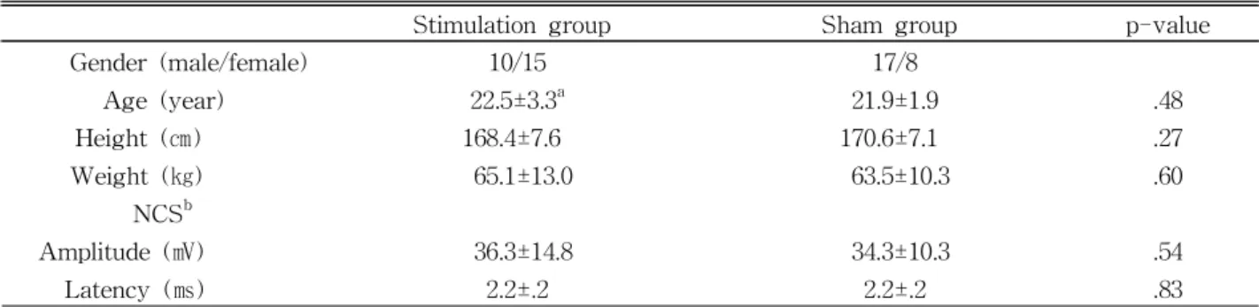
Stimulation group Sham group p-value
Gender (male/female) 10/15 17/8
Age (year) 22.5±3.3
a21.9±1.9 .48
Height (㎝) 168.4±7.6 170.6±7.1 .27
Weight (㎏) 65.1±13.0 63.5±10.3 .60
NCS
bAmplitude (㎷) 36.3±14.8 34.3±10.3 .54
Latency (㎳) 2.2±.2 2.2±.2 .83
a
mean±standard deviation,
bnerve conduction study.
Table 1. Demographic and general characteristics of the subjects (N=50) but they are relieved soon after stimulation and do
not continue (Antal et al, 2007; Poreisz et al, 2007).
Recent, research into decision making (Fecteau et al, 2007), language (Flöel et al, 2008), memory (Fregni et al, 2005), and pain (Fregni et al, 2007) has inves- tigated the clinical application of tDCS. These re- searchers have reported the effects of cerebral cortex control through diverse neural networks. In partic- ular, tDCS is used as an excellent means for en- hancing mood and anxiety in patients suffering from depression, and also to control chronic pain (Boggio et al, 2008) in patients with traumatic spinal cord in- jury (Fregni et al, 2006b), fibromyalgia (Fregni et al, 2006a), and cancer (Silva et al, 2007). There has been much research, in various fields, into the effects of applying tDCS, but most of the research into sen- sory functions, dealing with pain and its mecha- nisms, has not been verified. Boggio et al (2008) ap- plied anodal tDCS to different cerebral cortex areas of healthy adults, and reported that the perception and pain threshold in the primary motor cortex (M1) and the pain threshold only in the dorsolateral pre- frontal cortex (DLPFC) increased.
The current perception threshold (CPT) test is a quantitative sensory function test and may be applied to patients without discomfort and within a relatively short time compared to other existing tests. This test selectively stimulates the peripheral nervous fi- bers; the large myelinated nerve Aβ, small myeli- nated nerve Aδ, and unmyelinated nerve C in the form of a sine curve at 2000 ㎐, 250 ㎐, and 5 ㎐. It
is possible to quantify the sensory threshold by elec- trical stimulation through the skin with three differ- ent frequencies, and therefore the test is used for di- agnosis of various neuropathies including peripheral neuropathy (Katims et al, 1987). Kodama et al (2009) examined changes in the thresholds of Aβ, Aδ, and C by applying the CPT test to the M1, and the so- matosensory evoked potentials (SEPs) test to the primary sensory cortex (S1), using low frequency rTMS. According to the CPT test of the M1, thresholds of Aβ, Aδ, and C all increased, and excit- ability of the S1 was inhibited in the SEPs. To date, diverse studies have measured sensory changes after the application of tDCS, but there has been no study that investigated changes in each sensory nerve as Kodama et al (2009) did. Therefore, this study ap- plied tDCS to the M1 of the cerebral cortex, and measured changes in the peripheral sensory nerves, thereby clarifying the effects of tDCS on sensory nerves and providing evidential material for its clin- ical application.
Methods
Subjects
The subjects were healthy, right-handed adults
who did not have a history of brain damage or neu-
rological abnormality, and did not exhibit any prob-
lem in electroneurography. The number of subjects
was 50 (male: 27, female: 23) and they were equally
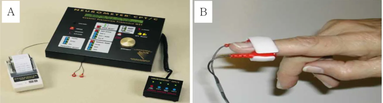
A B
Figure 1. A: Method of current perception threshold test, B: A Neurometer
Ⓡcurrent perception threshold (CPT)/C was used to measure CPT values at frequencies of 2000, 250, and 5 ㎐ in the right finger to assess the tactile sense, fast pain, and slow pain, respectively.
and randomly assigned to either a tDCS group or a sham tDCS group. Demographic data are shown in Table 1. Sufficient explanation was given to them and a written consent was obtained from them.
Electroneurography
All the subjects received electroneurography (Viking IV, Nicolet Co., Kennewick, USA). electro- neurography was conducted prior to the CPT test in order to verify whether the subjects’ right upper ex- tremity sensory nerves were normal. For the electro- neurography, median nerves among the right upper extremity sensory nerves were measured in an ex- amination room, where the temperature was main- tained at between 26 and 28 ℃ (skin temperature: 30 to 32 ℃), according to the method presented by Liverson and Ma (Nische and Paulus, 2001).
Amplitudes and latencies of the sensory nerves were recorded.
Current perception threshold (CPT) test
CPT values of all subjects were calculated prior to the application of tDCS. The CPT test was con- ducted with a Neurometer
Ⓡ(Neurotron Inc., Baltimore, USA). The subjects sat comfortably on a chair, a thin layer of conductive gel was applied, and then a pair of gold electrodes was attached with an unstretched tape to the distal part of the distal inter- phalangeal joint of the second finger (Figure 1). The
subjects were randomly and equally assigned to a control group or to an experimental group, and then the CPT values were measured in a single blind-method and in manual mode. A current with frequencies of 2000 ㎐, 250 ㎐, 5 ㎐ was applied to the subjects with an intensity of stimulation starting from .001 ㎃, until the subjects felt the electrical current for the first time. The stimulation intensity ranged from .001 ㎃ to 9.99 ㎃. When the subjects felt the electrical current, the stimulation was turned off. The intensity was then lowered to 100 ㎂, an- other stimulation was given, and the threshold values were checked. Stimulation was given again within an error margin of 20 ㎂ to measure the threshold values. CPT values were repetitively measured to obtain a constant result. When the same result oc- curred twice, consecutively, the value was considered as the threshold of the subject. After applying tDCS to all the subjects, CPT values were measured again, using the method described above.
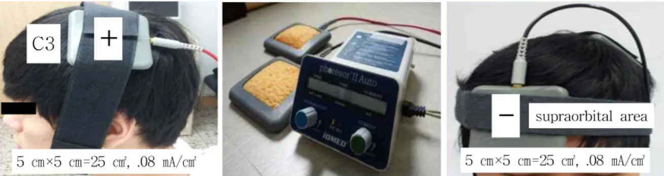
Transcranial direct current stimulation (tDCS)
The tDCS device, Phoresor
ⓇⅡ Auto (PM850, IOMED
Ⓡ, Utah, USA) was used. The size of the two sponge electrodes attached to the scalp was 25 ㎠ (5 ㎝×5 ㎝) and their current density was .08 ㎃/㎠.
The electrodes were soaked with .9% physiological
saline and attached to the subjects as tightly as
possible, but to an extent at which the subjects did
C3 +
5 ㎝×5 ㎝=25 ㎠, .08 ㎃/㎠
-
5 ㎝×5 ㎝=25 ㎠, .08 ㎃/㎠
supraorbital area
Figure 2. The equipment for the transcranial direct current stimulation and the stimulation targets (C3: central 3).
Stimulation group Sham group
2000 ㎐ 250 ㎐ 5 ㎐ 2000 ㎐ 250 ㎐ 5 ㎐
Pre 261.20±84.32
a109.10±51.75 142.60±56.00 315.50±62.38 130.40±65.67 189.40±123.03 Post 310.60±91.96 175.50±78.99 207.50±94.49 299.50±53.59 122.40±65.13 126.70±54.52
p value .000* .002* .008* .285 .552 .062
a