Biomedical Science Letters 2017, 23(4): 310~320 https://doi.org/10.15616/BSL.2017.23.4.310 eISSN : 2288-7415
Inhibitory Effects of Total Saponin Korean Red Ginseng on Thromboxane A2 Production and P-Selectin Expression via
Suppressing Mitogen-Activated Protein Kinases
Jung-Hae Shin1,§, Hyuk-Woo Kwon2,§, Man Hee Rhee2,† and Hwa-Jin Park1,†
1Department of Biomedical Laboratory Science, College of Biomedical Science and Engineering, Inje University, Gimhae 50834, Korea
2Laboratory of Veterinary Physiology and Signaling, College of Veterinary Medicine, Kyungpook National University, Daegu 41566, Korea
Ginseng has been widely used for traditional medicine in eastern Asia and is known to have inhibitory effects on cardiovascular disease (CVD) such as thrombosis, atherosclerosis, and myocardial infarction. Because, platelet is a crucial mediator of CVD, many studies are focusing on inhibitory mechanism of platelet functions. Among platelet activating molecules, thromboxane A2 (TXA2) and P-selectin play a central role in CVD. TXA2 leads to intracellular signaling cascades and P-selectin plays an important role in platelet-neutrophil and platelet-monocyte interactions leading to the inflammatory response. In this study, we investigated the inhibitory mechanisms of total saponin fraction from Korean red ginseng (KRG-TS) on TXA2 production and P-selectin expression. Thrombin-elevated TXA2 production and arachidonic acid release were decreased by KRG-TS dose (25 to 150 μg/mL)-dependently via down regulation of microsomal cyclooxygenase-1 (COX-1), TXA2 synthase (TXAS) activity and dephosphorylation of cytosolic phospholipase A2
(cPLA2). In addition, KRG-TS suppressed thrombin-activated P-selectin expression, an indicator of granule release via dephosphorylation of mitogen-activated protein kinases (MAPK). Taken together, we revealed that KRG-TS is a beneficial novel compound inhibiting TXA2 production and P-selectin expression, which may prevent platelet aggregation-mediated thrombotic disease.
Key Words: Total saponin fraction, MAPKs. Thromboxane A2, Arachidonic acid, P-selectin
INTRODUCTION
Platelets are activated at the sites of vascular injury. Upon activation, inositol-1,4,5-trisphosphate (IP3) is released from
plasma membrane (Berridge and Irvine, 1984) and released IP3 mobilizes Ca2+ from endoplasmic reticulum into cyto- plasm. Intracellular Ca2+ stimulates translocation of cytosolic phospholipase A2 (cPLA2) from cytosol to membranes and cPLA2 is phosphorylated at Ser505 by p38 mitogen-activated
Original Article
*Received: August 20, 2017 / Revised: September 21, 2017 / Accepted: September 26, 2017
§These authors contributed equally to this work.
†These corresponding authors contributed equally to this work.
†Corresponding author: Man Hee Rhee. Laboratory of Veterinary Physiology and Signaling, College of Veterinary Medicine, Kyungpook National University, Daegu 41566, Korea.
Tel: +82-53-950-5967, Fax: +82-53-950-5955, e-mail: [email protected]
†Corresponding author: Hwa-Jin Park. Department of Biomedical Laboratory Science, College of Biomedical Science and Engineering, Inje University, 197, Inje-ro, Gimhae, Gyungnam 50834, Korea.
Tel: +82-55-320-3538, Fax: +82-55-334-3426, e-mail: [email protected]
○CThe Korean Society for Biomedical Laboratory Sciences. All rights reserved.
○CCThis is an Open Access article distributed under the terms of the Creative Commons Attribution Non-Commercial License (http://creativecommons.org/licenses/by-nc/3.0/) which permits unrestricted non-commercial use, distribution, and reproduction in any medium, provided the original work is properly cited.
protein kinase (p38 MAPK) for full catalytic activity. The activated cPLA2 hydrolyzes the sn-2 acyl bond of phos- pholipids and releases arachidonic acid (AA) (Kramer et al., 1996; McNicol and Shibou, 1998). The AA is a precursor of thromboxane A2 (TXA2) (Hamberg et al., 1975) and the TXA2 biosynthesis is triggered by cyclooxygenase-1 (COX- 1) and TXA2 synthase (TXAS) (Needleman et al., 1976;
Patrignani et al., 1999). TXA2 is released from platelets and interacts with membrane receptor of other platelets in an autacoidal reaction, which acts as a positive promoter on acti- vated platelets and resting platelets simultaneously (Jennings, 2009). It is well known that aspirin and ozagrel have anti- thrombotic effects by inhibiting the TXA2 production. The mechanisms are concerned with COX-1 and TXAS (Patrono, 1994).
Selectins are expressed by activation or inflammatory response on various vascular cells, including platelets, leuko- cytes and endothelial cells (Kansas, 1996; Ley, 2003). L- selectin is present on leukocytes and E-selectin is present on endothelial cells and P-selectin is expressed on endothelial cells and platelets. The P-selectin in platelets is released by agonists from α-granule, and is re-expressed to the surface (Zarbock et al., 2007). P-selectin plays an important role in interactions with immune cells (von Hundelshausen and Weber, 2007).
Ginseng, the root of Panax ginseng Meyer, is known to have various pharmacological activities (Ernst, 2010; Kim and Park, 2011). Recently, it is reported that KRG-TS has an effect on CVD through reduction of blood pressure (Chung, 2010), anti-coagulation effects (Wee et al., 2010), endothe- lium relaxation (Jung et al., 2011), and inhibition of hyper- cholesterolemia-induced platelet aggregation (Hwang et al., 2008). In our previous report, we demonstrated that KRG- TS was involved in increase of cAMP level and subsequent reduction of [Ca2+]i mobilization in thrombin-induced rat platelet aggregation (Lee et al., 2013). However, the inhibi- tory mechanism by KRG-TS is not fully understood. Thus, we demonstrated that the modulatory mechanism of TXA2
production and P-selectin expression by KRG-TS on human platelets for prevention of CVD.
MATERIALS AND METHODS Materials
KRG-TS was obtained from Korea Ginseng Corporation (Daejeon, Korea). Thrombin was purchased from Chrono- Log Corporation (Havertown, PA, USA). Aspirin was pur- chased from Sigma Chemical Corporation (St. Louis, MO, USA). TXB2 EIA kit, COX-1 fluorescent activity assay kit, ozagrel, and Prostaglandin H2 were purchased from Cayman Chemical (Ann Arbor, MI, USA). Anti-phosphor-cPLA2
(Ser505), anti-phosphor-p38, anti-p38, anti-phosphor-ERK1/
2, anti-ERK1/2, anti-phosphor-JNK1, and anti-rabbit IgG- horseradish peroxidase conjugate (HRP) and lysis buffer were purchased from Cell Signaling (Beverly, MA, USA).
Anti-β-actin, anti-COX-1 and anti-TXAS were purchased from Santa Cruz Biotechnology (Santa Cruz, CA, USA).
PVDF membrane and ECL solution were purchased from GE Healthcare (Piseataway, NJ, USA). Human arachidonic release kit was purchased from (Cusabio, Wuhan, Hubei, China). CD62P (P-selectin) antibody was purchased from Biolegend (San Diego, CA, USA).
Preparation of washed human platelets
Human platelet-rich plasma (PRP) with ACD solution (0.8% citric acid, 2.2% sodium citrate, 2.45% glucose) was supplied from Korean Red Cross Blood Center (Changwon, Korea). To remove red blood cells, it was centrifuged for 10 min at 250 g, then centrifuged for 10 min at 1,300 g. The platelets were washed using washing buffer (138 mM NaCl, 2.7 mM KCl, 12 mM NaHCO3, 0.36 mM NaH2PO4, 5.5 mM glucose, and 1 mM Na2EDTA, pH 6.5), then resuspended in suspension buffer (138 mM NaCl, 2.7 mM KCl, 12 mM NaHCO3, 0.36 mM NaH2PO4, 0.49 mM MgCl2, 5.5 mM glucose, 0.25% gelatin, pH 6.9). The human platelets in suspending buffer was adjusted to a final concentration of 5 × 10⁸/mL and aforementioned procedures were per- formed at 25℃ to maintain platelet activity. The approval (PIRB12-072) for these experiments was received from National Institute for Bioethics Policy Public Institutional Review Board (Seoul, Korea).
Determination of platelet aggregation
Human Platelets (108/mL) were preincubated with or without KRG-TS in the presence of 2 mM of CaCl2 for 2 min at 37℃, then, stimulated with thrombin (0.05 U/mL).
The platelet aggregation rate was determined as an increase in light transmission for 5 min using an aggregometer (Chrono-Log Corporation, Havertown, PA, USA).
Measurement of TXB2
TXA2 is unstable and quickly converted to thromboxane B2 (TXB2) in platelets. Thus, the amounts of TXA2 were evaluated by measuring TXB2 concentration (Hamberg et al., 1975). Thrombin-induced platelet aggregation was ter- minated by adding both ice-cold 5 mM EDTA and 0.2 mM indomethacin to inhibit subsequent metabolism of arach- idonic acid to TXA2. The amounts of TXB2 were determined using a TXB2 EIA kit according to the procedure described by the manufacturer (Cayman Chemical Co, Ann Arbor, MI, USA).
Isolation of microsomal fraction
Human platelets (108/mL) containing suspending buffer (pH 7.4) with 1% protease inhibitor was sonicated at sen- sitivity 100% for 20 sec, 1 cycles, and 10 times on ice to obtain platelet homogenates using a sonicator (Bandelin, HD2070, Germany). And then, homogenates were ultracen- trifuged at 105,000 g for 1 h at 4℃ to obtain microsomal fraction containing endoplasmic reticulum (ER) membrane (Lagarde et al., 1981).
COX-1 activity assay
For the measurement of COX-1 activity, the microsomal fraction of platelets was pre-incubated with a positive con- trol, aspirin (500 μM), and with various concentrations of KRG-TS at 37℃ for 30 min. COX-1 activity was assayed with COX-1 fluorescent assay kit (Cayman Chemical Co, Ann Arbor, MI, USA).
Thromboxane A synthase activity
For the measurement of TXAS, microsomal fraction was preincubated with a positive control, ozagrel (11 nM, IC50),
and with various concentrations of KRG-TS at 37℃ for 5 min. The reaction is initiated by adding prostaglandin H2, and incubated at 37℃ for 1 min. The reactions are termi- nated by the addition of 1 M citric acid and neutralized by 1 N NaOH, the amount of TXB2 was determined by using TXB2 EIA kit according to the procedure described by manufacturer (Cayman Chemical Co, Ann Arbor, MI, USA).
Arachidonic release
Thrombin-induced platelet aggregation was terminated, and centrifuged with 200 g at 4℃ for 10 min, and super- natants were used for the assay of AA release EIA kit (Cusabio, Wuhan, Hubei, China). AA release was measured at 450 nm using a Synergy HT multi-Model Microplate Reader (BioTek Instruments, Winoosku, VT, USA).
Western blot for analysis of COX-1, TXAS, cPLA2-, and MAPKs-phosphorylation
The platelet aggregation was terminated by adding a lysis buffer (20 mM Tris-HCl, 150 mM NaCl, 1 mM Na2EDTA, 1 mM EGTA, 1% Triton X-100, 2.5 mM sodium pyrophos- phate, 1 mM β-glycerophosphate, 1 mM ATPase, 1 mM Na3VO4, 1 μg/mL leupeptin, and 1 mM phenylmethanesul- fonyl fluoride) then, platelet protein and microsomal frac- tion were measured using a BCA protein assay kit (Pierce Biotechnology, IL, USA). Protein (15 μg) and microsomal fraction (30 μg) were separated by SDS-PAGE (6%, 1.5 mm), then PVDF membrane was used for protein transfer. The dilutions for 1st antibody and 2nd antibody were 1:1,000 and 1:10,000, respectively. The membranes were visualized using ECL solution. The degrees of phosphorylation were analyzed using the Quantity One, Ver. 4.5 (BioRad, Hercules, CA, USA).
Determination of P-selectin release
After platelet aggregation, the platelets were resuspended by ice-cold PBS (pH 7.4) and cells were incubated with Alexa Fluor 488 anti-human CD62P for 60 min at 4℃
under the dark condition. Next, platelets were washed three times by ice-cold PBS to reduce unbounded antibody and fixed using 0.5% paraformaldehyde. Alexa Fluor 488 anti- human CD62P binding to platelets were determined using
flow cytometry (BD Biosciences, San Diego, CA, USA) and data were analyzed using CellQuest software.
Statistical analyses
The experimental results are presented as the mean ± standard deviation accompanied by the number of obser- vations. Data were determined by analysis of variance (ANOVA). If this analysis showed significant differences among the group means, then each group was compared by the Newman-Keuls method. Statistical analysis was carried out according to the SPSS 21.0.0.0 (SPSS, Chicago, IL, USA). P<0.05 was considered to be statistically significant.
RESULTS
Effects of KRG-TS on thrombin-induced human platelet aggregation
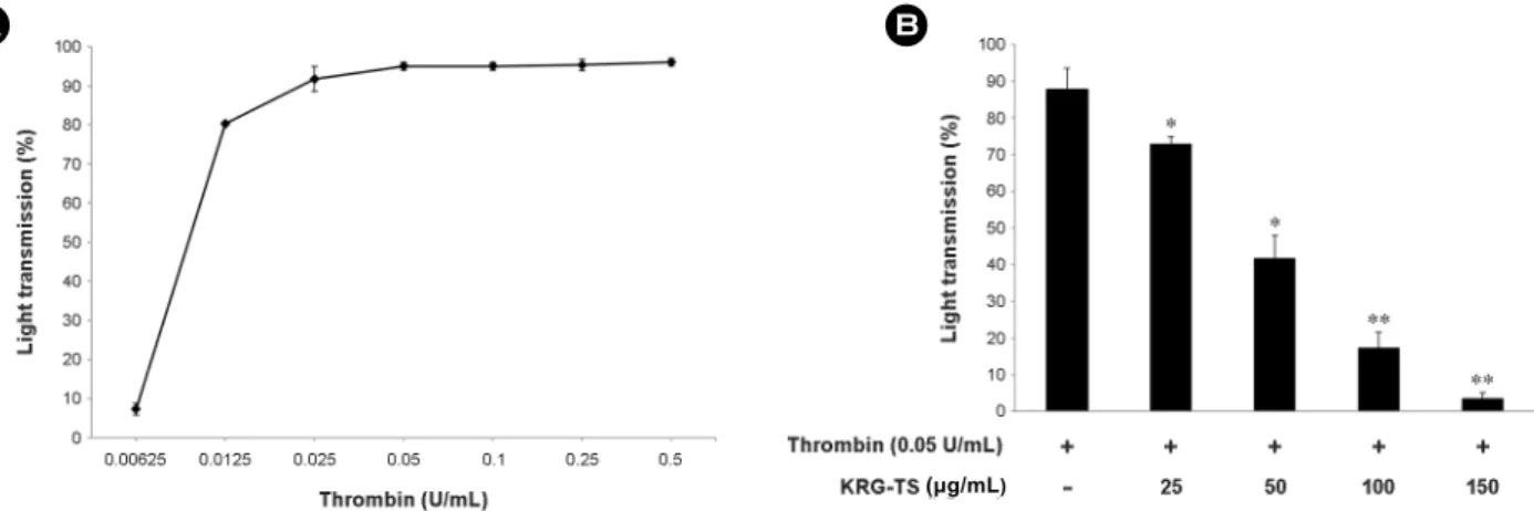
The concentration of thrombin-elevated maximal human platelet aggregation was approximately 0.05 U/mL (Fig.
1A). Thus, thrombin (0.05 U/mL) was used as an agonist in this study. Thrombin increased human platelet aggregation rate was 87.8 ± 5.7% However, KRG-TS strongly reduced thrombin-induced platelet aggregation dose-dependently (Fig.
1B). In our previous report showed that various concentra- tion of KRG-TS (20, 50, 100, 150 μg/mL) did not influence on unstimulated human platelet (Kwon et al., 2016).
Effects of KRG-TS on TXA2 production and COX-1, TXAS activity
Next, we investigated TXA2 production. The amount of TXA2 was markedly increased by thrombin from 1.1 ± 0.1, resting platelets, to 55.2 ± 2.2 ng/108 platelets. However, KRG-TS reduced the TXA2 production (Fig. 2). To deter- mine whether KRG-TS involves in inhibition of COX-1 and TXAS, we needed enzyme sources having COX-1 or TXAS.
We determined which fraction in platelets expresses COX-1 and TXAS in homogenates fraction, microsome fraction, Fig. 1. Effects of KRG-TS on thrombin-induced human platelet aggregation. (A) The concentration threshold of thrombin on human platelet aggregation. (B) The effects of KRG-TS on thrombin-induced human platelet aggregation. Platelet aggregation was carried out as described in "Materials and Methods" section. The data are expressed as the mean ± standard deviation (n=4). *P<0.05 versus the thrombin- stimulated human platelets, **P<0.01 versus the thrombin-stimulated human platelets.
Fig. 2. Effects of KRG-TS on TXA2 production. Effects of KRG- TS on TXA2 production. Determination of TXA2 production was carried out as described in "Materials and Methods" section. The data are expressed as the mean ± standard deviation (n=3). **P<
0.01 versus the thrombin-stimulated human platelets.
A B
(μg/mL)
(μg/mL)
and cytosols fraction. As the result, high expressed COX-1 (70 kDa) and TXAS (58 kDa) were observed in microsomal fraction (Fig. 3A). Thus, we used to determine the activity
of COX-1 and TXAS using microsomal fraction. As shown in Fig. 3B, COX-1 activity of microsomal fraction in the absence of KRG-TS (control) was 2.3 ± 0.04 nmol/protein- mg/min. However, 50, 100, and 150 μg/mL of KRG-TS inhibited COX-1 activity to 1.8 ± 0.10, 1.5 ± 0.11, and 1.4
± 0.14 ng/protein-mg/min respectively. 500 μM of aspirin, a positive control, inhibited COX-1 activity to 1.3 ± 0.05 nmol/protein-mg/min (Fig. 3B).
To determine whether KRG-TS is involved in TXAS, cell-free enzyme assay method with microsomal fraction of platelets was also used. In microsomal fraction (control), TXAS activity was 200.1 ± 1.8 ng/protein-mg/min (Fig. 3C).
However, 50, 100, and 150 μg/mL of KRG-TS inhibited TXAS activity to 182.0 ± 2.2, 166.6 ± 2.1, and 150.2 ± 1.8 ng/protein-mg/min respectively. In addition, 11 nM of ozagrel as a positive control was used, which inhibited TXAS activity to 130.2 ± 1.1 ng/protein-mg/ min (Fig. 3C).
Effects of KRG-TS on Arachidonic acid release and cPLA2 phosphorylation
In order to verify the inhibitory mechanism of KRG-TS on TXA2 production, we investigated AA release. Thrombin- induced AA release increased to 1925.5 ± 22.2, but KRG- TS dose (25, 50, 100, 150)-dependently inhibited the AA release (Fig. 4A). Moreover, because cPLA2 acts as a key mediator to regulate the AA release in human platelets, we investigated whether KRG-TS inhibited the phosphorylation of cPLA2. As shown in Fig. 4B, the cPLA2 was strongly phosphorylated by thrombin (Fig. 4B, lane 2), but KRG-TS inhibited the cPLA2-phosphorylation dose-dependently (Fig.
4B, lanes 3 to 5). These results indicated that KRG-TS inhibited TXA2 production was due to the down regulation of cPLA2/AA release pathway.
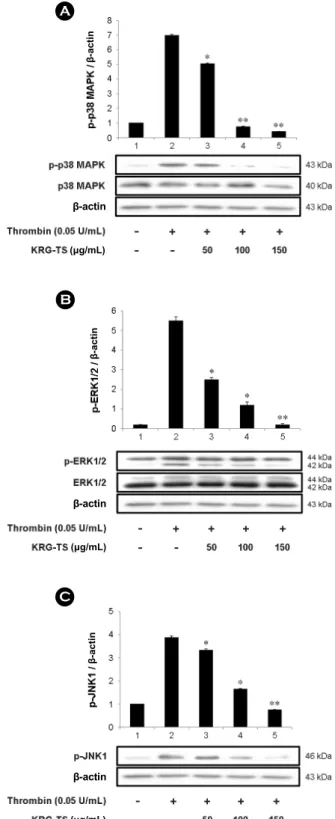
Effects of KRG-TS on MAPKs phosphorylation The MAPKs are divided into three subgroups: extra- cellular signal-regulated kinases (ERKs), c-Jun N-terminal kinases (JNKs), and p38 MAPK. It is reported that ERK2, JNK1 and p38 MAPK detected in human platelet and phos- phorylated by thrombin (Bugaud et al., 1990; Kramer et al., 1995; Fadal-Wollbold et al., 2002). It is well known that the enzyme activity of cPLA2 is achieved by its phosphory- Fig. 3. Effects of KRG-TS on COX-1, and TXAS activity. (A)
Determination of the effects of the enzyme sources on COX-1 and TXAS activities. (B) Effects of KRG-TS on COX-1 activity. (C) Effects of KRG-TS on TXAS activity. Western blot and COX-1 and TXAS activities were determined as described in "Materials and Methods" section. The data are expressed as the mean ± standard deviation (n=4). *P<0.05 versus the thrombin-stimulated human platelets.
B A
C
β-actin
(μg/mL) (μg/mL) (500 μM)
lation and p38 MAPK phosphorylates cPLA2 (Ser505). The p38 MAPK is also activated by its phosphorylation. Thus, we evaluated that KRG-TS has inhibitory effect on p38 MAPK phosphorylation. As shown in Fig. 5A, KRG-TS inhibited thrombin-elevated the phosphorylation of p38 MAPK dose (50 to 150 μg/mL)-dependently.
It has been revealed that ERK2 is involved in the TXA2
production (Yacoub et al., 2006; Garcia et al., 2007). The specific inhibitors of ERK1/2 and p38 MAPK, PD98059 and SB203580, blocked COX-1 directly, and SB203580 inhibited the conversion of PGH2 to TXA2 (Börsch-Haubold et al., 1998), which means that MAPKs may influence on endo-
Fig. 5. Effects of KRG-TS on MAPKs-phosphorylation. (A) Effects of KRG-TS on p38 MAPK phosphorylation. (B) Effects of KRG-TS on ERK1/2 phosphorylation. (C) Effects of KRG-TS on JNK1 phosphorylation. Western blot was determined as described in "Materials and Methods" section. The data are expressed as the mean ± standard deviation (n=3). *P<0.05 versus the thrombin- stimulated human platelets, **P<0.01 versus the thrombin-stimulated human platelets.
A
β-actin
(μg/mL)
β-actin
C
β-actin
(μg/mL)
β-actin
B
β-actin
β-actin
(μg/mL)
Fig. 4. Effects of KRG-TS on AA release and cPLA2-phos- phorylation. (A) Effects of KRG-TS on AA release. (B) Effects of KRG-TS on cPLA2-phosphorylation. AA release assay and Western blot were determined as described in "Materials and Methods"
section. The data are expressed as the mean ± standard deviation (n=3). *P<0.05 versus the thrombin-stimulated human platelets,
**P<0.01 versus the thrombin-stimulated human platelets.
A
B
(μg/mL)
(μg/mL) β-actin
β-actin
genous enzyme activity leading to TXA2 production.
It is also known that phosphorylation of ERK is involved in extracellular Ca2+ influx (Rosado and Sage, 2001; Rosado and Sage, 2002) and the study in JNK1-deficient mice showed that JNK1 is involved in granule secretion and TXA2
production (Adam et al., 2010). Thus, we investigated the
effect of KRG-TS on dephosphorylation of ERK2 and JNK1.
As shown in Fig. 5B, thrombin potently phosphorylated ERK2 (42 kDa) of ERK1 (44 kDa) and ERK2 (42 kDa) (Fig.
5B, lane 2) as compared with those by control, unstimulated platelets (Fig. 5B, lane 1), and phosphorylated JNK1 (46 kDa) (Fig. 5C, lane 2) as compared with those by control
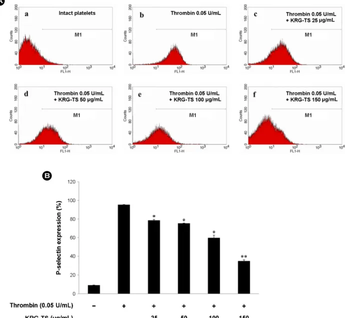
Fig. 6. Effects of KRG-TS on P-selectin expression. (A) The flow cytometry histograms on P-selectin expression. a, Intact platelets (base);
b, Thrombin (0.05 U/mL); c, Thrombin (0.05 U/mL) + KRG-TS (25 μg/mL); d, Thrombin (0.05 U/mL) + KRG-TS (50 μg/mL); e, Thrombin (0.05 U/mL) + KRG-TS (100 μg/mL); f, Thrombin (0.05 U/mL) + KRG-TS (150 μg/mL). (B) Effects of KRG-TS on thrombin-induced P-selectin expression (%). Determination of P-selectin expression was carried out as described in "Materials and Methods" section. The data are expressed as the mean ± standard deviation (n=3). *P<0.05 versus the thrombin-stimulated human platelets, **P<0.01 versus the thrombin-stimulated human platelets.
A
B
μg/mL
μg/mL μg/mL
μg/mL
(μg/mL)
(Fig. 5C, lane 1). However, KRG-TS dose (50 to 150 μg/
mL)-dependently inhibited thrombin-induced the phosphory- lation of ERK2 and JNK1 (Fig. 5B, lanes 3 to 5), (Fig. 5C lanes 3 to 5).
Effects of KRG-TS on P-selectin expression
Compounds in α-granule of platelets are known to involve in inflammation, coagulation and angiogenesis (Broos et al., 2011), these are also Ca2+-dependently released by various platelet agonists. In special, P-selectin is involved in inflam- mation by binding to the P-selectin glycoprotein ligand-1 receptor on monocyte (von Hundelshausen and Weber, 2007;
Zarbock et al., 2007). Therefore, we investigated the effect of KRG-TS on P-selectin expression on human platelet surface.
Thrombin elevated the expression of P-selectin (Fig. 6A-B) as compared with that by unstimulated platelets (Fig. 6A-a).
However, KRG-TS dose (25 to 150 μg/mL)-dependently inhibited thrombin-induced the expression of P-selectin (Fig.
6A-c~f, 6B). The expression of P-selectin is achieved by Ca2+ dependent kinase (Nishikawa et al., 1980), and ERK is involved in increase of intracellular Ca2+ concentration when platelets are activated (Rosado and Sage, 2002). In addition, JNK1 knockout mouse showed decreased granule release (Adam et al., 2010). Thus, it is thought that KRG- TS suppressed the P-selectin expression is achieved by dephosphorylation of ERK2 and JNK1.
DISCUSSION
Aspirin has a clear inhibition of TXA2 production in platelets through the inactivation of COX-1. Several clin- ical trials have shown that inhibition of platelet COX-1 activity by aspirin leads the prevention of myocardial in- farction and ischemic stroke. Aspirin reduces the risk of serious vascular events, but aspirin has a bleeding problem (Trialists'Collaboration, 2002). Therefore, a compound that can inhibit TXA2 production has a potential for application as an anti-thrombotic agent.
KRG-TS decreased thrombin-elevated platelet aggregation dose-dependently (Fig. 1B), which is accordance with that KRG-TS inhibited thrombin-induced TXA2 production (Fig.
2). However, it is not insufficient to understand an inhibitory
action of KRG-TS on TXA2 production. Thus, we tried to explain its inhibitory mechanism by KRG-TS on TXA2
production by assaying the activities of TXA2 production- associated COX-1, TXAS, cPLA2 and p38 MAPK. Because COX-1 and TXAS are localized in endoplasmic reticulum (Patrignani et al., 1999; Needleman et al., 1976), we isolated microsomes from cytosol in platelets, and confirmed the abundant expression of COX-1 (70 kDa) and TXAS (58 kDa) in microsomal fraction (Fig. 3A). We determined effects of KRG-TS on the activities of both enzymes by using microsomal fraction. As the results, KRG-TS inhibited COX- 1 and TXAS activity in the presence of KRG-TS (50, 100, 150 μg/mL) directly like aspirin and ozagrel (Fig. 3B, 3C).
Next, we focused on thrombin-induced AA release, upstream signaling molecule of TXA2 and as shown in Fig. 4A, thrombin-induced AA release was decreased by KRG-TS.
The AA release-associated signaling molecule, cPLA2 and p38 MAPK are also diminished by KRG-TS (Fig. 4B, 4C).
Comparing the inhibitory ratio of TXA2 production and its associated enzymes (COX-1, TXAS) by KRG-TS, the in- hibitory degree of TXA2 production by KRG-TS (150 μg/
mL) is very high as compared with inhibitory degrees of COX-1 and TXAS by KRG-TS (150 μg/mL). These results mean that the strong inhibition of TXA2 production by KRG- TS is due to the various inhibitory effects such as inhibition of COX-1, TXAS, AA release and dephosphorylation of cPLA2 and p38 MAPK.
The progression of atherosclerosis is triggered by inflam- matory cell such as monocytes, neutrophils, and macro- phages (Phillips et al., 2005). Thus, P-selectin is an important molecule for inflammatory response leading atherosclerosis.
Although KRG-TS has antiplatelet effects through inhibition of TXA2 production, if KRG-TS does not inhibit inflam- mation, atherosclerosis lesion would be generated at injury site of vascular wall. ATP and serotonin released from dense body are known to involve in amplification of platelet aggre- gation (Mustard and Packham, 1970; Holmsen and Day, 1970) and P-selectin released from α-granule is known to involve in causing of inflammation (von Hundelshausen and Weber, 2007; Zarbock et al., 2007). Because KRG-TS inhibited the P-selectin expression, it is thought that KRG- TS may inhibit aggregation-amplification and inflammation.
In real, many studies about anti-inflammatory activity by KRG-TS have been reported (Byeon et al., 2009; Park and Cho, 2009; Lee et al., 2014).
About dietary of a ginseng extract, It has reported that long-term (4 to 5 years) intake of red ginseng products (i.e.
water extract, tea, drink) inhibit platelet aggregation, blood coagulation and hyperlipidemia, an index of atherosclerosis, and their effects were well also sustained in the subjects who have obesity, hyperlipidemia, and hypertension (Lee and Park, 1998; Park et al., 2000). Moreover, it has reported that dietary water-extract of Korean red ginseng (KRG-WE) suppressed rabbit platelet aggregation under hypercholeste- rolemia (Hwang et al., 2008) and oral administration (250 to 500 μg/kg-BW) of KRG-WE significantly inhibited platelet aggregation ex vivo, and KRG-WE (300, 500 μg/mL) in- hibited washed rabbit platelet aggregation in vitro (Yang et al., 2015). With regard to the effects of ginsenosides on platelet aggregation, it is reported that ginsenoside Rg3 has inhibi- tory effect on collagen-induced blood platelet aggregation (Matsuda et al., 1985) and thromboxane A2 production, ATP release and [Ca2+]i mobilization (Lee et al., 1997). Further- more, ginsenoside Rg3-enriched red ginseng extract showed inhibitory effects on ERK 1/2, JNK and p38 MAPK (Jeong et al., 2017). In our previous study, we also checked the inhibitory effects of G-Rg3 (20S, 20R) in thrombin induced human platelet (Shin et al., 2015). Moreover, we revealed the inhibitory effects of ginsenoside Ro on vasodilator- stimulated phosphoprotein and clot retraction (Shin et al., 2016). These reports suggest that inhibitory effects of KRG- TS are achieved by ginsenoside Rg3 and ginsenoside Ro.
However, the inhibitory mechanism of ginsenosides is not fully understood.
In conclusion, we have revealed that anti-platelet effects of KRG-TS on TXA2 production and P-selectin expression.
KRG-TS suppressed the phosphorylation of cPLA2, and p38 MAPK leading TXA2 production. Simultaneously, KRG-TS decreased the phosphorylation of ERK2 and JNK1 leading P-selectin expression. These results show that the anti-platelet effects by KRG-TS are achieved through down regulation of MAPKs phosphorylation, which is useful to understand the anti-platelet effects of ginseng saponin we eat. There- fore, it is thought that KRG-TS would be a great potentiality
as a functional food in the therapy and prevention of CVD associated with platelet aggregation.
ACKNOWLEDGEMENTS
This research was supported by the National Research Foundation of Korea Grant funded by the Korean govern- ment (Grant no. 2015R1D1A1A09057204).
CONFLICT OF INTEREST
The authors declare no conflict of interest.
REFERENCES
Adam F, Kauskot A, Nurden P, Sulpice E, Hoylaerts MF, Davis RJ, Bryckaert M. Platelet JNK1 is involved in secretion and thrombus formation. Blood. 2010. 115: 4083-4092.
Berridge MJ, Irvine RF. Inositol trisphosphate, a novel second messenger in cellular signal transduction. Nature. 1984. 312:
315-321.
Börsch-Haubold AG, Pasquet S, Watson SP. Direct inhibition of cyclooxygenase-1 and-2 by the kinase inhibitors SB 203580 and PD 98059 SB 203580 also inhibits thromboxane synthase.
Journal of Biological Chemistry. 1998. 273: 28766-28772.
Broos K, Feys HB, De Meyer SF, Vanhoorelbeke K, Deckmyn H.
Platelets at work in primary hemostasis. Blood Reviews. 2011.
25: 155-167.
Bugaud F, Nadal-Wollbold F, Lévy-Toledano S, Rosa JP, Bryckaert M. Regulation of c-jun-NH2 terminal kinase and extracellular- signal regulated kinase in human platelets. Blood. 1990. 94:
3800-3805.
Byeon SE, Choi WS, Hong EK, Lee J, Rhee MH, Park HJ, Cho JY. Inhibitory effect of saponin fraction from Codonopsis lanceolata on immune cell-mediated inflammatory responses.
Archives of Pharmacal Research. 2009. 32: 813-822.
Chung IM, Lim JW, Pyun WB, Kim H. Korean red ginseng im- proves vascular stiffness in patients with coronary artery disease.
Journal of Ginseng Research. 2010. 34: 212-218.
Ernst E. Panax ginseng: an overview of the clinical evidence.
Journal of Ginseng Research. 2010. 34: 259-263.
Fadal-Wollbold F, Pawlowski M, Lévy-Toledano S, Berrou E, Rosa JP, Bryckaert M. Platelet ERK2 activation by thrombin is dependent on calcium and conventional protein kinases C but not Raf-1 or B-Raf. FEBS Letters. 2002. 531: 475-482.
Garcia A, Shankar H, Murugappan S, Kim S, Kunapuli SP. Regu-
lation and functional consequences of ADP receptor-mediated ERK2 activation in platelets. Biochemical Journal. 2007. 404:
299-308.
Hamberg M, Svensson J, Samuelsson B. Thromboxanes: a new group of biologically active compounds derived from prosta- glandin endoperoxides. Proceedings of the National Academy of Sciences of the United States of America. 1975. 72: 2994 -2998.
Holmsen H, Day HJ. The selectivity of the thrombin-induced platelet release reaction: subcellular localization of released and retained constituents. The Journal of Laboratory and Clinical Medicine.
1970. 75: 840-855.
Hwang SY, Son DJ, Kim IW, Kim DM, Sohn SH, Lee JJ, Kim SK. Korean red ginseng attenuates hypercholesterolemia- enhanced platelet aggregation through suppression of diacyl- glycerol liberation in high-cholesterol-diet-fed rabbits. Phyto- therapy Research. 2008. 6: 778-783.
Jennings LK. Role of platelets in atherothrombosis. The American Journal of Cardiology. 2009. 103: 4A-10A.
Jeong D, Irfan M, Kim SD, Kim S, Oh JH, Rhee MH. Ginsenoside Rg3-enriched red ginseng extract inhibits platelet activation and in vivo thrombus formation. Journal of Ginseng Research.
2017. 41: 548-555.
Jung YH, Park KY, Jeon JH, Kwak YS, Song YB, Wee JJ, Rhee MH, Kim TW. Red ginseng saponin fraction A isolated from Korean red ginseng by ultrafiltration on the porcine coronary artery. Journal of Ginseng Research. 2011. 35: 325-330.
Kansas GS. Selectins and their ligands: current concepts and con- troversies. Blood. 1996. 88: 3259-3287.
Kim SK, Park JH. Trends in ginseng research in 2010. Journal of Ginseng Research. 2011. 35: 389-398.
Kramer RM, Roberts EF, Strifler BA, Johnstone EM. Thrombin induces activation of p38 MAP kinase in human platelets.
Journal of Biological Chemistry. 1995. 270: 27395-27398.
Kramer RM, Roberts EF, Um SL, Börsch-Haubold AG, Watson SP, Fisher MJ, Jakubowski JA. p38 mitogen-activated protein kinase phosphorylates cytosolic phospholipase A2 (cPLA2) in thrombin-stimulated platelets. evidence that proline-directed phosphorylation is not required for mobilization of arachidonic acid by cPLA2. Journal of Biological Chemistry. 1996. 271:
27723-27729.
Kwon HW, Shin JH, Cho HJ, Rhee MH, Park HJ. Total saponin from Korean red ginseng inhibits binding of adhesive proteins to glycoprotein IIb/IIIa via phosphorylation of VASP (Ser 157) and dephosphorylation of PI3K and Akt. Journal of Ginseng
Research. 2016. 40: 76-85.
Lagarde M, Menashi S, Crawford N. Localisation of phospholipase A2 and diglyceride lipase activities in human platelet intra- cellular membranes. FEBS Letter. 1981. 124: 23-26.
Lee DH, Cho HJ, Kim HH, Rhee MH, Ryu JH, Park HJ. In- hibitory effects of total saponin from Korean red ginseng via vasodilator-stimulated phosphoprotein-Ser157 phosphorylation on thrombin-induced platelet aggregation. Journal of Ginseng Research. 2013. 37: 176-186.
Lee JH, Park HJ. Effects of intaking of red ginseng products on human platelet aggregation and blood lipids. Journal of Gin- seng Research. 1998. 22: 173-180.
Lee SR, Park JH, Kim ND, Choi KJ. Inhibitory effects of ginse- noside Rg3 on platelet aggregation and its mechanism of action. Journal of Ginseng Research. 1997. 21: 132-140.
Lee YJ, Han JY, Lee CG, Heo K, Park SI, Park YS, Kim JS, Yang KM, Lee KJ, Rhee MH, Kim SD. Korean red ginseng saponin fraction modulates radiation effects on lipopolysaccharide- stimulated nitric oxide production in RAW264. 7 macrophage cells. Journal of Ginseng Research. 2014. 38: 208-214.
Ley K. The role of selectins in inflammation and disease. Trends in Molecular Medicine. 2003. 9: 263-268.
Matsuda H, Kubo M, Tani T, Arichi S, Kitagawa I. Pharma- cological study on panax ginseng CA MEYER V.: effects of red ginseng on the experimental disseminated intravascular coagulation (4). on ginsenoside-Rg3, Rh1 and Rh2. The Japanese Society of Pharmacognosy. 1985. 39: 123-125.
McNicol A, Shibou TS. Translocation and phosphorylation of cytosolic phospholipase A2 in activated platelets. Thrombosis Research. 1998. 92: 19-26.
Mustard JF, Packham MA. Factors influencing platelet function:
adhesion, release, and aggregation. Pharmacological Reviews.
1970. 22: 97-187.
Needleman P, Moncada S, Bunting S, Vane JR, Hamberg M, Samuelsson B. Identification of an enzyme in platelet micro- somes which generates thromboxane A2 from prostaglandin endoperoxides. Nature. 1976. 261: 558-560.
Nishikawa M, Tanaka T, Hidaka H. Ca2+-calmodulin-dependent phosphorylation and platelet secretion. Nature. 1980. 287: 863 -865.
Park HJ, Lee JH, Lee SJ, Ham HS, Cho HJ, Lim CR, Park KH.
Effects of intaking of red ginseng products on correlationship between obesity and blood lipids. Korean Journal of Clinical Laboratory Science. 2000. 6: 253-260.
Park JS, Cho JY. Anti-inflammatory effects of ginsenosides from
panax ginseng and their structural analogs. African Journal of Biotechnology. 2009. 8: 3682-3690.
Patrignani P, Sciulli MG, Manarini S, Santini G, Cerletti C, Evangelista V. COX-2 is not involved in thromboxane bio- synthesis by activated human platelets. Journal of Physiology and Pharmacology. 1999. 50: 661-667.
Needleman P, Moncada S, Bunting S, Vane JR, Hamberg M, Samuelsson B. Identification of an enzyme in platelet micro- somes which generates thromboxane A2 from prostaglandin endoperoxides. Nature. 1976. 261: 558-560.
Patrono C. Aspirin as an antiplatelet drug. New England Journal of Medicine. 1994. 330: 1287-1294.
Phillips DR, Conley PB, Sinha U, Andre P. Therapeutic approaches in arterial thrombosis. Journal of Thrombosis and Haemostasis.
2005. 3: 1577-1589.
Rosado JA, Sage SO. Role of the ERK pathway in the activation of store-mediated calcium entry in human platelets. Journal of Biological Chemistry. 2001. 276: 15659-15665.
Rosado JA, Sage SO. The ERK cascade, a new pathway involved in the activation of store-mediated calcium entry in human platelets. Trends in Cardiovascular Medicine. 2002. 12: 229 -234.
Shin JH, Kwon HW, Cho HJ, Rhee MH, Park HJ. Inhibitory effects of total saponin from Korean red ginseng on [Ca2+]i mobilization through phosphorylation of cyclic adenosine monophosphate-dependent protein kinase catalytic subunit and inositol 1, 4, 5-trisphosphate receptor type I in human platelets.
Journal of Ginseng Research. 2015. 39: 354-364.
Shin JH, Kwon HW, Cho HJ, Rhee MH, Park HJ. Vasodilator- stimulated phosphoprotein-phosphorylation by ginsenoside Ro inhibits fibrinogen binding to αIIb/β3 in thrombin-induced
human platelets. Journal of Ginseng Research. 2016. 40: 359 -365.
Trialists'Collaboration A. Collaborative meta-analysis of rando- mised trials of antiplatelet therapy for prevention of death, myocardial infarction, and stroke in high risk patients. British Medical Journal. 2002. 324: 71-86.
von Hundelshausen P, Weber C. Platelets as immune cells. Circu- lation Research. 2007. 100: 27-40.
Wee JJ, Kim YS, Kyung JS, Song YB, Do JH, Kim DC, Lee SD.
Identification of anticoagulant components in korean red ginseng. Journal of Ginseng Research. 2010. 34: 355-362.
Yacoub D, Théorêt JF, Villeneuve L, Abou-Saleh H, Mourad W, Allen BG, Merhi Y. Essential role of protein kinase Cδ in platelet signaling, αIIbβ3 activation, and thromboxane A2 release. Journal of Biological Chemistry. 2006. 281: 30024 -30035.
Yang Y, Lee J, Rhee MH, Yu T, Baek KS, Sung NY, Cho JY.
Molecular mechanism of protopanaxadiol saponin fraction- mediated anti-inflammatory actions. Journal of Ginseng Re- search. 2015. 39: 61-68.
Zarbock A, Polanowska-Grabowska RK, Ley K. Platelet-neutrophil- interactions: linking hemostasis and inflammation. Blood Reviews. 2007. 21: 99-111.
https://doi.org/10.15616/BSL.2017.23.4.310
Cite this article as: JH Shin, HW Kwon, MH Rhee, HJ Park. Inhibitory Effects of Total Saponin Korean Red Ginseng on Thromboxane A2 Production and P-Selectin Expression via Suppressing Mitogen-Activated Protein Kinases. Biomedical Science Letters. 2017. 23: 310-320.