DOI 10.17480/psk.2016.60.2.92
6-Hydroxydopamine으로 유도된 질소적 세포 사멸에 대한 고려홍삼 추출물의 보호효과
이찬 · 장정희* · 박규환#
경북대학교 약학대학 약학연구소, *계명대학교 의과대학
(Received February 17, 2016; Revised March 23, 2016; Accepted March 24, 2016)
Protective Effect of Korean Red Ginseng against 6-Hydroxydopamine-induced Nitrosative Cell Death via Fortifying Cellular Defense System
Chan Lee, Jung-Hee Jang* and Gyu Hwan Park#
Institute of Pharmaceutical Sciences, College of Pharmacy, Kyungpook National University, Daegu 41566, Korea
*School of Medicine, Keimyung University, Daegu 42601, Korea
Abstract — Parkinson’s disease (PD) is one of the representative neurodegenerative movement disorders with the selec- tive loss of dopaminergic neurons in the substantia nigra. 6-Hydroxydopamine (6-OHDA) is widely used as an experimental model system to mimic PD and has been reported to cause neuronal cell death via oxidative and/or nitrosative stress. There- fore, daily intake of dietary or medicinal plants which fortifies cellular antioxidant capacity can exert neuroprotective effects in PD. In the present study, we have investigated the protective effect of Korean red ginseng (KRG) against 6-OHDA- induced nitrosative death in C6 glioma cells. Treatment of C6 cells with 6-OHDA decreased cell viability and increased expression of inducible nitric oxide synthase, production of nitric oxide as well as peroxynitrite, and formation of nitroty- rosine. 6-OHDA led to apoptotic cell death as determined by decreased Bcl-2/Bax, phosphorylation of JNK, activation of caspase-3, and cleavage of PARP. Conversely, pretreatment of C6 cells with KRG attenuated 6-ODHA-induced cytotoxicity, apoptosis, and nitrosative damages. To further elucidate the molecular mechanism of KRG protection against 6-OHDA- induced nitrosative cell death, we have focused on the cellular self-defense molecules against exogenous noxious stimuli.
KRG treatment up-regulated heme oxygenase-1 (HO-1), a key antioxidant enzyme essential for cellular defense against oxi- dative and/or nitrosative stress via activation of Nrf2. Taken together, these findings suggest KRG may have preventive and/
or therapeutic potentials for the management of PD.
Keywords □ Korean red ginseng, 6-hydroxydopamine, C6 glioma cells, heme oxygenase-1, nitrosative stress
파킨슨씨병은 진행형 퇴행성 뇌질환으로 중뇌의 흑색질 치밀 부(substantia nigra pars compacta, SNpc) 도파민성 신경세포의 선택적인 사멸에 의한 도파민의 분비 저하가 주요 원인으로 인 식되며, 경직(rigidity), 안정시 진전(resting tremor), 서동 (bradykinesia), 균형유지 및 보행장애 등의 증상을 나타낸다.1)병 리적인 현상으로는 오접힘(misfolding) 단백질의 응집(aggregation) 에 의한 루이소체(Lewy body)들이 사후 부검한 인간의 중뇌 흑
색질 치밀부에서 관찰되고 있으며, 이러한 오접힘은 산화적 및 질소적 스트레스에 의한 것으로 보고 되고 있다.2)
6-Hydroxydopamine(6-OHDA)은 카테콜아민성 신경독소로 파 킨슨씨병의 in vitro 및 in vivo 실험 모델 유도에 광범위하게 사 용되고 있으며 과산화수소(hydrogen peroxide, H2O2), superoxide anion, hydroxyl radical 등 활성산소종(reactive oxygen species, ROS)의 생성, 칼슘 항상성 교란 및 미토콘드리아 complex I과 IV의 기능 억제를 통해 산화적 스트레스를 유발하는 것으로 보 고되었다.3,4) 실제 파킨슨씨병 환자의 뇌에서 전이금속의 수치 및 지질과산화, 산화적 DNA 손상이 높게 나타났으며, 반대로 내인 성 항산화 물질인 글루타치온(glutathione, GSH)량과 항산화 효 소의 발현은 낮은 것으로 알려졌다.5)한편, 최근 6-OHDA는 nitric oxide(NO)와 같은 활성질소종(reactive nitrogen species, RNS)
#
Corresponding Author Gyu Hwan Park
Institute of Pharmaceutical Sciences, College of Pharmacy, Kyung- pook National University, Daegu 41566, Korea
Tel.: 053-950-8576 Fax.: 053-950-8557 E-mail: [email protected]
Short Report
종설의 생성도 증가시키는 것으로 보고되었다.6) 생성된 NO는 superoxide anion과 결합하여 더욱 강력한 활성질소종인 peroxynitrite를 생성하며, 이는 생체내 거대분자인 지질, 단백질, DNA의 산화적 손상을 통해 도파민성 신경세포의 선택적 세포사 멸을 유발하는 것으로 in vitro 및 in vivo 실험, 파킨슨씨병 환 자의 뇌 연구를 통해서 검증되었다.7)
성상세포(astrocyte)는 중추신경계에서 세포내 · 외의 이온농도 유지, 구조적 지지, 신경전달물질의 합성과 분비, 신경세포에 영 양물질 제공 등 신경세포의 발달, 분화, 성장, 복구, 재생의 과정 에서 중요한 역할을 한다고 알려져 있다.8)특히 성상세포로부터 분비되는 gilal cell line-derived neurotrophic factor(GDNF)는 도파민계 신경세포 모델에서 H2O2와 6-OHDA로 야기되는 세포 독성을 억제하였으며,9,10) 6-OHDA로 유도된 파킨슨씨병 마우스 동물모델에서 흑질부의 도파민성 신경세포 손상을 감소시켰다.11) 따라서, 6-OHDA로 인한 성상세포 사멸을 억제하고 그 기능을 강화시킨다면 파킨슨씨병에서 도파민성 신경세포의 손상에 대해 보호효과를 가질 것으로 기대되며, 이러한 일환으로 최근 식품 또는 약용식물에 존재하는 신경세포 보호 천연소재에 대한 관심 이 높아지고 있다.
현재까지 파킨슨씨병 치료제로 사용되는 약물에는 L-DOPA 제 제, 도파민 수용체 효능약물, 항콜린성 약물, monoamine oxidase (MAO) 저해제 등이 있으며, 이 약물들은 도파민의 전구체 또는 도파민 수용체에 직접 작용하는 물질들로 파킨슨씨병의 치료에 효과를 가지고 있지만, 장기간 사용 시 오심, 구토, 식욕감퇴 등 의 부작용을 유발한다. 따라서 세포내 신경세포 방어기전을 강 화할 수 있는 새로운 치료방법의 개발은 도파민성 신경세포를 활 성화하여 도파민의 생성 및 분비를 촉진시키고, 현재 치료제로 사용되는 약물들의 사용을 감소시켜 부작용을 경감할 수 있을 것 이다.
인삼은 한국, 중국, 일본 등 동양의학에서 일찍이 다양한 질환 의 예방 및 치료 목적으로 활용된 약용식물이다. 인삼 사포닌 진 세노사이드(ginsenoside)가 주요 약리 활성성분으로 항암작용, 항 당뇨작용, 면역기능 조절 작용 및 뇌기능 개선 작용 등의 다양한 약효를 가지고 있어 건강보조식품으로 널리 이용되고 있다.12)사 포닌 성분 중 ginsenoside Rg1는 파킨슨씨병의 세포실험 모델인 methyl-4-phenyl-1, 2, 3, 6-tetra-pyridine(MPTP), 1-methyl-4- phenylpyridinium(MPP+), 6-OHDA에 의해 유도된 도파민성 신 경세포의 산화적 세포사멸 대해 보호효과가 있는 것으로 보고되 었다.13-15)
특히, 고려홍삼은 6년근 인삼의 뿌리를 쪄서 말린 것으로 고 려홍삼추출물은 암, 고혈압, 당뇨, 자연적 상처치유, 간독성, 류 마티스 관절염, 비만 등의 다양한 질환에서 약리 작용을 가지고 있는 것으로 알려져 있다.16)뇌질환에서의 약리효과로는 N- methyl-D-aspartate(NMDA) 및 베타아밀로이드(Aβ25-35)로 유도
되는 신경세포 사멸에 대한 보호 작용,17)치매 환자에서의 기억 력 증진,18) 우울증19) 및 주의력 결핍 과잉 행동 장애20) 환자에 서 임상증상 개선 효과 등이 보고되었다. 고려홍삼의 뇌신경 보 호 효과에 대해 최근 다양한 뇌질환 모델에서 제시하고 있지만 파킨슨씨병에 대한 효능검토는 다소 부족한 실정이다, 따라서 본 연구에서는 파킨슨씨병 모델 유도에 사용되어지고 있는 6-OHDA 을 성상세포에 처리하여 특히 6-OHDA에 의해 유도되는 질소적 스트레스에 대한 고려홍삼의 신경보호 효과 및 작용기전을 규명 하고자 하였다.
실험 방법
실험 재료
본 실험에 사용한 랫트 성상세포주 C6 glioma 세포는 ATCC (Rockville, MD, USA)로부터 분양받아 사용하였으며, 세포배양 을 위한 Dulbecco’s modified Eagel’s medium(DMEM) 배지, fetal bovine serum(FBS) 및 항생제(penicillin/streptomycin)는 Gibco(Grand Island, NY, USA)사 제품을 사용하였다. 6- Hydroxydopamine(6-OHDA), MTT[3-(4,5-dimethylthiazol-2-yl)- 2,5-diphenyltetrazolium bromide] 및 그 외 일반 시약들은 Sigma 제품(St. Louis, MO, USA)을 이용하였다. 고려홍삼추출 물(Korean red ginseng extract, KRG) 시료(홍삼정 201-9091) 는 한국인삼공사(대전, 대한민국)로부터 제공받아 사용하였다.
세포배양 및 시약처리
C6 glioma 세포는 10% FBS, penicillin(10,000 IU/ml) 및 streptomycin(10,000 g/ml)을 포함하는 DMEM 배지를 이용하여, 5% CO2, 37oC 조건의 배양기를 사용하여 배양하였다. 세포배양 배지는 2일마다 새로운 배지로 갈아주었으며, 실험을 위하여 각 실험에서 요구되는 수의 세포를 도포한 후, 24시간 뒤 세포가 안 정적으로 부착되면 고려홍삼추출물(KRG)을 1시간 전처리 후 6- OHDA을 24시간 처리하였다.
세포생존율 측정
세포생존율 측정을 위하여 MTT 분석법을 이용하였다. MTT 분석법은 노란색의 tetrazolium 시약이 살아있는 세포의 미토콘 리아성 환원 효소에 의해 보라색의 formazan으로 변환되는 원리 를 이용하여 측정하는 분석법이다. C6 glioma 세포를 48-well plate에 5×10⁴cells/300μl로 도포한 후 세포가 안정적으로 부착 되면 KRG을 1시간 전처리하고 6-OHDA을 가하여 24시간 동안 추가 배양하였다. 이후 MTT 시약이 첨가된 새로운 배지로 갈아 준 뒤 37oC 배양기에서 2시간 동안 배양하였다. 상등액을 제거 한 후 DMSO 200 μl를 가하여 formazan을 완전히 용해한 다음, microplate reader(Emax, Molecular device, USA)를 사용하여
570 nm에서 흡광도를 측정하여 대조군의 흡광도를 100% 기준 으로 상대적인 세포생존율(%)을 산출하였다.
TUNEL 염색법
TUNEL 분석법은 세포사멸 과정에서 일어나는 DNA 분절 (fragmentation)을 측정하는 방법이다. C6 glioma 세포를 4-well slide에 도포한 다음, 24시간 뒤 세포가 안정적으로 부착되면 KRG를 30분간 전처리 후 6-OHDA을 처리하였다. 24시간 뒤 10% neutral buffered formalin을 이용하여 30분간 실온에서 고 정시킨 다음 인산완충용액(phosphate-buffered saline, PBS)으로 3회 세척하였으며, 3% H2O2을 함유하는 메탄올을 이용하여 4oC 에서 1시간 동안 배양하여 내인성 peroxidase의 활성을 차단하 였다. 이후, permeabilization solution(0.1% Triton X-100 in 0.1% sodium acetate)을 사용하여 4oC에서 10분간 배양하였으며, PBS로 3회 세척 후 Roche사(Mannheim, Germany)의 terminal deoxynucleotidyl transferase(TdT)와 digoxigenin-labeled nu- cleotides를 이용하여 37oC에서 1시간 배양하여 표지시켰다. PBS 세척 후 peroxidase-conjugated anti-goat antibody를 가하여 37°oC에서 30분 동안 반응시킨 다음 Vector사(Burlingame, CA, USA)의 diamino-benzidine(DAB) 시약을 이용하여 발색시킨 뒤 50% glycerol(v/v)을 사용하여 봉입 후 현미경으로 형태를 관찰 하였다.
Western blot analysis
6-OHDA 및 KRG를 C6 glioma 세포에 처리 한 후, Pierce사 (Rockford, IL, USA)의 RIPA buffer를 이용하여 세포들을 용해 시켰다. 이후 4oC 원심분리기를 이용하여 14,000 g에서 15분간 원심분리하여 상층액으로부터 단백질을 추출하였다. 단백질 정 량은 Pierce사의 bicinchoninic acid(BCA) 단백질 정량 시약을 사용하였으며, 정량 후 30 μg의 단백질을 10-12% polyacrylamide gel(SDS-PAGE)로 전기영동한 다음 polyvinylidene fluoride (PVDF) 멤브레인(membrane)으로 300 mA에서 4시간 동안 트랜 스퍼 하였다. 멤브레인을 0.1% tween-20을 포함하는 5% nonfat dry milk를 이용하여 실온에서 1시간 동안 blocking한 다음, 1차 항체들로 4oC에서 overnight 배양하였다. PBST(PBS, 0.1%
Tween-20)로 10분간 3회 멤브레인을 세척 후, 2차 항체를 실온에 서 1시간 배양한 다음, Pierce사의 enhanced chemoluminescence (ECL) 시약을 사용하여 발색시켰으며, LAS4000 image(Fujifilm, Japan) 장비를 사용하여 촬영하였다.
Nitric oxide 생성 측정
Nitric oxide 유래의 nitrite-nitrate의 측정을 위하여 C6 glioma 세포에 KRG를 30분간 전처리 한 후 6-OHDA와 함께 24시간 배양한 다음 세포배양액 150 μl와 Griess 시약[0.1% N-(naphthyl)
ethylenediamine dihydrochloride and 1% sulfanilamide in 5%
phosphoric acid]을 동량 첨가한 후 실온에서 30분간 반응시켰다.
이후 microplate reader를 사용하여 540 nm에서 흡광도를 측정 하였다.
Peroxynitrite 생성 측정
Peroxynitrite의 측정을 위하여 C6 glioma 세포에 KRG를 30 분간 전처리 한 후 6-OHDA 가하여 24시간 배양하였다. 이후 50μM dihydrorhodamine 123(DHR123, Invitrogen, Carlsbad, CA, USA)을 이용하여 37oC에서 30분간 배양하였다. 30분 뒤 PBS로 세척 후, DMSO를 사용하여 세포를 용해시킨 다음 microplate reader(Gemini XS, Molecular devices Co.)를 이용 하여 485 nm(excitation)와 535 nm(emission)에서 형광을 측정하 였다.
면역세포화학염색법
Nitrotyrosine의 측정을 위하여, 6×10⁴cells/600μl의 C6 세포 를 4-well 슬라이드에 도포한 다음, KRG를 30분간 전처리 한 후 24시간 6-OHDA을 처리하였다. PBS로 세척 후 10% neutral buffered formalin를 사용하여 실온에서 30분 동안 고정 뒤, 1%
bovine serum albumin(BSA)로 상온에서 1시간 동안 배양하여 비특이적 결합을 차단(blocking)하였다. 1차 항체인 anti- nitrotyrosine 항체로 4oC에서 overnight 배양한 후, PBS로 세척 한 다음 biotinylation 된 2차 항체로 1시간 동안 실온에서 배양 하였다. 다음 avidin이 결합되어 있는 biotinylated HRP enzyme reagent(Santa Cruz)로 30분간 배양 후 Vector사의 DAB 발색시 약을 이용하여 발색한 다음 50% glycerol(v/v)을 사용하여 봉입 후 현미경으로 형태를 관찰하였다.
통계처리
실험결과는 평균±표준편차로 표기하였다. 그룹간의 통계적 유 의성은 SPSS 프로그램을 사용하여 ANOVA로 검정한 후, 사후 분석으로 Turkey’s test를 실시하여 p<0.05 일 때 유의성이 있 다고 판정하였다.
실험 결과 및 고찰
6-OHDA로 유도된 성상세포 독성에 대한 KRG의 보호효과를 검토하기 위하여, 다양한 농도(0, 0.25, 0.5, 1 mg/ml)의 KRG를 C6 glioma 세포에 한 시간 전처리하고 이후 6-OHDA(200 μM) 와 함께 24시간 동안 처리하여 MTT reduction assay로 세포생 존률을 측정하였다. 6-OHDA의 세포독성에 의한 세포생존율은 45.4%로 대조군에 비해 현저한 감소를 나타내었으며, 반면 KRG 를 전처리 한 그룹에서는 56.4%, 66.3%, 82.6%로 농도 의존적
으로 유의성 있게 세포생존율이 증가하였다(Fig. 1). 파킨슨씨병 세포 및 동물 모델에서 고려홍삼의 보호효능을 검토한 연구결과 들은 거의 없었지만, 인삼추출물의 경우 MPP+로 유발된 신경세 포 독성에 대한 보호 효과가 일부 보고된 바 있다.21,22)
이후 실험에서는 6-OHDA로 인한 세포사멸의 형태를 규명하 기 위하여 아폽토시스(apoptosis)의 대표적인 측정 지표 중의 하 나인 DNA 단편화(DNA fragmentation)을 in situ nick-end labeling(TUNEL staining)으로 관찰하였다. 아폽토시스 과정에서 endonuclease의 활성화에 의해 DNA는 180~200 bp의 단위로 분 절화 되며 TUNEL staining은 분절된 DNA의 3'-OH 위치에 표 식된 dUTP를 결합시켜 효소-기질 발색반응을 이용하여 측정하 게 된다. 6-OHDA(200 μM)를 C6 glioma 세포에 24시간 처리한 결과 TUNEL staining에 의해 갈색으로 염색된 세포의 수가 증 가되었으며, 대조군과 비교하여 59.8% 가량의 세포가 아폽토시 스로 확인되었다(Fig. 2). 한편, 이는 KRG를 전처리 함으로써 특 히 1 mg/ml의 농도에서 아폽토시스가 효과적으로 억제되었다 (Fig. 2).
아폽토시스를 조절하는데 관여하는 여러 가지 유전자 중에서 대표적인 것이 Bcl-2 gene family이며, 이는 기능적인 면에서 크 게 두 가지로 나누어지고, 양자의 상대적인 비가 세포의 생존과 사멸을 결정짓는데 중요한 역할을 하게 된다. 즉, Bcl-2와 Bcl- XL은 아폽토시스를 억제하는 anti-apoptotic한 기능을 가지며, Bax, Bad, Bak, Bid, Bik, Bcl-XS등은 아폽토시스를 매개하는 proapoptotic한 기능을 가져, Bcl-2 또는 Bcl-XL의 비가 상대적으 로 높아지는 경우 세포는 생존으로 반대의 경우 사멸로 진행을 하게 된다.23) 6-OHDA(200μM)를 C6 glioma 세포에 처리한 결
과 anti-apoptotic 한 Bcl-2의 발현이 감소되고, proapoptotic 한 Bax의 발현이 증가되는 것을 Western blot을 통해 관찰할 수 있 었으며, 이러한 변동은 KRG를 전처리 함으로써 다시 정상상태 로 회복되었다(Fig. 3A).
이러한 Bcl-2 family 단백질들은 주로 미토콘드리아 막에 존재 하며, 이러한 미토콘드리아 단백질들의 발현 조절에는 c-Jun N- terminal kinase(JNK) 활성이 중요한 역할을 한다. 실제 6- OHDA(200μM)를 처리하는 경우 JNK에 인산화가 일어나 활성 이 증가됨을 Western blot을 통하여 관찰하였으며, 이는 KRG를 전처리 함으로써 농도 의존적으로 저해되었다(Fig. 3B). 이러한 아폽토시스 관련 신호전달은 궁극적으로 caspase 활성화와 PARP 의 절단(PARP cleavage)을 유발하며, DNA fragmentation을 일 으키게 된다. C6 glioma 세포에서 6-OHDA 처리에 의해 caspase-3와 PARP가 활성화되어, 절단된 형태의 cleaved caspase-3와 cleaved PARP의 양이 증가되었고, KRG를 처리한 경우 농도의존적으로 감소되었다(Fig. 3C~D). 이상의 결과를 요
Fig. 2 − Amelioration of 6-OHDA-induced apoptotic cell death by KRG. TUNEL staining was performed to measure apoptotic cell death by analysing DNA fragmentation in situ. A.
Representative images of TUNEL staining were indicated.
a: vehicle-treated control, b: 6-OHDA (200 μM), c: 6-OHDA (200 μΜ)+KRG (0.5 mg/ml), d: 6-OHDA (200 μM)+KRG (1 mg/ml). B. Quantitative data were derived from repre- sentative images from three independent experiments.
Data are shown as mean±S.D. (n=3).
##p<0.01: vehicle- treated control vs. 6-OHDA-treated alone groups, **p<
0.01: 6-OHDA vs. 6-OHDA+KRG groups.
Fig. 1 − Protective effect of KRG (Korean red ginseng extract) against 6-OHDA-induced cytotoxicity in C6 glioma cells.
MTT reduction assay was conducted to measure cee viability. C6 cells were pretreated with KRG (0, 0.25, 0.5, 1 mg/ml) for 1 h and further incubated with 6-OHDA (200 μM). Data are shown as mean±S.D. (n=3).
##p<0.01:
vehicle-treated control vs. 6-OHDA-treated alone groups,
**p<0.01: 6-OHDA vs. 6-OHDA+KRG groups.
약하면 6-OHDA이 유도하는 C6 glioma 세포사멸에 대한 KRG 의 신경보호효과는 Bcl-2 발현 증가, Bax 발현 감소, JNK 인산 화 억제, caspase-3 및 PARP 활성화 억제를 통해 매개되었다.
이전의 연구 결과에서 인삼추출물은 SH-SY5Y 세포에서 MPP+ 로 유도되는 아폽토시스 과정에서 Bax/Bc/-2의 증가, cytochrome c 방출, caspase-3 활성화를 억제함이 보고된 바 있다.21)
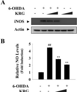
다음으로, 6-OHDA로 유도된 세포사멸에 대한 KRG의 보호 기전을 규명하기 위한 일환으로 질소적 스트레스 확인하기 위하 여 iNOS의 발현과 NO 생성을 측정하였다. 실험결과 6-OHDA 처리에 의해 iNOS의 발현과 NO의 생성이 모두 증가되었고 이 는 KRG를 전처리 함으로써 감소되었다(Fig. 4). 이전의 연구에 서도 다양한 신경세포 모델을 활용하여 6-OHDA가 iNOS의 발 현 증가를 통해 NO 생성을 유발하고, 천연물질을 처리하여 이 를 억제하는 경우 신경세포 보호 작용을 가짐을 보고하였다.24,25) 특히, 최근 6-OHDA 및 대표적 염증유발물질인 lipopolysaccharide 로 유도된 성상세포의 DNA 손상 및 아폽토시스 과정에서 iNOS 발현 증가를 통한 NO 생성과 이후 미토콘드리아 기능이상이 중 요한 역할을 하는 것으로 알려졌다.26)
이후 실험에서는 superoxide anion과 NO의 결합으로 생성되 고 더욱 반응성이 강한 산화물질인 peroxynitrite의 양을 DHR123 시약을 사용하여 측정하였다. C6 glioma 세포에 6-OHDA(200 μM) 처리시 peroxynitrite의 생성이 증가됨을 형광강도를 측정 함으로 확인했으며, 이는 KRG의 전처리로 감소되었다(Fig. 5A).
Peroxynitrite는 높은 반응성으로 인해 중요한 생체내 거대분자 들 산화시켜 구조 및 활성을 변형시키며 그 대표적인 산물 중의 하나가 nitrotyrosine이다. 면역세포염색법을 통해 확인한 결과, 6-OHDA를 처리 후 nitrotyrosine 생성량이 현저히 증가되었고, KRG를 함께 전처리한 세포에서는 상대적으로 생성량이 낮은 것 을 관찰하였다(Fig. 5B). 관련된 이전의 연구에서는 특히 인삼의 열처리 가공에 의해 항산화 성분의 함량이 증가되며 peroxynitrite 와 활성산소종의 소거능이 강화됨이 제시된 바 있다.27,28)
한편, KRG가 6-OHDA로 유도되는 질소적 신경세포 사멸에 대해 보호작용을 갖는 작용기전을 규명하기 위하여 생체내에 존 재하는 대표적인 항산화 효소인 HO-1의 발현을 Western blot으 로 측정하였다. 생체내에는 활성산소종 및 활성질소종에 의한 세 포사멸에 대하여 다양한 항산화 방어 시스템이 존재한다. 최근 이들에 의한 신경세포 보호 작용이 강조되고 있으며 이러한 일환 으로 HO-1에 대한 관심이 점차 높아지고 있다. HO-1은 산화-환 원 반응에 민감한 일종의 스트레스 단백질로, 다양한 조직 및 세 포에 있어, 항산화, 항염증, 세포방어, 세포사멸 억제 작용을 갖는
Fig. 4 − Effect of KRG on 6-OHDA-induced nitrosative stress. Cells were incubated with 6-OHDA for 24 h with or without KRG pretreatment. A. Protein expression of iNOS was monitored by Western blot analysis using anti-iNOS antibody. B. NO production was measured by Griess assay detecting the amount of nitrite released into media. Data are presented as mean±S.D. (n=3).
##p<0.01: vehicle-treated control vs. 6- OHDA-treated alone groups, *p<0.05 and **p<0.01: 6- OHDA vs. 6-OHDA+KRG groups.
Fig. 3 − Effect of KRG on 6-OHDA-induced apoptotic signaling
pathways in C6 glioma cells. Cells were pretreated with
KRG for 1 h and then exposed to 6-OHDA for 24 h. Protein
samples were isolated and prepared for Western blot
analysis. Protein expression of pro-apoptotic Bax and anti-
apoptotic Bcl-2 (A), phosphorylated and unphosphorylated
forms of JNK (B), and cleaved forms of caspase-3 (C) as
well as PARP (D) was measured. Actin levels were
monitored to confirm the equal amount of protein loading.
것으로 알려져 있기 때문에, 산화적 및 염증적 과정이 매개하는 퇴행성뇌질환의 새로운 치료표적으로 큰 주목을 받고 있다.29)특 히, HO-1의 작용에 의한 heme의 분해 산물들, 즉 일산화탄소 (carbon monoxide, CO), 빌리루빈(bilirubin), 빌리버딘(biliverdin) 등이 이러한 보호 효과를 매개하는 것으로 추정되고 있다.
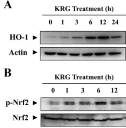
본 연구에서 C6 세포에 KRG를 처리하고 시간에 따른 HO-1 단백질의 변화를 Western blot으로 확인한 결과 시간의존적인 증가를 보였으며, 특히 12시간에 최고로 발현되어 지속적으로 유 지되었다(Fig. 6A). 이러한 HO-1의 발현은 상위 전사인자인 Nrf2 에 의해 조절을 받는 것으로 알려져 있으며, 본 연구에서도 KRG 를 C6 세포에 처리했을 때 인산화를 통한 Nrf2의 활성화가 6시 간대에 최대로 유도되었다(Fig. 6B). KRG 처리에 의해서 Nrf2 의 단백발현수준에는 유의적인 변화가 없었기 때문에, KRG는 C6 세포에서 Nrf2의 발현증가보다는 인산화를 통해 신경보호기
능 관련유전자의 전사활성을 증가시키는 것으로 사료된다. 이상 의 결과는 KRG가 HO-1-Nrf2로 이어지는 세포내 항산화 방어 기전을 높게 유지하여 질소적 스트레스로 인한 세포사멸에 대하 여 높은 저항성을 가짐을 시사한다. 최근 연구결과에 따르면 KRG 는 인체 혈관 내피 세포(human umbilical vein endothelial cells, HUVECs)와 PC12 세포에서 각각 과산화수소30)로 또는 내분비 계 교란물질인 polychlorinated biphenyls(PCB)31)로 유도된 세포 독성에 대해 Nrf2 활성화 및 HO-1 발현 증가를 통해 보호효과 를 나타냈다. 인삼과 홍삼의 활성성분 중 하나인 진세노사이드 Rb1은 SH-SY5Y 세포에서 HO-1 발현 증가를 통해 6-OHDA로 유 도되는 세포사멸을 억제하였으며, 이는 에스트로겐 수용체와 관 련된 PI3K/Akt/Nrf2 경로에 의해 매개되는 것으로 보고되었다.32)
결 론
6-OHDA로 유도된 C6 glioma 세포 사멸에 대한 KRG의 보호 효과 및 기전을 검토하여 다음과 같은 결론을 도출하였다.
1) 성상세포 모델인 C6 glioma 세포에서 KRG는 6-OHDA로 유도된 신경세포 독성을 보호하였다. 특히, 아폽토시스 과정에서 Bcl-2/Bax 발현을 정상화 시키고, JNK, caspase-3, PARP 활성화 의 신호전달을 억제하였다.
2) KRG는 6-OHDA로 야기되는 신경독성 및 세포사멸 과정에 서 질소적 손상 즉, iNOS의 발현 증가, NO 및 peroxynitrite 등 활성질소종의 생성, nitrotyrosine 형성을 효과적으로 억제하였다.
3) KRG는 특히 Nrf2 활성화 및 하위 항산화 유전자인 HO-1
Fig. 6 − Effect of KRG on the cellular antioxidnat defense system.
Cells were treated with indicated time with KRG and total proteins were extracted with RIPA buffer. Protein expression of antioxidant enzyme HO-1 (A) and activation of redox- sensitive transcription factor Nrf2 (B) were assessed by Western blot analysis using anti-HO-1 and anti-p-Nrf2 specific antibodies. The levels of actin and Nrf2 was compared as loading controls.
Fig. 5 − Attenuation of 6-OHDA-induced peroxynitrite formation and subsequent nitrosative damages by KRG. C6 cells were pretreated with KRG for 1 h and 6-OHDA was added to the media for additional 24 h. A. Intracellular peroxynitrite levels were measured by using DHR123 dye. Relative fluorescence intensity was calculated regarding control as 100%. B. Formation of nitrotyrosine was examined by immunocytochemistry using anti-nitrotyrosine antibody.
The representative images (left panel) and quantitative data (right panel) were indicated. Data are indicated as mean±
S.D. (n=3).
##p<0.01: vehicle-treated control vs. 6-OHDA- treated alone groups, *p<0.05 and **p<0.01: 6-OHDA vs.
6-OHDA+KRG groups. a: vehicle-treated control, b: 6-
OHDA (200 μM), c: 6-OHDA (200 μM)+KRG (0.5 mg/ml),
d: 6-OHDA (200 μM)+KRG (1 mg/ml).
의 발현을 증가시켜 세포 내 방어체계 강화를 통해 6-OHDA로 인한 질소적 스트레스 및 사멸로부터 성상세포를 보호했으며, 파 킨슨씨병 예방 및 치료를 위한 기능성 천연소재로서 KGR의 활 용가능성을 제시하였다.
이상의 결론은 신경세포의 적응반응을 이용한 신경보호기술을 개발하고, 질소적 손상을 다중치료표적에 포함시키는 등 새로운 파킨슨씨병 조절전략들을 수립하는데 과학적 근거를 제공한다.
감사의 말씀
이 논문은 2012학년도 경북대학교 신임교수정착연구비에 의 하여 연구되었으며, 이에 감사드립니다.
References
1) Protter, D., Lang, C. and Cooper, A. A. : α-Synuclein and mitochondrial dysfunction: A pathogenic partnership in Parkinson’s disease? Parkinson’s Dis. 2012, 829207 (2012).
2) Wilkaniec, A., Strosznajder, J. B. and Adamczyk, A. : Toxicity of extracellular secreted alpha-synuclein: Its role in nitrosative stress and neurodegeneration. Neurochem. Int. 62, 776 (2013).
3) Cohen, G. and Heikkila, R. E. : The generation of hydrogen peroxide, superoxide radical, and hydroxyl radical by 6- hydroxydopamine, dialuric acid, and related cytotoxic agents. J.
Biol. Chem. 249, 2447 (1974).
4) Glinka, Y. Y. and Youdim, M. B. : Inhibition of mitochondrial complexes I and IV by 6-hydroxydopamine. Eur. J. Pharmacol.
292, 329 (1995).
5) Youdim, M. B., Drigues, N. and Mandel, S. : Oxidative stress indices in Parkinson’s disease : biochemical determination.
Methods Mol. Med. 62, 137 (2001).
6) Henze, C., Earl, C., Sautter, J., Schmidt, N., Themann, C., Hartmann, A. and Oertel, W. H. : Reactive oxidative and nitrogen species in the nigrostriatal system following striatal 6-hydroxydopamine lesion in rats. Brain Res. 1052, 97 (2005).
7) Gupta, S. P., Yadav, S., Singhal, N. K., Tiwari, M. N., Mishra, S. K. and Singh, M. P. : Does restraining nitric oxide biosynthesis rescue from toxins-induced parkinsonism and sporadic Parkinson’s disease? Mol. Neurobiol. 49, 262 (2014).
8) Garzón, D., Cabezas, R., Vega, N., Ávila-Rodriguez, M., Gonzalez, J., Gómez, R. M., Echeverria, V., Aliev, G. and Barreto, G. E. : Novel Approaches in Astrocyte Protection:
from Experimental Methods to Computational Approaches. J.
Mol. Neurosci. Jan 23. [Epub ahead of print] (2016).
9) Gharib, E., Gardaneh, M. and Shojaei, S. : Upregulation of glutathione peroxidase-1 expression and activity by glial cell line-derived neurotrophic factor promotes high-level protection
of PC12 cells against 6-hydroxydopamine and hydrogen peroxide toxicities. Rejuvenation Res. 16, 185 (2013).
10) Sandhu, J. K., Gardaneh, M., Iwasiow, R., Lanthier, P., Gangaraju, S., Ribecco-Lutkiewicz, M., Tremblay, R., Kiuchi, K. and Sikorska, M. : Astrocyte-secreted GDNF and glutathione antioxidant system protect neurons against 6OHDA cytotoxicity. Neurobiol. Dis. 33, 405 (2009).
11) Voutilainen, M. H., Bäck, S., Pörsti, E., Toppinen, L., Lindgren, L., Lindholm, P., Peränen, J., Saarma, M. and Tuominen, R. K. : Mesencephalic astrocyte-derived neurotrophic factor is neurorestorative in rat model of Parkinson’s disease. J.
Neurosci. 29, 9651 (2009).
12) Jang, J. H. and Surh, Y. J. : Therapeutic potential of ginseng for the prevention and treatment of neurological disorders. In:
Functional Foods of the East, Taylor and Francis Group, LLC, p.
147 (2010).
13) Chen, X. C., Zhou, Y. C., Chen, Y., Zhu, Y. G., Fang, F. and Chen, L. M. : Ginsenoside Rg1 reduces MPTP-induced substantia nigra neuron loss by suppressing oxidative stress.
Acta Pharmacol. Sin. 26, 56 (2005).
14) Xu, H., Jiang, H., Wang, J. and Xie, J. : Rg1 protects the MPP+-treated MES23.5 cells via attenuating DMT1 up- regulation and cellular iron uptake. Neuropharmacol. 58, 488 (2010).
15) Xu, H., Jiang, H., Wang, J. and Xie, J. : Rg1 protects iron- induced neurotoxicity through antioxidant and iron regulatory proteins in 6-OHDA-treated MES23.5 cells. J. Cell. Biochem.
111, 1537 (2010).
16) Lee, S. M., Bae, B. S., Park, H. W., Ahn, N. G., Cho, B. G., Cho, Y. L. and Kwak, Y. S. : Characterization of Korean Red Ginseng (Panax ginseng Meyer): History, preparation method, and chemical composition. J. Ginseng Res. 39, 384. (2015).
17) Kim, S., Lee, Y. and Cho, J. : Korean red ginseng extract exhibits neuroprotective effects through inhibition of apoptotic cell death. Biol. Pharm. Bull. 37, 938 (2014).
18) Heo, J. H., Lee, S. T., Oh, M. J., Park, H. J., Shim, J. Y., Chu, K. and Kim, M. : Improvement of cognitive deficit in Alzheimer’s disease patients by long term treatment with korean red ginseng. J. Ginseng Res. 35, 457 (2011).
19) Jeong, H. G., Ko, Y. H., Oh, S. Y., Han, C., Kim, T. and Joe, S. H. : Effect of Korean Red Ginseng as an adjuvant treatment for women with residual symptoms of major depression. Asia Pac. Psychiatry. 7, 330 (2015).
20) Lee, S. H., Park, W. S. and Lim, M. H. : Clinical effects of korean red ginseng on attention deficit hyperactivity disorder in children: an observational study. J. Ginseng Res. 35, 226 (2011).
21) Hu, S., Han, R., Mak, S. and Han, Y. J. : Protection against 1- methyl-4-phenylpyridinium ion (MPP+)-induced apoptosis by
water extract of ginseng (Panax ginseng C.A. Meyer) in SH- SY5Y cells. J. Ethnopharmacol. 135, 34 (2011).
22) Kim, E. H., Jang, M. H., Shin, M. C., Shin, M. S. and Kim, C. J. : Protective effect of aqueous extract of Ginseng radix against 1-methyl-4-phenylpyridinium-induced apoptosis in PC12 cells. Biol. Pharm. Bull. 26, 1668 (2003).
23) Siddiqui, W. A., Ahad, A. and Ahsan, H. : The mystery of BCL2 family: Bcl-2 proteins and apoptosis: an update. Arch. Toxicol.
89, 289 (2015).
24) Zhang, L. Q., Sa, F., Chong, C. M., Wang, Y., Zhou, Z. Y., Chang, R. C., Chan, S. W., Hoi, P. M. and Lee, S. M. Y. : Schisantherin A protects against 6-OHDA-induced dopaminergic neuron damage in zebrafish and cytotoxicity in SH-SY5Y cells through the ROS/NO and AKT/GSK3β pathways. J. Ethnopharmacol.
170, 8 (2015).
25) Gao, K., Liu, M., Cao, J., Yao, M., Lu, Y., Li, J., Zhu, X., Yang, Z. and Wen, A. : Protective effects of Lycium barbarum polysaccharide on 6-OHDA-induced apoptosis in PC12 cells through the ROS-NO pathway. Molecules. 20, 293 (2014).
26) Gupta, S., Goswami, P., Biswas, J., Joshi, N., Sharma, S., Nath, C. and Singh, S. : 6-Hydroxydopamine and lipopolysaccharides induced DNA damage in astrocytes: involvement of nitric oxide and mitochondria. Mutat. Res. Genet. Toxicol. Environ.
Mutagen. 778, 22 (2015).
27) Kang, K. S., Tanaka, T., Cho, E. J. and Yokozawa, T. : Evaluation of the peroxynitrite scavenging activity of heat- processed ginseng. J. Med. Food 12, 124 (2009).
28) Kang, K. S., Kim, H. Y., Pyo, J. S. and Yokozawa, T. : Increase in the free radical scavenging activity of ginseng by heat- processing. Biol. Pharm. Bull. 29, 750 (2006).
29) Chen, J. : Heme oxygenase in neuroprotection: from mechanisms to therapeutic implications. Rev. Neurosci. 25, 269 (2014).
30) Yang, H., Lee, S. E., Jeong, S. I., Park, C. S., Jin, Y. H. and Park, Y. S. : Up-regulation of heme oxygenase-1 by Korean red ginseng water extract as a cytoprotective effect in human endothelial cells. J. Ginseng Res. 35, 352 (2011).
31) Park, S. H., Jang, J. H., Chen, C. Y., Na, H. K. and Surh, Y. J. : A formulated red ginseng extract rescues PC12 cells from PCB-induced oxidative cell death through Nrf2-mediated upregulation of heme oxygenase-1 and glutamate cysteine ligase. Toxicol. 278, 131 (2010).
32) Hwang, Y. P. and Jeong, H. G. : Ginsenoside Rb1 protects against 6-hydroxydopamine-induced oxidative stress by increasing heme oxygenase-1 expression through an estrogen receptor-related PI3K/Akt/Nrf2-dependent pathway in human dopaminergic cells. Toxicol. Appl. Pharmacol. 242, 18 (2010).