Application of Single-nucleotide Polymorphism and Mycobacterial Interspersed Repetitive Units-Variable Number of Tandem Repeats Analyses to Clinical Mycobacterium tuberculosis Isolates from Korea
Go Eun Choi, M.S.1, Mi Hee Jang, B.S.1, Hyun-Jung Cho, M.D.2, Sun Min Lee, M.D.1, Jongyoun Yi, M.D.1, Eun Yup Lee, M.D.1, Chulhun L. Chang, M.D.1, Yeong Dae Kim, M.D.3, and Moon-Bum Kim, M.D.4,5 Department of Laboratory Medicine1, Pusan National University, Busan; Department of Laboratory Medicine2, Konyang University Hospital, College of Medical Science, Konyang University, Daejeon; Departments of Thoracic and Cardiovascular Surgery3 and Dermatology4, School of Medicine,
and Medical Research Institute5, Pusan National University, Busan, Korea
Background: Single-nucleotide polymorphism (SNP) analysis is a powerful strategy for large-scale molecular population studies examining phylogenetic relationships among bacterial strains. Mycobacterial interspersed repetitive units-variable number of tan- dem repeats (MIRU-VNTR) can be easily digitized to share data among laboratories. This study applied SNP and MIRU-VNTR analy- ses for molecular strain typing of Mycobacterium tuberculosis isolates collected throughout Korea.
Methods: We studied 102 clinical M. tuberculosis isolates, including 6 paired strains, collected from 11 university hospitals in Korea in 2008 and 2009. SNPs were detected using hairpin primer assays, and then, MIRU-VNTR analysis was performed.
Results: Thirty-five SNPs contained polymorphisms that helped differentiate the 96 tested isolates. The isolates were classified into 15 clusters. The Beijing family strains were distributed within closely related clusters in the SNP dendrogram. For MIRU-VNTR analy- sis, the 96 isolates were divided into 12 groups. The discriminatory index in 8 of these groups (MIRU-10, -23, -26, and -31; ETR-A, -B, -C, and -F) was high (Hunter–Gaston diversity index > 0.6). Unlike the SNP method, MIRU-VNTR analysis did not identify any nota- ble localizations of Beijing or non-Beijing family isolates in specific clusters.
Conclusions: SNP and MIRU-VNTR analyses are surrogate molecular strain-typing methods for M. tuberculosis in Korea where Bei- jing family isolates are predominant.
Key Words: Mycobacterium tuberculosis, Single-nucleotide polymorphism, Mycobacterial interspersed repetitive units-variable number of tandem repeats
Received: September 28, 2010 Manuscript No: KJLM-10-141 Revision received: November 9, 2010
Accepted: November 24, 2010
Corresponding author: Moon-Bum Kim, M.D.
Department of Dermatology, Pusan National University School of Medicine, 1-10 Ami-dong, Seo-gu, Busan 602-739, Korea
Tel: +82-51-240-7272, Fax: +82-51-245-9467 E-mail: [email protected]
ISSN 1598-6535 © The Korean Society for Laboratory Medicine.
This is an Open Access article distributed under the terms of the Creative Commons Attribution Non-Commercial License (http://creativecommons.org/licenses/by-nc/3.0) which permits unrestricted non-commercial use, distribution, and reproduction in any medium, provided the original work is properly cited.
INTRODUCTION
Molecular typing methods have been extensively applied to understand the genetic variations in and phylogenetic re- lationships among various Mycobacterium tuberculosis strains, particularly the strains responsible for large-scale
infection outbreaks and the drug-resistant strains, and to differentiate re-infection from relapse [1-3].
Detection of the IS6110-restriction fragment length poly- morphism (RFLP) is considered the gold standard for ge- notyping M. tuberculosis, because this RFLP has high dis- criminatory power [4]. However, RFLP has some drawbacks;
a long culture time is required to obtain enough DNA, and it is time- and labor-intensive [5]. Unlike the IS6110 genotyp- ing method, spoligotyping, which is based on polymorphisms of direct repeat loci, is simple and rapid because it is PCR- based [6, 7]. One limitation of this method is its inability to discriminate Beijing family strains. This renders the method unsuitable for strain typing as an epidemiologic tool in Ko- rea where the Beijing family strains are predominant [8, 9].
Recently, genome sequence data of 4 M. tuberculosis com- plex strains have become available, and this has enabled the development of a molecular method using single-nucleotide
polymorphisms (SNPs) to identify M. tuberculosis strains [10, 11]. These SNPs can be used as a precise tool in phylo- genetic studies [12-14]. SNP analysis also provides a power- ful strategy for large-scale molecular population studies ex- amining phylogenetic relationships among bacterial strains [15]. However, there is no standard SNP analysis method for estimating genetic relationships between strains isolated during large-scale outbreaks of infection or drug-resistant strains.
Mycobacterial interspersed repetitive units-variable num- ber of tandem repeats (MIRU-VNTR), another strain-typ- ing method, has been developed in the past decade [2]. This method is considerably faster, requires only small amounts of DNA, and can be easily digitized to share data among laboratories [5, 16]. Thus, SNP and MIRU-VNTR analyses are good alternative methods for M. tuberculosis molecular strain typing.
This study applied SNP and MIRU-VNTR analyses to M.
tuberculosis isolates collected throughout Korea.
MATERIALS AND METHODS
1. Bacterial isolates
We studied 102 clinical M. tuberculosis isolates collected from 11 university hospitals in Korea in 2008 and 2009.
Among the isolated strains, 96 were collected from single cultures performed in 96 patients, and 6 were collected from different cultures performed in the same patients. Epidemi- ologic distribution of the 96 isolates had already been char- acterized by IS6110-RFLP, and all isolates were collected from epidemiologically unrelated patients [17]. The 6 paired strains were used to evaluate the reproducibility of the strain- typing methods used in this study.
2. SNP analysis
We selected 45 SNPs previously demonstrated to provide high resolution between clinical isolates [18]. Positions of each of the 45 SNPs and its nucleotides in M. tuberculosis H37Rv are presented in Table 1. SNPs were detected using hairpin primer (HP) assays as described previously [19].
DNA including a target nucleotide in each primer was am- plified using the HP assay. A wild-type HP primer and dif- ferent mutant HP primer were used in a complementary fashion. Primer sequences are published in a previous paper and are not repeated here [19]. In brief, the amplification protocol was as follows: stage 1, 95°C for 10 min, 70°C for 30 sec; stage 2, 72°C for 30 sec, 95°C for 20 sec, 69°C for 30 sec, lowering 1° in the last step for every cycle during 10 cy- cles; and stage 3, 72°C for 30 sec, 95°C for 20 sec, and 60°C
for 30 sec; this was repeated 40 times. PCR products were analyzed on a 2% agarose gel.
3. MIRU-VNTR
MIRU-VNTR was performed as described previously [20, 21]. Briefly, isolates were genotyped by PCR amplifica- tion of the 12 MIRU-VNTR loci (MIRU-02, -04, -10, -16, -20,-23, -24, -26, -27, -31, -39, and -40) and 4 exact tandem repeat (ETR) loci (ETR-A, -B, -C, -F). The amplification protocol consisted of 30 cycles of 30 sec at 95°C, 30 sec at 61°C, and 1 min at 72°C. PCR products were analyzed on a 2% agarose gel, and the number of tandem repeats was cal- culated.
4. IS6110-RFLP
The IS6110-RFLP patterns of the 96 isolates have been published previously [17]. An additional 6 stains were sub- jected to IS6110-RFLP analysis, which was performed using an internationally standardized method [4].
5. Phylogenetic analysis
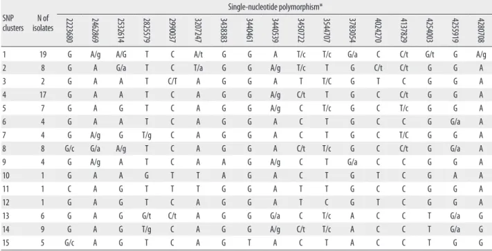
Phylogenetic analysis was performed using the complete Table 1. Position of 45 single-nucleotide polymorphisms and identification of their nucleotides in Mycobacterium tuberculosis H37Rv
Position Nucleotide Position Nucleotide
37031 C 2223680 G
43943 A 2376133 A
92197 T 2462869 G
220048 C 2532614 G
311611 G 2627946 A
519804 G 2825579 T
797736 C 2891265 C
909164 C 2990037 C
918314 T 3207247 A
923063 T 3438383 G
949219 C 3440461 T
1068149 T 3440539 A
1163132 T 3450722 T
1191859 T 3455683 G
1294396 C 3544707 T
1477596 C 3783054 G
1548147 G 4024270 T
1692067 A 4119243 T
1692683 C 4137829 C
1884695 G 4254003 G
1892015 T 4255919 A
1952599 C 4280708 G
2158580 G
data of 96 isolates for SNP analysis at 45 loci and for MIRU- VNTR analysis at 16 loci. A dendrogram of SNP and MIRU- VNTR analyses was constructed from the UPGMA cluster based on the overall similarity of each nucleotide polymor- phism (http://genomes.urv.cat/UPGMA/). For discrimina- tion analysis of MIRU-VNTR, the Hunter–Gaston diversity index (HGDI) was calculated as described elsewhere [22]
and used for comparison of the discriminatory power of each VNTR locus.
RESULTS
1. Evaluation and phylogenetic analysis of SNPs
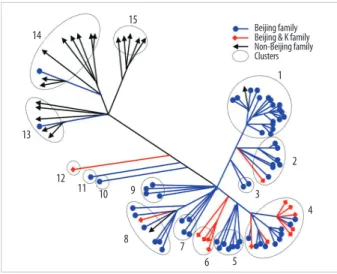
Among the 45 SNPs, 35 had polymorphisms that could be used to differentiate the 96 tested isolates. The other 10 SNPs did not contain polymorphisms. Based on SNP results, isolates were classified into 15 clusters (Fig. 1). Cluster 1, the most populated cluster, contained 19 isolates, followed by cluster 4 with 17 isolates. Three clusters consisted of only 1 isolate each (Table 2). As shown in Fig. 1, nearly all Beijing family strains, which are defined and identified by IS6110 RFLP data [17], were distributed within closely related clus- ters in the SNP dendrogram. Seventy-four of the 76 Beijing family strains were allocated to SNP clusters 1 through 12.
The remaining 2 isolates were allocated to clusters 13 and 14. Nearly all 14 isolates of the K family, a subfamily of the Beijing family identified in a previous study [17], were allo- cated to clusters 4 and 6.
2. Phylogenetic analysis of VNTR
The 96 isolates showed amplification products from 16 VNTR loci and contained at least 1 copy of each locus. Re- sults of allelic diversity and HGDI testing are summarized in Table 3. Among the 16 loci, the discriminatory index in 8 (MIRU-10, -23, -26 and -31; ETR-A, -B, -C, and -F) was high (HGDI > 0.6) according to the definition of Sola et al. [23].
Six loci (MIRU-4, -16, -24, -27, -39 and -40) were dispersed and discriminated moderately well (0.3 ≤ HGDI ≤ 0.6). Two loci (MIRU-2 and -20) were poorly discriminated (HGDI
< 0.3). The complete data were divided into 12 groups. The largest group was group 2, which included 15 isolates. Group 11 contained the only unique pattern (Fig. 2). Unlike the SNP method, MIRU-VNTR did not show notable localiza- tions of Beijing or non-Beijing family isolates in specific clusters.
3. Reproducibility of SNP and VNTR analyses
The 6 paired isolates produced exactly the same results in IS6110-RFLP, SNP, and VNTR patterns for a given patient (data not shown), demonstrating that the 2 methods are re- producible.
DISSCUSION
SNP analysis was developed after whole-genome sequence data became available and has been for genome-scale analy- sis to estimate overall chromosomal relationships among isolates [24, 25]. SNP analysis is a powerful strategy for ex-
Beijing family 14
15
13
12 11 10
8 9
7
6 5
4 3
2 1 Beijing & K family Non-Beijing family Clusters
Fig. 1. Distance-based neighbor joining phylogenetic tree of 96 Mycobacte- rium tuberculosis isolates based on 35 single-nucleotide polymorphisms re- veals 15 clusters (indicated by dotted circles). Each IS6110-restriction frag- ment length polymorphism type is indicated by a closed circle, quadrangle, or arrow.
1
10
8 7 6
8 9
5
4 12
11
2
3 Beijing family
Beijing & K family Non-Beijing family Clusters
Fig. 2. Distance-based neighbor joining phylogenetic tree of 96 Mycobacte- rium tuberculosis isolates based on based on mycobacterial interspersed re- petitive units-variable number of tandem repeats of 16 loci reveals 12 clus- ters (dotted circles). Each IS6110-restriction fragment length polymorphism type is indicated by a closed circle, quadrangle, or arrow.
amining phylogenetic relationships among bacterial strains and is easy to perform [26, 27]. Among various methodolo- gies described previously, we chose the HP assay for this study to improve PCR specificity by decreasing nucleotide mismatching [19, 28]. Additionally, the material costs are lower for this method than for a sequencing-based method [19]. Previous studies have demonstrated that all tested M.
tuberculosis strains were distinguishable by SNP analysis, and therefore, the authors proposed that SNP analysis was an ideal tool to investigate biological variations among clin- ical M. tuberculosis isolates [15, 18]. Filliol et al. [18] identi- fied a minimal number of 45 highly informative SNPs by analyzing 212 SNPs against 323 clinical M. tuberculosis and Mycobacterium bovis isolates. Our SNP analysis was based on this set of 45 SNPs. In this study, however, only 35 SNP loci could be used to categorize our 96 M. tuberculosis iso- lates. Interestingly, nearly all Beijing family strains, includ- ing those of the K family, were allocated to closely related clusters. Our results indicate that SNP analysis using 35 loci is useful for phylogenetic analysis of clinical M. tuberculosis isolates from Korea. This method is effective, easy, and eco- nomical. In the 96 M. tuberculosis isolates examined, 10 of 45 SNPs were identical. We think that this difference was due to the difference in strain distribution between the current and previous studies [18]. Our study included a larger pro- portion of Beijing family isolates, whereas only 15 Beijing family isolates, among the 246 tested isolates, were included
in the previous study [18]; moreover, those 15 isolates were classified as a single SNP group. Therefore, some SNPs proven to be polymorphic between strains isolated in 1 re- gion may not be suitable to discriminate strains originating in other regions.
The MIRU-VNTR typing method is technically more flex- ible and faster than IS6110-RFLP typing, and the results are expressed as numerical codes, which are easy to compare and exchange [29, 30]. A previous report demonstrated that MIRU-VNTR analysis using well-selected loci is useful for analyzing clinical M. tuberculosis isolates from Korea, where the Beijing family is predominant [31]. In the current MIRU- VNTR study using 96 isolates, we identified 2 interesting findings. First, in contrast to SNP results, MIRU-VNTR patterns were not related to IS6110-RFLP patterns. As shown in Fig. 2, isolates of Beijing and non-Beijing families were distributed in almost all clusters. This means that changes in MIRU-VNTR loci developed independently of changes in IS6110 insertion. Hanekom et al. [32] described discor- dant results between MIRU-VNTR and IS6110 RFLP and suggested that the degree of discordance was dependent on the genetic distance between isolates. However, SNP analy- sis resulted in clusters closely related to IS6110-RFLP pat- terns. Filliol et al. [18] described similar close clustering of Beijing family strains by both the SNP and spoligotyping methods. We could not locate M. tuberculosis molecular strain-typing data that had been obtained by simultaneously SNP
clusters N of isolates
Single-nucleotide polymorphism*
37031 43943 92197 311611 519804 797736 909164 918314 1068149 1191859 1294396 1477596 1548147 1692067 1692683 1884695 1892015 1952599
1 19 G/c A/g G T G T/c T/c T C T C/t C/t G A C/a G/a T/c C
2 8 G A G T G T T T C T C T/C G A C G T T/c
3 2 G A G T G/A T T T C T C T G A C G T C
4 17 G A G T G T C T C T/c C T/c G A C/a G C/t C
5 7 G/c A G T/g G T C T C T C T/c G A C G C C
6 4 G G G T G T T/C T C T C C G A C G T/c T/c
7 4 G A G T G T T T C T C C/T G/a A C/a G T C
8 8 G/c A G T/g G T C/t T C T/c C T G A/g C/a G T C
9 4 G A G T/g G T T/c T C T/C C T/c G A C G C/t C
10 1 G A G T G T T T C T C T G A C G T C
11 1 G A G T G T T T C C C C G A A G T C
12 1 C A G G G T T T C T C C G G C G C C
13 6 C/g A/g G T G/a C C/t T/c C/t T C C A/g A C G/a T C/t
14 9 G A T/g T/g G C/t C/t T/c C/t T C/t C G/a A/g C/a G T C/t
15 5 G/c A/g G/t G/t G/a C C T/c C T C C G A C G/a T C
(Continued to the next page)
using IS6110-RFLP, SNPs, and MIRU-VNTR. Here, we de- scribe a close relationship between SNP and IS6110-RFLP and independent results for MIRU-VNTR and IS6110-RFLP.
This means that, unlike IS6110-RFLP, spoligotyping, or SNP, MIRU-VNTR shows high discriminatory power for typing M. tuberculosis strains in areas where the Beijing family is highly prevalent. Second, the discriminatory power of MIRU-VNTR in some loci varied from that described in a previous study conducted with 81 isolates from Korea [31].
In that study, Yun et al. [31] reported that MIRU-23 and ETR-A, -B, and -C had low discriminatory power (HGDI
< 0.3). In our study, however, these 4 loci were highly dis- criminatory (HGDI > 0.6), with MIRU-23 having the sec- ond highest discriminatory power (HGDI = 0.75). The high- est discriminatory power was shown by MIRU-26 in both studies (HGDI > 0.80). The discriminatory power of a spe- cific locus is inevitably affected by the selection of isolates, and the selection criteria were different between the 2 stud- ies. In the previous study, all isolates were collected from Pusan National University Hospital or provided by the Ko- rean Institute of Tuberculosis, so the distribution of patients’
place of residences might have been restricted [31]. In this study, all 96 isolates were collected from 11 university hos- pitals located in various areas, and the number of isolates from each hospital was roughly proportional to the recorded
SNP
clusters N of isolates
Single-nucleotide polymorphism*
2223680 2462869 2532614 2825579 2990037 3207247 3438383 3440461 3440539 3450722 3544707 3783054 4024270 4137829 4254003 4255919 4280708
1 19 G A/g A/G T C A/t G G A T/c T/c G/a C C/t G/t G A/g
2 8 G A G/a T C T/a G G A/g T/c T G C/t C/t G G A
3 2 G A A T C/T A G G A T T/C G T C G G A
4 17 G A A T C A G G A/g C/t T G C C/t G G A
5 7 G A G T C A G G A/g C T/c G C T/c G G A
6 4 G A A T C A G G A C T G C C G G/a A
7 4 G A/g G T/g C A G G A C T G C T/C G G A
8 8 G/c G/a A/g T C A G G A C/t T/c G C C/t G G/a A
9 4 G A/g A T C A A G A/g C T G/a C C G G A
10 1 G A A G T T A G A C T G T C G A A
11 1 C A G T T T G G A T T G C C G G A
12 1 G A G T C A G G A T C G T C G G A
13 6 G A G G/t C/t A G G G/a C T/c A C C T G/a G
14 9 G A G T/g C A G G A/g C/t T/c A C C T G/a G
15 5 G/c A G T C A G T A C T A C C T G G
*When 2 nucleotides are specified, a capital letter indicates the major population, a lower case indicates the minor population, and 2 capital letters indicate 2 major populations.
Abbreviation: SNP, single-nucleotide polymorphism.
Table 3. Variable repeat numbers in each VNTR locus and allelic diversity among 96 clinical isolates of Mycobacterium tuberculosis collected through- out Korea
VNTR loci N of VNTR repeats
HGDI*
1 2 3 4 5 6 7 8 9
MIRU-2 88 9 1 0.25
MIRU-4 54 36 8 0.58
MIRU-10 65 8 20 0.62
MIRU-16 31 62 4 1 0.59
MIRU-20 90 8 0.12
MIRU-23 6 4 41 24 9 14 0.75
MIRU-24 66 17 8 0.58
MIRU-26 22 3 34 5 11 11 9 1 2 0.86
MIRU-27 67 19 12 0.59
MIRU-31 17 42 18 14 7 0.72
MIRU-39 61 32 5 0.59
MIRU-40 66 21 8 3 0.60
ETR-A 1 27 51 15 4 0.72
ETR-B 49 14 20 15 0.71
ETR-C 1 20 36 26 15 0.73
ETR-F 18 31 29 20 0.62
*Underlining indicates loci showing high discriminatory power, as defined by Sola et al. [23].
Abbreviations: VNTR, variable number of tandem repeats; HGDI, Hunter–
Gaston diversity index; MIRU, mycobacterial interspersed repetitive units;
ETR, exact tandem repeat.
patient numbers in the area served by that institution. There- fore, our study represents MIRU-VNTR performance more accurately. Combining the results of both the studies, we found that MIRU-10, -26, -31, and ETR-F show a high dis- criminatory power for molecular strain typing of M. tuber- culosis isolates from Korea. Our assessment of the clinical utility of other loci, such as ETR-A, -B, and -C, that showed good discriminatory power in only 1 study is reserved until further data are obtained.
We confirmed the reproducibility of SNP and MIRU- VNTR analyses using 6 pairs of strains from different cul- tures of the same patients. Both SNP and MIRU-VNTR can discriminate clinical M. tuberculosis isolates in Korea, where Beijing family strains are highly prevalent. Both methods are PCR-based and early application to clinical specimens is possible. Strain typing is often requested to detect outbreaks or contamination, and rapid results are required to control these phenomena. Although IS6110-RFLP has high discrim- inatory power, it is not useful for these purposes. Therefore, SNP and MIRU-VNTR can be used as surrogate molecular strain-typing methods for M. tuberculosis in Korea.
Authors’ Disclosures of Potential Conflicts of Interest No potential conflict of interest relevant to this article was reported.
Acknowledgement
This work was supported by the Korea Research Founda- tion Grant funded by the Korean Government (Ministry of Education and Human Resources Development, Basic Re- search Promotion Fund) (KRF-2008-1-E00318).
REFERENCES
1. Gopaul KK, Brown TJ, Gibson AL, Yates MD, Drobniewski FA.
Progression toward an improved DNA amplification-based typing technique in the study of Mycobacterium tuberculosis epidemiol- ogy. J Clin Microbiol 2006;44:2492-8.
2. Van Soolingen D. Molecular epidemiology of tuberculosis and other mycobacterial infections: main methodologies and achieve- ments. J Intern Med 2001;249:1-26.
3. Seidler A, Nienhaus A, Diel R. The transmission of tuberculosis in the light of new molecular biological approaches. Occup Environ Med 2004;61:96-102.
4. van Embden JD, Cave MD, Crawford JT, Dale JW, Eisenach KD, Gicquel B, et al. Strain identification of Mycobacterium tuberculosis by DNA fingerprinting: recommendations for a standardized methodology. J Clin Microbiol 1993;31:406-9.
5. Barlow RE, Gascoyne-Binzi DM, Gillespie SH, Dickens A, Qamer S, Hawkey PM. Comparison of variable number tandem repeat and IS6110-restriction fragment length polymorphism analyses
bacterium tuberculosis isolates. J Clin Microbiol 2001;39:2453-7.
6. Gori A, Bandera A, Marchetti G, Degli Esposti A, Catozzi L, Nardi GP, et al. Spoligotyping and Mycobacterium tuberculosis. Emerg Infect Dis 2005;11:1242-8.
7. Gori A, Esposti AD, Bandera A, Mezzetti M, Sola C, Marchetti G, et al. Comparison between spoligotyping and IS6110 restriction fragment length polymorphisms in molecular genotyping analysis of Mycobacterium tuberculosis strains. Mol Cell Probes 2005;19:
236-44.
8. Kamerbeek J, Schouls L, Kolk A, van Agterveld M, van Soolingen D, Kuijper S, et al. Simultaneous detection and strain differentia- tion of Mycobacterium tuberculosis for diagnosis and epidemiol- ogy. J Clin Microbiol 1997;35:907-14.
9. Kremer K, van Soolingen D, Frothingham R, Haas WH, Hermans PW, Martin C, et al. Comparison of methods based on different molecular epidemiological markers for typing of Mycobacterium tuberculosis complex strains: interlaboratory study of discrimina- tory power and reproducibility. J Clin Microbiol 1999;37:2607-18.
10. Cole ST, Brosch R, Parkhill J, Garnier T, Churcher C, Harris D, et al. Deciphering the biology of Mycobacterium tuberculosis from the complete genome sequence. Nature 1998;393:537-44.
11. Fleischmann RD, Alland D, Eisen JA, Carpenter L, White O, Peter- son J, et al. Whole-genome comparison of Mycobacterium tuber- culosis clinical and laboratory strains. J Bacteriol 2002;184:5479-90.
12. Kersulyte D, Mukhopadhyay AK, Velapatino B, Su W, Pan Z, Gar- cia C, et al. Differences in genotypes of Helicobacter pylori from different human populations. J Bacteriol 2000;182:3210-8.
13. Schork NJ, Fallin D, Lanchbury JS. Single nucleotide polymorphisms and the future of genetic epidemiology. Clin Genet 2000;58:250-64.
14. Gutacker MM, Smoot JC, Migliaccio CA, Ricklefs SM, Hua S, Cousins DV, et al. Genome-wide analysis of synonymous single nucleotide polymorphisms in Mycobacterium tuberculosis com- plex organisms: resolution of genetic relationships among closely related microbial strains. Genetics 2002;162:1533-43.
15. Gutacker MM, Mathema B, Soini H, Shashkina E, Kreiswirth BN, Graviss EA, et al. Single-nucleotide polymorphism-based popula- tion genetic analysis of Mycobacterium tuberculosis strains from 4 geographic sites. J Infect Dis 2006;193:121-8.
16. Frothingham R and Meeker-O’Connell WA. Genetic diversity in the Mycobacterium tuberculosis complex based on variable num- bers of tandem DNA repeats. Microbiology 1998;144 ( Pt 5):1189- 17. Choi GE, Jang MH, Song EJ, Jeong SH, Kim JS, Lee WG, et al. 96.
IS6110-restriction fragment length polymorphism and spoligo- typing analysis of Mycobacterium tuberculosis clinical isolates for investigating epidemiologic distribution in Korea. J Korean Med Sci 2010;25:1716-21.
18. Filliol I, Motiwala AS, Cavatore M, Qi W, Hazbón MH, Bobadilla del Valle M, et al. Global phylogeny of Mycobacterium tuberculosis based on single nucleotide polymorphism (SNP) analysis: insights into tuberculosis evolution, phylogenetic accuracy of other DNA fingerprinting systems, and recommendations for a minimal stan- dard SNP set. J Bacteriol 2006;188:759-72.
19. Hazbón MH and Alland D. Hairpin primers for simplified single- nucleotide polymorphism analysis of Mycobacterium tuberculosis and other organisms. J Clin Microbiol 2004;42:1236-42.
20. Supply P, Allix C, Lesjean S, Cardoso-Oelemann M, Rüsch-Gerdes
cobacterial interspersed repetitive unit-variable-number tandem repeat typing of Mycobacterium tuberculosis. J Clin Microbiol 2006;44:4498-510.
21. Supply P, Mazars E, Lesjean S, Vincent V, Gicquel B, Locht C. Vari- able human minisatellite-like regions in the Mycobacterium tuber- culosis genome. Mol Microbiol 2000;36:762-71.
22. Hunter PR and Gaston MA. Numerical index of the discriminatory ability of typing systems: an application of Simpson’s index of di- versity. J Clin Microbiol 1988;26:2465-6.
23. Sola C, Filliol I, Legrand E, Lesjean S, Locht C, Supply P, et al. Ge- notyping of the Mycobacterium tuberculosis complex using MI- RUs: association with VNTR and spoligotyping for molecular epi- demiology and evolutionary genetics. Infect Genet Evol 2003;3:
125-33.
24. Hirsh AE, Tsolaki AG, DeRiemer K, Feldman MW, Small PM. Sta- ble association between strains of Mycobacterium tuberculosis and their human host populations. Proc Natl Acad Sci U S A 2004;101:
4871-6.
25. Brosch R, Gordon SV, Marmiesse M, Brodin P, Buchrieser C, Eigl- meier K, et al. A new evolutionary scenario for the Mycobacterium tuberculosis complex. Proc Natl Acad Sci U S A 2002;99:3684-9.
26. Kaboev OK, Luchkina LA, Tret’iakov AN, Bahrmand AR. PCR hot start using primers with the structure of molecular beacons (hair- pin-like structure). Nucleic Acids Res 2000;28:E94.
27. Nazarenko I, Lowe B, Darfler M, Ikonomi P, Schuster D, Rashtchian
with a single fluorophore. Nucleic Acids Res 2002;30:e37.
28. Bouakaze C, Keyser C, de Martino SJ, Sougakoff W, Veziris N, Dabernat H, et al. Identification and genotyping of Mycobacterium tuberculosis complex species by use of a SNaPshot Minisequenc- ing-based assay. J Clin Microbiol 2010;48:1758-66.
29. Supply P, Lesjean S, Savine E, Kremer K, van Soolingen D, Locht C.
Automated high-throughput genotyping for study of global epide- miology of Mycobacterium tuberculosis based on mycobacterial interspersed repetitive units. J Clin Microbiol 2001;39:3563-71.
30. Kwara A, Schiro R, Cowan LS, Hyslop NE, Wiser MF, Roahen Harrison S, et al. Evaluation of the epidemiologic utility of second- ary typing methods for differentiation of Mycobacterium tubercu- losis isolates. J Clin Microbiol 2003;41:2683-5.
31. Yun KW, Song EJ, Choi GE, Hwang IK, Lee EY, Chang CL. Strain typing of Mycobacterium tuberculosis isolates from Korea by my- cobacterial interspersed repetitive units-variable number of tan- dem repeats. Korean J Lab Med 2009;29:314-9.
32. Hanekom M, van der Spuy GD, Gey van Pittius NC, McEvoy CR, Hoek KG, Ndabambi SL, et al. Discordance between mycobacte- rial interspersed repetitive-unit-variable-number tandem-repeat typing and IS6110 restriction fragment length polymorphism ge- notyping for analysis of Mycobacterium tuberculosis Beijing strains in a setting of high incidence of tuberculosis. J Clin Microbiol 2008;46:3338-45.