110
pISSN 2288-6575 • eISSN 2288-6796 https://doi.org/10.4174/astr.2017.92.2.110 Annals of Surgical Treatment and Research
CASE REPORT
Fulminant course in a case of malignant phyllodes tumor
Young Woo Chang, Hwan Soo Kim, Deok Woo Kim1, Gil Soo Son
Department of Breast Endocrine Surgery, Korea University College of Medicine, Seoul, 1Department of Plastic and Reconstructive Surgery, Korea University College of Medicine, Seoul, Korea
INTRODUCTION
Phyllodes tumors are rare and account for 0.3 to 1% of all breast malignancies [1]. Malignant phyllodes tumors are classified according to the degree of stromal cellular atypia and represent 10% to 30% of all phyllodes tumors. There are some reports on malignant phyllodes tumors with an excellent prognosis, but few reports on aggressive phyllodes tumors that lead to death. We present a case of malignant phyllodes tumor with a fulminant course.
CASE REPORT
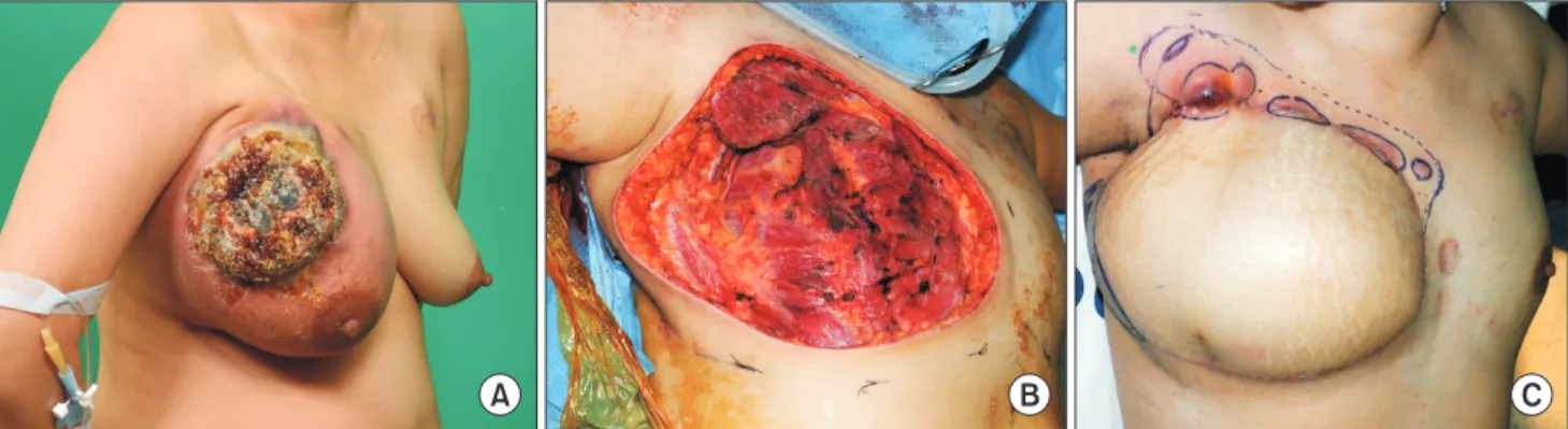
A 31yearold woman from Uzbekistan presented with an inflammatory and ulcerative mass in her right breast that had been there since 2011. The mass had grown rapidly over the past 3 months and it was extremely painful with a huge ulcerative wound (Fig. 1A). Breast ultrasonography and a core needle biopsy were performed initially in an outside breast clinic and the biopsy result was a diagnosis of invasive carcinoma of no specific type. She was referred to Korea University Medical Center, Ansan in January 2016, 2 weeks after being diagnosed.
Additional imaging workup was performed to check the primary breast mass and metastatic lesion, including a MRI of the breast; a CT scan of the chest and abdomen; a bone scan;
and a PET scan. Breast MRI revealed an irregular necrotic 120
mm mass in the right upper outer quadrant that had invaded the skin and pectoralis major muscle. Several lymph node (LN) metastases at the right axillary level I were suggested on the CT and PET scans, but there was no evidence of distant metastasis.
She underwent neoadjuvant chemotherapy with doxorubicin and cyclophosphamide for reduction of the mass and systemic therapy. After the first cycle of neoadjuvant chemotherapy, a core needle biopsy was performed again in our hospital, since our pathologist had suggested the possibility of a phyllodes tumor through review of the outside slides. The rebiopsy result was a diagnosis of phyllodes tumor, and a right modified radical mastectomy (MRM) and transverse rectus abdominis myocutaneous (TRAM) flap were performed (Fig. 1B). The right breast mass weighed 2,212 g at the time of excision.
The final pathology confirmed a malignant phyllodes tumor measuring 125 mm × 120 mm. The tumor showed brisk stromal overgrowth with lymph and vascular tumor emboli, and the mitotic count was 46 per 10 high power fields. All We present the case of a 31-year-old woman with an inflammatory and ulcerative malignant phyllodes tumor in her right breast. A right modified radical mastectomy and transverse rectus abdominis myocutaneous (TRAM) flap were performed.
A month after the initial operation, several masses recurred at the superior margin and deep margin of the TRAM flap.
Wide excision was performed, but masses recurred at the inferior margin and in both lung fields 2 weeks after the second operation. Six weeks after the second operation, the patient died due to progression of dyspnea and respiratory failure.
[Ann Surg Treat Res 2017;92(2):110-112]
Key Words: Phyllodes tumor, Phyllodes tumor, Breast neoplasms
Received July 19, 2016, Reviewed July 26, 2016, Accepted August 4, 2016 Corresponding Author: Gil Soo Son
Department of Breast Endocrine Surgery, Korea University Ansan Hospital, Korea University College of Medicine, 123 Jeokgeum-ro, Danwon-gu, Ansan 15355, Korea
Tel: +82-31-412-5992, Fax: +82-31-412-4827 E-mail: [email protected]
Copyright ⓒ 2017, the Korean Surgical Society
cc Annals of Surgical Treatment and Research is an Open Access Journal. All articles are distributed under the terms of the Creative Commons Attribution Non- Commercial License (http://creativecommons.org/licenses/by-nc/4.0/) which permits unrestricted non-commercial use, distribution, and reproduction in any medium, provided the original work is properly cited.
Annals of Surgical Treatment and Research 111 resection margins were negative and we obtained margins
of more than 10 mm except for the deep resection margin which was less than 1 mm. Immunohistochemical staining for estrogen receptor (ER) and progesterone receptor (PR) were positive, but for human epidermal growth factor receptor 2 was negative. There was no metastasis in the 18 retrieved LNs.
A month after the initial operation, several masslike pro
truding lesions appeared at the superior margin of the TRAM flap (Fig. 1C). Breast MRI showed recurrent tumors in the deep margin of the TRAM flap as well as the superior margin. The recurrent tumor in the deep margin measured 84 mm × 48 mm and had invaded the pectoralis minor and intercostal muscles (Fig. 2A, B). A wide excision was performed and a su perior negative margin was obtained but a negative deep margin could not be achieved.
Two weeks after the second operation, masses appeared again at the inferior margin and a chest CT scan revealed new round opacities in both lung fields that were thought to be metastases (Fig. 2C). Despite palliative radiation therapy, bilateral pleural effu sion increased gradually and the patient suffered from dysp nea. Six weeks after the second operation, the patient died
due to progression of dyspnea and respiratory failure.
DISCUSSION
Phyllodes tumors are rare fibroepithelial breast tumors that usually present as a rapidly growing breast lump. The tu
mors are classified as benign, borderline, or malignant, but distinguishing among these histotypes is difficult. The classi
fi cation is based on semiquantitative evaluation of mitotic acti
vity, infiltrative as compared to circumscribed tumor margins, and the presence of stromal overgrowth [2]. The variable biologic behavior of phyllodes tumors has led to many debates about the treatment options.
A complete surgical excision is generally the treatment of choice for a phyllodes tumor [3]. Obtaining a negative his
tologic margin of at least 1 cm is suggested [4], but a recent retrospective study demonstrated that it was important to obtain a negative margin regardless of the length [5]. In our case, all histologic margins were negative for more than 1 cm except the deep margin at the initial operation. Nevertheless, the tumor recurred on the superior and inferior margin of Young Woo Chang, et al: Fulminant course of malignant phyllodes tumor
A B C
Fig. 1. (A) An inflammatory and ulcerative mass in the right breast. (B) Post modified radical mastectomy image showing en bloc removal of the tumor including the invaded pectoralis muscle. (C) Masslike protruding lesions that appeared at the superior margin of the transverse rectus abdominis myocutaneous flap a month after the initial operation.
A B C
Fig. 2. (A, B) Breast MRI showing recurrent tumor (84 mm × 48 mm) at the deep margin of the transverse rectus abdominis myocutaneous flap invading the pectoralis minor and intercostal muscles, a month after the initial operation. (C) A chest CT scan for both lung fields showing new round opacities and suggesting metastases (arrows).
112
Annals of Surgical Treatment and Research 2017;92(2):110112
the TRAM flap as well as the deep margin. This means that recurrence of aggressive malignant phyllodes tumors can be influenced by not only a negative margin, but also histologic grade, such as stromal overgrowth, high mitotic index, sarcomatous stroma, and an infiltrative margin.
Axillary LN metastases of phyllodes tumor are only rarely reported and axillary LN dissection is usually not required [3].
Although our patient was diagnosed with a phyllodes tumor, axillary LN dissection was performed because LN metastases were strongly suggested based on the chest CT and PET scan. No metastasis was found among the 18 retrieved LNs despite the aggressive activity of the disease. Thus, axillary LN dissection for phyllodes tumors should be carefully considered.
Radiation therapy decreases local recurrence of phyllodes tumor in the adjuvant setting [6]. However, it is difficult to directly assess the effectiveness of radiation therapy since pre
vious studies enrolled patients with a negative resection margin at the initial operation. Furthermore, there has been no study on metastatic phyllodes treated with radiation therapy because of its rarity. In our case, the recurrent tumors after the second operation were not controlled by radiation therapy. The benefit of radiation therapy for aggressive malignant phyllodes tumors is hence questionable.
Moreover, the effectiveness of chemotherapy is controversial.
A 10year observational study of adjuvant chemotherapy for malig nant phyllodes tumor demonstrated that there was no benefit for patients from treatment with doxorubicin and dacarbazine [7]. However, according to a study of soft tissue sar coma, neoadjuvant chemotherapy including ifosfamide is fea sible to treat sarcoma [8]. Our patient could not undergo chemo therapy because of her poor general condition caused by pro gressive dyspnea.
Hormone therapy is not effective for malignant phyllodes tu mors even when they are positive for ER and PR [9] because hormone receptors in phyllodes tumor are located mainly in the epithelial component, not in the stromal component [10]. In our case, the patient did not undergo hormone therapy despite being positive for ER and PR.
In conclusion, aggressive malignant phyllodes tumor remains a therapeutic challenge despite extensive research, and further studies are required. Physicians should be aware that malignant phyllodes tumor can progress to a fulminant course.
CONFLICTS OF INTEREST
No potential conflict of interest relevant to this article was reported.
1. Geisler DP, Boyle MJ, Malnar KF, McGee JM, Nolen MC, Fortner SM, et al. Phyllo
des tumors of the breast: a review of 32 cases. Am Surg 2000;66:3606.
2. Kim S, Kim JY, Kim DH, Jung WH, Koo JS.
Analysis of phyllodes tumor recurrence according to the histologic grade. Breast Cancer Res Treat 2013;141:35363.
3. Macdonald OK, Lee CM, Tward JD, Chappel CD, Gaffney DK. Malignant phyl
lodes tumor of the female breast: asso
cia tion of primary therapy with cause
spe cific survival from the Surveillance, Epidemiology, and End Results (SEER) program. Cancer 2006;107:212733.
4. Rowell MD, Perry RR, Hsiu JG, Barranco SC. Phyllodes tumors. Am J Surg 1993;
165:3769.
5. Jang JH, Choi MY, Lee SK, Kim S, Kim J, Lee J, et al. Clinicopathologic risk factors for the local recurrence of phyllodes tu
mors of the breast. Ann Surg Oncol 2012;
19:26127.
6. Barth RJ Jr, Wells WA, Mitchell SE, Cole BF.
A prospective, multiinstitutional study of adjuvant radiotherapy after resection of malignant phyllodes tumors. Ann Surg Oncol 2009;16:228894.
7. MoralesVasquez F, GonzalezAngulo AM, Broglio K, LopezBasave HN, Gallardo D, Hortobagyi GN, et al. Adjuvant chemo
therapy with doxorubicin and dacarbazine has no effect in recurrencefree survival of malignant phyllodes tumors of the breast. Breast J 2007;13:5516.
8. Gortzak E, Azzarelli A, Buesa J, Bramwell
VH, van Coevorden F, van Geel AN, et al.
A randomised phase II study on neoadju
vant chemotherapy for ‘highrisk’ adult softtissue sarcoma. Eur J Cancer 2001;37:
1096103.
9. Telli ML, Horst KC, Guardino AE, Dirbas FM, Carlson RW. Phyllodes tumors of the breast: natural history, diagnosis, and treatment. J Natl Compr Canc Netw 2007;
5:32430.
10. Tse GM, Lee CS, Kung FY, Scolyer RA, Law BK, Lau TS, et al. Hormonal recep tors expression in epithelial cells of mam
mary phyllodes tumors correlates with pathologic grade of the tumor: a mul ti
center study of 143 cases. Am J Clin Pathol 2002;118:5226.