ISSN 2234-3806 • eISSN 2234-3814
https://doi.org/10.3343/alm.2017.37.4.349 www.annlabmed.org 349
Ann Lab Med 2017;37:349-351
https://doi.org/10.3343/alm.2017.37.4.349
Letter to the Editor
Clinical Microbiology
Robinsoniella peoriensis Bacteremia: a Second Case in Korea
Sangeun Lim, M.D.1, Hee Jae Huh, M.D.2, Nam Yong Lee, M.D.2, Eun-Jeong Joo, M.D.3, Joon-Sup Yeom, M.D.3, Seungjun Lee, M.D.1, Hee-Yeon Woo, M.D.1, Hyosoon Park, M.D.1, and Min-Jung Kwon, M.D.1
Department of Laboratory Medicine1, Kangbuk Samsung Hospital, Sungkyunkwan University School of Medicine, Seoul; Department of Laboratory Medicine and Genetics2, Samsung Medical Center, Sungkyunkwan University School of Medicine, Seoul; Division of Infectious Diseases3, Department of Internal Medicine, Kangbuk Samsung Hospital, Sungkyunkwan University School of Medicine, Seoul, Korea
Dear Editor,
Robinsoniella peoriensis is a gram-positive, spore-forming, an- aerobic rod originally isolated from swine manure storage pits [1]. Most R. peoriensis strains have been isolated from environ- mental sources, but this organism was isolated from human blood, abdominal fluid, wound, muscle hematoma puncture, or necrotic tissue in nine cases [2-6]. Here, we describe a second Korean case of R. peoriensis bacteremia, which was confirmed via 16S rRNA sequencing.
A 63-yr-old man with diabetes mellitus and multiple lacunar infarctions was admitted for scheduled chemotherapy. He had been diagnosed as having small cell lung cancer six months prior to admission. He lived in an urban area of Seoul and de- nied having traveled or having had any animal contact in the past year. His initial vital signs on admission were normal, and initial laboratory test results were as follows: Hb, 67 g/L; white blood cell count, 6.3×10⁹/L (neutrophils, 78.4%); and platelets, 87 ×109/L. The serum C-reactive protein (CRP) level was ele- vated to 108.10 nmol/L, and the lactate dehydrogenase (LDH) level was elevated to 13.83 µkat/L. The patient was suspected of having aspiration pneumonia because of haziness in the right middle and lower lobe fields on chest X-ray. Blood culture sets (two aerobic and one anaerobic) were taken from three separate
vein sites. The patient was initially treated with 4 g Tazocin via intravenous administration three times per day.
On day 3 after admission, the patient developed fever of 38.4°C.
On day 4 after admission, the body temperature was elevated to 39.5°C, the CRP level was elevated to 253.72 nmol/L, and chest X-ray revealed consolidations with increased patchiness in both lung fields.
After one day of incubation, one anaerobic blood culture showed growth of microorganisms; two days later, gray-white, smooth, and non-hemolytic colonies of unequal sizes were observed only on the anaerobic blood plate (Fig. 1A). Gram staining of a puri- fied colony indicated the presence of gram-positive, rod-shaped bacteria with ovoid-shaped spores located centrally (Fig. 1B).
The BD BACTEC FX (BD Diagnostics, Heidelberg, Germany) and VITEK2 (bioMérieux, Marcy l’Etoile, France) systems indi- cated the presence of Clostridium clostridioforme (92%), with a questionable level of confidence owing to its low biofrequency.
An antimicrobial susceptibility test of the isolate could not be performed. On day 5 after admission, antibiotic therapy with Taz- ocin was switched to imipenem at 500 mg intravenous (IV) three times per day. No organisms were grown on follow-up blood cul- ture. However, the patient died from progression of pneumonia and respiratory failure. The etiologic diagnosis was established
Received: September 19, 2016 Revision received: January 4, 2017 Accepted: March 8, 2017
Corresponding author: Min-Jung Kwon
Department of Laboratory Medicine, Kangbuk Samsung Hospital, Sungkyunkwan University School of Medicine, 29 Saemunan-ro, Jongno- gu, Seoul 03181, Korea
Tel: +82-2-2001-5211, Fax: +82-2-757-0711, E-mail: [email protected]
© Korean Society for Laboratory Medicine.
This is an Open Access article distributed under the terms of the Creative Commons Attribution Non-Commercial License (http://creativecommons.org/licenses/by-nc/4.0) which permits unrestricted non-commercial use, distribution, and reproduction in any medium, provided the original work is properly cited.
1 / 1 CROSSMARK_logo_3_Test
2017-03-16 https://crossmark-cdn.crossref.org/widget/v2.0/logos/CROSSMARK_Color_square.svg
Lim S, et al.
Robinsoniella peoriensis bacteremia
350 www.annlabmed.org https://doi.org/10.3343/alm.2017.37.4.349 post-mortem by means of molecular analysis. To identify the bac-
teria, molecular identification was performed by DNA amplifica- tion and sequencing analysis of the 16S rRNA gene [8]. A Gen- Bank BLAST search revealed that the 16S rRNA gene sequence of the isolate showed 99.17% homology for 1,316 bp with the corresponding sequence of R. peoriensis (GenBank accession number NR_041882.1). Murimonas intestini, C. oroticum, and Hespellia porcina were the next best matches, with similarities of 94.60%, 94.53%, and 94.47%, respectively. When the se- quence was submitted to the Ez-Taxon database v2.1 (http://
www.ezbiocloud.net), the highest similarity was with R. peorien- sis (99.04%), followed by C. nexile, C. saccharolyticum, and M.
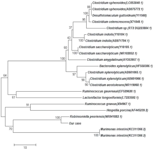
intestini, with similarities of 94.88%, 94.33%, and 94.26%, re- spectively. A phylogenetic tree was constructed by using the nei- ghbor-joining method in Molecular Evolutionary Genetics Analy- sis (MEGA) software version 6.0 (http://www.megasoftware.net;
Fig. 2).
Current phenotypic identification systems, including the VI- TEK2 system, are unable to identify R. peoriensis and may con- fuse it with other organisms such as Clostridium [4-7]. An incre- asing number of studies have recently emerged describing mi- croorganisms misidentified by conventional methods [9]. There- fore, identification of rare bacteria and bacteria with unusual phe-
notypic profiles by conventional methods needs to be confirmed by a reliable tool such as 16S rRNA sequencing [10].
We report our experience with the use of 16S rRNA sequenc- ing and various databases for the identification of an unknown anaerobic organism. This report represents the second case of R. peoriensis isolation from human blood in Korea. Most anaer- obic infections originate from the patient’s own microflora or an- other exogenous environmental source, and anaerobic bactere- mia may be serious or even life-threatening in immunocompro- mised patients. Although R. peoriensis is rarely reported as a pathogen, the possibility should be carefully evaluated depend- ing on the patient’s status. In the present case, the patient suf- fered from a chronic debilitation disorder with diabetes mellitus and lung cancer while undergoing chemotherapy. R. peoriensis isolated from a blood specimen was successfully identified by using molecular methods in a clinical laboratory.
Authors’ Disclosures of Potential Conflicts of Interest
No potential conflicts of interest relevant to this article were re- ported.
Fig. 1. Colonial and microscopic morphology of Robinsoniella peoriensis. (A) Dull white and flat colonies with mixed sizes and smooth edg- es on an anaerobic blood plate. (B) Gram-positive and rod-shaped bacteria with ovoid-shaped spores located centrally (×400).
A B
Lim S, et al.
Robinsoniella peoriensis bacteremia
https://doi.org/10.3343/alm.2017.37.4.349 www.annlabmed.org 351
Fig. 2. Unrooted neighbor-joining phylogenetic tree based on 16S rRNA sequences of Robinsoniella peoriensis and 21 other similar organ- isms.
REFERENCES
1. Cotta MA, Whitehead TR, Falsen E, Moore E, Lawson PA. Robinsoniella peoriensis gen. nov., sp. nov., isolated from a swine-manure storage pit and a human clinical source. Int J Syst Evol Microbiol 2009;59:150-5.
2. López P, Belda S, García M, Royo G. Infection of a spontaneous mus- cular hematoma due to Robinsoniella peoriensis, in a patient with alco- holic liver cirrhosis. Enferm Infecc Microbiol Clin 2010;28:565-7.
3. Cassir N, Laget L, Renvoisé A, Gennari JM, Drancourt M. Robinsoniella peoriensis infection following surgery for scoliosis: a case report. J Med Case Rep 2012;6:174.
4. Gomez E, Gustafson DR, Colgrove R, Ly T, Santana R, Rosenblatt JE, et al. Isolation of Robinsoniella peoriensis from four human specimens. J Clin Microbiol 2011;49:458-60.
5. Shen D, Chen R, Ye L, Luo Y, Tang YW. Robinsoniella peoriensis bacte- remia in a patient with pancreatic cancer. J Clin Microbiol 2010;48:3448-
50.
6. Jeon Y, Kim TS, Kim HB, Park KU, Song J, Kim EC. First Korean case of Robinsoniella peoriensis bacteremia in a patient with aspiration pneu- monia. Ann Lab Med 2012;32:370-4.
7. Ferraris L, Aires J, Butel MJ. Isolation of Robinsoniella peoriensis from the feces of premature neonates. Anaerobe 2012;18:172-3.
8. Fontana C, Favaro M, Pelliccioni M, Pistoia ES, Favalli C. Use of the Mi- croSeq 500 16S rRNA gene-based sequencing for identification of bac- terial isolates that commercial automated systems failed to identify cor- rectly. J Clin Microbiol 2005;43:615-9.
9. Petti CA, Polage CR, Schreckenberger P. The role of 16S rRNA gene sequencing in identification of microorganisms misidentified by conven- tional methods. J Clin Microbiol 2005;43:6123-5.
10. Woo PC, Lau SK, Teng JL, Tse H, Yuen KY. Then and now: use of 16S rDNA gene sequencing for bacterial identification and discovery of nov- el bacteria in clinical microbiology laboratories. Clin Microbiol Infect 2008;
14:908-34.