서 론
거골 골연골 병변(osteochondral lesion of the talus)은 거골의 관절 연골(articular cartilage) 및 연골하골(subchondral bone)을 포함하는 관절면(articular surface)에 분절(fragmentation)과 잠재적인 분리가 나타나는 질환이다.1,2) 6-11세 사이에 발생하기도 하나 12-19세 사이의 청소년기에 가장 호발하며 경쟁적인 스포츠 활동 참여와 발생 간에 연관성이 높은 것으로 알려져 있다.3) 관절면에서 분절 된 골연골편(osteochondral fragment)의 역학적 안정성(mechanical stability)과 크기가 중요한 예후인자로 알려져 있으며,4-7) 관절면 이 유지되고 병변이 전위되지 않은 경우에는 수술적 치료 없이 보존적 치료만으로 치유될 가능성이 높다.8-11) 하지만 역학적으로
불안정한 골연골편은 골연골 결손(osteochondral defect) 부위에서 전위되어 관절 내 유리체(intra-articular loose body)로 진행될 수 있으므로 수술적 치료가 반드시 필요한 것으로 보고되고 있다.12) 최근 체육활동의 증가로 인하여 소아청소년에서의 거골 골연 골 병변의 유병률도 증가하고 있는 추세이다. 이러한 소아청소년 거골 골연골 병변은 성인의 경우보다 더 좋은 예후를 보이며 골 관절염으로의 진행 가능성도 매우 낮은 것으로 보고되고 있다.10) 본 논문에서는 소아청소년 스포츠 손상의 일환으로 발목 손상의 대표적 병변인 거골 골연골 병변의 최신지견을 포함한 전반적인 내용에 대해 문헌고찰과 함께 보고하고자 한다.
본 론
1. 발생기전 및 원인
거골 골연골 병변의 발생기전은 명확히 알려져 있지는 않으나 외 상성, 허혈성 요인과 유전, 내분비 및 감염 연관성이 제시되고 있
다.13-16) Bauer 등9)은 외상과 연관하여 격렬하고 반복적인 고강도
신체 활동이 지속적인 미세 손상(microtrauma)을 일으켜 거골 골
Copyright © 2018 by The Korean Orthopaedic Association
“This is an Open Access article distributed under the terms of the Creative Commons Attribution Non-Commercial License (http://creativecommons.org/licenses/by-nc/4.0/) which permits unrestricted non-commercial use, distribution, and reproduction in any medium, provided the original work is properly cited.”
The Journal of the Korean Orthopaedic Association Volume 53 Number 3 2018 Received November 3, 2017 Revised January 11, 2018
Accepted February 22, 2018
Correspondence to: Mi Hyun Song, M.D.
Department of Orthopaedic Surgery, Korea University Guro Hospital, 148 Gurodong- ro, Guro-gu, Seoul 08308, Korea
TEL: +82-2-2626-3239 FAX: +82-2-2626-2486 E-mail: wwiiw@naver.com ORCID: https://orcid.org/0000-0003-4082-1455
Knowledge Updates in Pediatric Orthopedic Sports Medicine
소아청소년에서의 거골 골연골 병변
송미현
고려대학교 구로병원 정형외과
Osteochondral Lesion of the Talus in Children and Adolescents
Mi Hyun Song, M.D.
Department of Orthopaedic Surgery, Korea University Guro Hospital, Seoul, Korea
Osteochondral lesion of the talus (OLT) is a disease affecting the subchondral bone and articular cartilage of the talus, which may cause fragmentation and displacement of the osteochondral lesion. The stability of the joint and the size of the lesion are important prognostic factors. Conservative treatment is preferred in the initial treatment of OLT in skeletal immature patients because it has a more favorable prognosis than adult OLT in terms of the healing potential and improvement of symptoms. Surgery is recommended when the conservative outcome is unsatisfactory and the fragment is large or displaced. Surgical procedures, including marrow stimulation technique, autologous osteochondral transplantation, and autologous chondrocyte implantation, were performed depending on the condition of the osteochondral lesion.
Key words: osteochondral lesion, talus, ankle, children, adolescents
연골 병변으로 이어지는 것으로 추정하였다. 주로 과체중이거나 관절의 이완성이 있는 환자에서 회내(supination)/회외력(prona- tion)에 의한 족관절 염좌가 발생할 때 거골 골연골 병변이 동반 될 수 있는 것으로 알려져 있다. 위치에 따라서는 병변이 거골의 전외측에 위치할 경우에는 외상성일 가능성이 높고 후내측 병변 의 경우에는 비외상성 또는 허혈성일 가능성이 높다.17)
허혈성 손상도 거골 골연골 병변의 다른 원인으로 받아들여지 고 있다.13,14) 거골의 연골하골에 손상이 발생한 후 치유 과정이 불 안정하게 진행되면 거골에 경화성 조직(sclerotic tissue)이 발생되 고 혈액 공급이 제한되면서 이차적인 연골 병변으로 진행된다.
병변이 초자연골(hyaline cartilage) 표면까지 진행되면 유리체가 발생된다.12)
2. 임상적 평가
거골 골연골 병변은 아무런 증상을 보이지 않는 경우도 있고 발 생 수개월이 지나서야 경미한 증상이 시작되기도 한다. 90% 이상 의 환자에서 보행 시에 만성적인 경미한 통증을 느끼고 통증과 붓기가 주된 증상이며 그 외에도 관절의 불안정성(instability)과 발목의 운동제한을 보이기도 한다. 관절 내 유리체가 있는 경우 에는 잠김(locking) 현상을 호소하기도 한다. 간혹 파행(limp)이나 진통보행(antalgic gait)을 보이는 경우도 있다.
촉진으로 거골의 후내측 혹은 전외측면에서 압통을 확인할 수 있으며 발목 관절의 불안정성과 거골의 골연골 병변의 병발이 의 심될 때에는 전방 전위 검사(anterior drawer test)나 거골 기울기 검사(talar tilt test)의 수행이 요구된다.12)
3. 영상의학적 검사
통상적인 단순 방사선 영상에서는 병변이 보이지 않는 경우가 있 으므로 사면(oblique) 영상과 족저굴곡(plantarflexion) 영상을 추 가로 촬영하면 족관절 후방에 위치한 병변을 확인하는 데 유리하 다. 단순 방사선 영상에서 방사선투과성(radiolucency) 병변으로 나타나며 경화성 가장자리(sclerotic rim)를 동반한 골결손 부위로 관찰되기도 한다. 민감도는 68.5%로 그리 높지 않다.
진단에 가장 유용한 검사는 자기공명영상(magnetic resonance imaging, MRI) 촬영이다.2,5-7,18) MRI 영상에서는 병변의 크기와 위치, 연골면의 상태, 유리체의 존재 등을 명확히 평가할 수 있 기 때문에 병변의 진행 단계를 판단하는 데 유리하다. 또한 종 골의 스트레스 골절(stress fracture)이나 활액막염(synovitis)과 같 이 병발할 수 있는 다른 상태에 대해서도 확인이 가능하다는 장 점이 있다. 분리된 골연골편은 T1-강조영상(T1-weighted image) 에서 저 신호강도(low signal intensity)로 보이고,T2-강조영상 (T2-weighted image)에서는 중 등도 혹은 고 신호강도(high signal intensity)로 확인된다. MRI 소견은 관절경 소견과 일치하는 경향 (민감도 92%, 특이도 55%)을 보이나 골연골편의 불안정성 평가
에 있어서는 위양성(false positive) 소견이 관찰되기도 한다.2) 주로 Berndt와 Harty 분류 III단계에서 이러한 위양성 소견이 관찰된다.
골연골편의 안정성 평가를 위해서 T1-강조영상에 관절면의 균열 소견을 확인하는 것이 신뢰성을 높일 수 있는 방법으로 제시되고 있으며, magnetic resonance 관절조영술(arthrography)을 이용하여 표면의 연골의 상태와 골연골편의 분리 여부에 대해서 확인하는 방법이 보고되기도 하였다.19)
컴퓨터 단층촬영(computed tomography) 영상은 골병변을 정밀 히 확인하는 데에 유용한 검사로 연골하골의 상태와 골편의 분리 여부에 대해 확인할 수 있으나 어린 환아에서는 방사선 조사의 부담이 존재한다는 단점이 있다.20)
핵의학적 검사(scintigraphy)도 진단에 사용이 가능하나,21) 이 검 사만으로는 골연골편의 불안정성 여부를 판단할 수 없기 때문에 치료 방법을 결정하는 데에 있어서는 유용하지 않다.
4. 분류법
단순 방사선 영상을 이용한 Berndt와 Harty 분류법이 가장 기본 적으로 사용된다.22) Berndt와 Harty 분류 I단계에서는 연골하골에 압박 손상만이 확인되며, 이후 골연골편의 부분적인 분리가 관찰 되면 Berndt와 Harty 분류 II단계로 분류된다. 완전한 분리가 일 어났지만 전위가 일어나기 전 단계는 Berndt와 Harty 분류 III단 계로, 유리체가 관찰되면 Berndt와 Harty 분류 IV단계로 분류된 다. 기존의 분류법에 관절경 소견과 MRI 소견을 반영하여 수정된 Berndt와 Harty 분류법이 발표되기도 하였다.23-25)
그 외에 Dipaola 등26)은 MRI 소견을 기반으로 한 분류법(Table 1) 을 발표하였고, Vannini 등27)은 급성과 만성여부에 따라 병변을 분류(Table 2)하고 각 단계별로 가장 적절한 치료의 방법을 제시 하기도 하였다.
5. 비수술적 치료
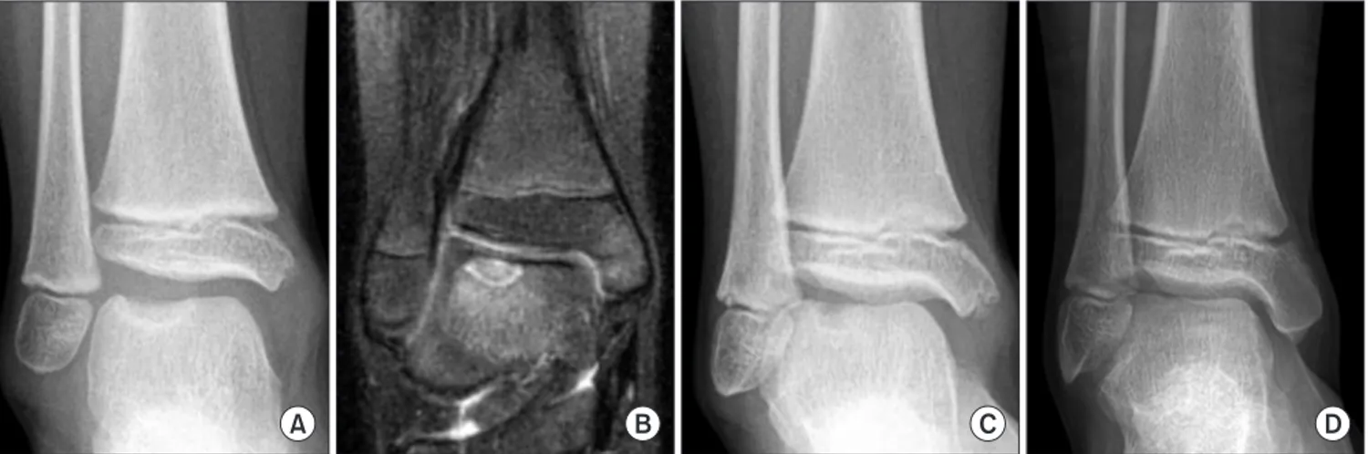
성인의 거골 골연골 병변에서 안정적인 병변(Berndt와 Harty 분 류 I-II단계)은 보존적 치료를 시도하고 병변이 불안정한 상태인 III-IV단계에서는 수술적 치료가 시행된다. 그러나 소아 및 청소 년에서 발생한 거골 골연골 병변의 경우에 성인보다 보존적 치료 로 병변이 치유되고 증상이 호전될 가능성이 높기 때문에 보존적 치료가 우선시되며 잔여 성장이 충분히 남은 환아의 경우에 특히 그러하다(Fig. 1).28)
보존적 치료의 방법으로는 석고고정을 통하여 움직임을 제한 하고 체중부하와 보행을 제한하는 방법이 있다.10) 환자들은 주로 4-8주간 석고고정을 하고 목발을 이용한 비체중부하 보행을 시
행한다.8,10,28) 그 이후에는 석고를 제거하고 보조기를 착용하면서
적극적인 관절운동을 시행하여 발목관절 운동범위의 회복을 얻
는다.8,28) 목발보행은 최소 6개월간 지속한다.10)
기존의 연구를 살펴보면 Higuera 등10)은 Berndt와 Harty 분류
I-III단계의 골연골 병변 환아들을 대상으로 68%에서 보존적 치 료를 시행하였으며 추시 도중 1예에서 수술적 치료가 필요하였 지만 이를 제외한 대부분의 환아들에서는 만족스러운 결과를 얻 을 수 있었다고 하였다. Kim 등28)의 연구에서도 비수술적으로 치 료한 환아군과 수술적 치료를 시행한 환아군의 치료 결과에서 통
계적 차이는 관찰되지 않았다. 그러나 Perumal 등8)은 초기 6개월 간 보존적 치료를 시행한 결과, 16%의 환자에서만 증상이 호전되 고 나머지 77%의 환자에서는 증상이 지속되거나 6%의 환자에서 는 증상이 악화되었으며 최종적으로는 42%의 환자에서 통증이 동반된 병변으로 인해 수술적 치료가 필요하였다고 보고하였다.
Table 1. Dipaola Staging System
Stage Arthroscopic Magnetic resonance imaging
I Irregularity and softening of articular cartilage, no definable fragment Thickening of articular cartilage and low signal changes II Articular cartilage breached, definable fragment, not displaceable Articular cartilage breached, low signal rim behind fragment
indicating fibrous attachment III Articular cartilage breached, definable fragment, displaceable,
but attached by some overlying articular cartilage
Articular cartilage breached, high signal changes behind fragment indicating synovial fluid between fragment and underlying subchondral bone
IV Loose body Loose body
Cited from the article of Dipaola et al. (Arthroscopy. 1991;7:101-4).26)
Table 2. Giannini’s Classification
Stage Extension Treatment
Acute injuries I <1 cm2 in diameter of lesions Debridement II >1 cm2 in diameter of lesions Fixation
Chronic injuries 0 Injuries with preserve the joint surfaces Retrograde drilling I <1.5 cm2 in diameter of lesions Microfracture II Lesions with >1.5 cm2 in diameter and <5 mm in depth Cartilage replacement
IIA Lesions with >1.5 cm2 in diameter and >5 mm in depth Cartilage replacement+bone graft
III Anatomy disruption Massive graft
Cited from the article of Vannini et al. (Orthop Clin North Am. 2012;43:237-44).27)
A B C D
Figure 1. Case of a 6-year-old boy treated conservatively. (A) Initial anteroposterior (AP) radiograph of the ankle joint showing a large osteolytic lesion with a sclerotic rim in the lateral aspect of the talar dome. (B) Coronal T2-weighted fat suppression magnetic resonance imaging showing an osteochondral fragment with high signal intensity in the medial aspect of the talar dome with intact articular cartilage. The lesion was classified as stage II according to Dipaola et al.26) (C) After a 2-year follow-up, the size of the lesion decreased on the AP radiograph. (D) Final AP radiograph at the 4-year follow-up showing complete healing of the lesion.
따라서 소아청소년기 거골 골연골 병변에서의 수술 적응증에 대해서는 아직 확립된 바가 없다. 대부분 초기 치료로는 보존적 치료가 우선시되지만 6개월 이상의 보존적 치료에도 통증이 지 속되거나 병변의 크기가 큰 경우와 전위가 발생한 경우에는 수술 적 치료가 권장된다.4,8,10-12,29) 관절 연골의 상태와 골연골편의 안정 성 여부도 치료 방법을 결정하는 데 중요한 기준으로 생각된다.30) 또한 방사선적으로 치유가 완료되기까지 오랜 시간이 걸리기 때 문에 실제 임상 증상은 없으나 방사선상으로는 병변이 계속 관찰 되는 경우를 볼 수 있다. 이러한 경우에는 임상적으로 증상의 지 속되는지 여부에 따라 보존적 요법과 수술적 요법 여부를 결정하 기도 한다.12)
6. 수술적 치료
수술적 치료를 요하는 경우에는 병변의 상태에 따라 골수 자극 술(marrow stimulation technique), 자가 골연골 이식술(autologous
osteochondral transplantation), 자가 연골세포 이식술(autologous chondrocyte implantation), 골수 유래 세포 이식술(bone mar- row-derived cells transplantation), 이종 골연골 이식술(allogenic osteochondral transplantation) 등을 시행하게 된다.
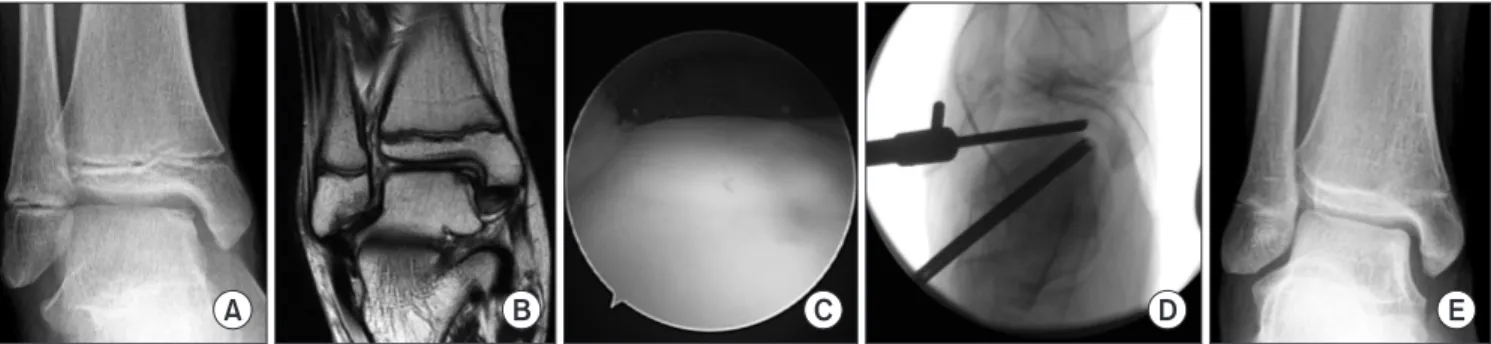
골수 자극술에는 천공술(drilling)과 미세골절술(microfracture) 등의 방법이 있다. 천공술은 골연골편이 안정적인 초기 병변에 주로 사용되는 방법이다. K-강선을 이용하여 거골의 연골하골까 지 천공을 시행하면 천공 부위에서 관절면으로 골모세포들이 새 어 나오면서 골연골편의 골유합과 신생혈관생성(neovasculariza- tion)을 촉진시키는 것으로 예상된다.29,31) 천공술은 K-강선이 삽 입되는 방향에 따라 순행성 천공술(antegrade drilling)과 역행성 천공술(retrograde drilling) (Fig. 2)로 나뉜다. 순행성 천공술은 핀 이 경골 내과(medial malleolus)를 관통하게 되므로 손상이 없었 던 경골의 관절면과 원위 경골 성장판을 손상시킬 수 있다는 단 점을 갖고 있다. 역행성 천공술은 거골두(head of the talus)에서 거
A B C D E
Figure 2. Case of an 11-year-old boy who underwent arthroscopic retrograde drilling and bone graft. (A) Preoperative anteroposterior (AP) radiograph of the ankle joint showing complete detachment of the osteochondral fragment in the medial aspect of the talar dome. This concurred with the Berndt and Harty stage III osteochondral lesion of the talus. (B) Coronal T1-weighted magnetic resonance imaging showing an iso-intense osteochondral fragment in the medial aspect of the talar dome with a low signal intensity lesion behind the fragment. The lesion was classified as stage II according to Dipaola et al.26) (C) Intraoperative arthroscopic image demonstrating an intact articular surface of the talar dome. (D) Intraoperative C-arm image performing retrograde drilling. (E) Postoperative 2-year AP radiograph showing complete healing of the lesion.
A B C D E
Figure 3. Case of a 17-year-old boy who underwent autologous osteochondral transplantation. (A) Preoperative anteroposterior (AP) radiograph of the ankle joint showing displacement of the osteochondral lesion in the medial aspect of the talar dome. The lesion was classified as a Berndt and Harty stage IV osteochondral lesion of the talus. (B) Coronal T2-weighted fat suppression magnetic resonance imaging showing the detachment of articular cartilage of the medial talar dome, which indicated that the lesion was classified as stage IV according to Dipaola et al.26) (C) Intraoperative gross photograph demonstrating a large-sized osteochondral lesion of the talar dome. (D) Postoperative gross photograph of the autologous osteochondral transplantation. (E) Postoperative 7-year AP radiograph showing complete healing of the lesion.
골 원개(talar dome)로 핀을 삽입하므로 이러한 단점은 없으나 기 술적으로 어렵고 골편의 전위를 유발할 수 있다는 단점이 있다.12) 천공술 이후 90% 정도에서 병변의 치유를 얻었다고 보고되고 있 다.31)
병변의 분리와 부분적인 전위가 진행된 경우에 단순 제거술 (excision)을 시행하기도 하지만 선호되는 방법은 아니다. 단순 제 거술은 단기 추시 결과는 좋으나 장기적으로는 골관절염 등의 후 유증을 남길 위험성이 높은 것으로 알려져 있다. 골연골편의 생 존력(viability)을 판단하여 고정이 가능하다면 K-강선, 압박 나사 못 등을 이용하여 골연골편을 고정하는 것이 관절면을 보존하는 데 더 유리하다.32) 하지만 이 역시 장기 추시 결과에 대해서는 보 고가 드물다.
미세절골술은 주로 작은 사이즈의 병변(<1.50 cm2)에서 치료 방법으로 사용되며, 손상된 관절 표면은 섬유연골(fibrocartilage) 로 회복된다.12) 수술방법이 쉽고 간단한 것이 장점이나 섬유연골 로 회복된 단면이 주위 정상 관절면과 일치하지 않으므로 병변이 큰 경우에는 적합하지 않다. 단기적으로는 만족스러운 결과가 보 고되었으나,33) 중기적인 추시 결과에서는 자가 골연골 이식술에 비하여 임상적으로나 MRI상으로 좋지 않은 결과를 보인다.34) 슬 관절 골연골 병변에서 미세절골술 후 결과를 확인해 보았을 때 41%의 환자에서 병변의 치유에 실패한 것으로 보고되었다.34) 1-5 cm2 사이즈의 병변에 대해서는 자가 골연골 이식술이 유용 하다(Fig. 3).34,35) 주로 대퇴골 외과의 비체중부하 부위에서 골연골 이식물(osteochondral plug)을 채취하여 거골의 병변에 삽입한다.
성인에서와 같이 내과 절골 도달법을 사용하므로 성장 종료 이후 수술을 시행하는 것이 적당하다. 미세절골술과 달리 골연골 이식 물이 삽입된 부위는 초자연골로 회복되는 것이 큰 장점이다. 하 지만 이식물 사이의 간격은 섬유연골이 생성된다. 공여 부위에서 얻을 수 있는 이식물의 크기가 제한되어 있어 크기가 큰 병변에 는 적합하지 않다는 점과 공여 부위에 합병증이 발생할 수 있다 는 점도 단점이다. 이식된 골연골편이 돌출되면 관절 운동 시에 충돌을 일으킬 수 있으므로 임펙터(impacter)를 이용하여 표면을 다듬으며 안정적으로 삽입한다.12) 술기가 까다로우므로 전용 기 구들을 이용하는 것이 좋고 주로 관혈적 접근법을 통하여 수술한 다.
넓은 면적의 병변에서는 자가 연골세포 이식술을 시행하기도 한다.36) 슬관절의 골연골 병변에서 시도되었던 이 술식은 병변 부 위를 초자연골과 섬유연골이 혼재되어 있는 형태로 재생시키는 것으로 알려져 있다. 임상적으로 만족스러운 결과들이 보고되고 있으나 장기 추시에서 얼마나 유용한지는 아직 입증되지 않았다.
최근에는 골수 유래 세포 이식술도 시도되고 있다.26,37) 골수 유 래 세포가 갖고 있는 다기능성(multipotential ability)으로 인하여 병변이 재생되는 것으로 알려져 있으나 역시 장기 추시 결과는 보고된 바가 없다.
이종 골연골 이식술은 병변의 크기가 큰 소아청소년기의 슬관 절 골연골 병변에서 유용성이 보고기도 하였으나 소아청소년기 거골 골연골 병변에서는 아직 유용성이 확인되지 않고 있다.38)
결 론
거골 골연골 병변은 거골의 관절연골과 연골하골에 분절과 분리 가 나타나는 질환이다. 관절면에서 분절된 골연골편의 안정성과 크기에 따라 치료 방법과 예후가 결정된다. 소아 및 청소년의 거 골 골연골 병변은 성인의 경우보다 병변이 치유되고 증상이 호전 될 가능성이 높기 때문에 보존적 치료가 우선 고려된다. 하지만 보존적 치료에 실패하거나, 병변의 크기가 큰 경우, 또한 전위가 발생한 경우에는 병변의 상태에 따라 골수 자극술, 자가 골연골 이식술 등을 시행하게 된다.
CONFLICTS OF INTEREST
The author has nothing to disclose.
REFERENCES
1. Bruns JBP. Osteochondrosis dissecans. Arthroskopie. 1998;
11:166-76.
2. Heywood CS, Benke MT, Brindle K, Fine KM. Correlation of magnetic resonance imaging to arthroscopic findings of stability in juvenile osteochondritis dissecans. Arthroscopy.
2011;27:194-9.
3. Kessler JI, Weiss JM, Nikizad H, et al. Osteochondritis disse- cans of the ankle in children and adolescents: demographics and epidemiology. Am J Sports Med. 2014;42:2165-71.
4. Wall E, Von Stein D. Juvenile osteochondritis dissecans. Or- thop Clin North Am. 2003;34:341-53.
5. Wall EJ, Vourazeris J, Myer GD, et al. The healing potential of stable juvenile osteochondritis dissecans knee lesions. J Bone Joint Surg Am. 2008;90:2655-64.
6. Hughes JA, Cook JV, Churchill MA, Warren ME. Juvenile osteochondritis dissecans: a 5-year review of the natural history using clinical and MRI evaluation. Pediatr Radiol.
2003;33:410-7.
7. Samora WP, Chevillet J, Adler B, Young GS, Klingele KE.
Juvenile osteochondritis dissecans of the knee: predictors of lesion stability. J Pediatr Orthop. 2012;32:1-4.
8. Perumal V, Wall E, Babekir N. Juvenile osteochondritis disse-
cans of the talus. J Pediatr Orthop. 2007;27:821-5.
9. Bauer M, Jonsson K, Lindén B. Osteochondritis dissecans of the ankle. A 20-year follow-up study. J Bone Joint Surg Br.
1987;69:93-6.
10. Higuera J, Laguna R, Peral M, Aranda E, Soleto J. Osteochon- dritis dissecans of the talus during childhood and adoles- cence. J Pediatr Orthop. 1998;18:328-32.
11. Letts M, Davidson D, Ahmer A. Osteochondritis dissecans of the talus in children. J Pediatr Orthop. 2003;23:617-25.
12. Vannini F, Cavallo M, Baldassarri M, et al. Treatment of juve- nile osteochondritis dissecans of the talus: current concepts review. Joints. 2015;2:188-91.
13. Steinhagen J, Niggemeyer O, Bruns J. Etiology and pathogen- esis of osteochondrosis dissecans tali. Orthopade. 2001;30:20- 7.
14. Petrie PW. Aetiology of osteochondritis dissecans. Failure to establish a familial background. J Bone Joint Surg Br.
1977;59:366-7.
15. Suckel A, Hoyer M, Raab C, Wünschel M. Osteochondrosis dissecans and osteochondral lesions of the talus: clinical and biochemical aspects. Sportverletz Sportschaden. 2012;26:91- 9.
16. Uozumi H, Sugita T, Aizawa T, Takahashi A, Ohnuma M, Itoi E. Histologic findings and possible causes of osteochondritis dissecans of the knee. Am J Sports Med. 2009;37:2003-8.
17. Roden S, Tillegard P, Unanderscharin L. Osteochondritis dissecans and similar lesions of the talus: report of fifty-five cases with special reference to etiology and treatment. Acta Orthop Scand. 1953;23:51-66.
18. Pill SG, Ganley TJ, Milam RA, Lou JE, Meyer JS, Flynn JM.
Role of magnetic resonance imaging and clinical criteria in predicting successful nonoperative treatment of osteochon- dritis dissecans in children. J Pediatr Orthop. 2003;23:102-8.
19. O’Connor MA, Palaniappan M, Khan N, Bruce CE. Osteo- chondritis dissecans of the knee in children. A comparison of MRI and arthroscopic findings. J Bone Joint Surg Br.
2002;84:258-62.
20. Moktassi A, Popkin CA, White LM, Murnaghan ML. Im- aging of osteochondritis dissecans. Orthop Clin North Am.
2012;43:201-11.
21. Cahill BR, Berg BC. 99m-Technetium phosphate compound joint scintigraphy in the management of juvenile osteochon- dritis dissecans of the femoral condyles. Am J Sports Med.
1983;11:329-35.
22. Berndt AL, Harty M. Transchondral fractures (osteochondri- tis dissecans) of the talus. J Bone Joint Surg Am. 1959;41:988- 1020.
23. Pritsch M, Horoshovski H, Farine I. Arthroscopic treatment of osteochondral lesions of the talus. J Bone Joint Surg Am.
1986;68:862-5.
24. Taranow WS, Bisignani GA, Towers JD, Conti SF. Retrograde drilling of osteochondral lesions of the medial talar dome.
Foot Ankle Int. 1999;20:474-80.
25. Hepple S, Winson IG, Glew D. Osteochondral lesions of the talus: a revised classification. Foot Ankle Int. 1999;20:789-93.
26. Dipaola JD, Nelson DW, Colville MR. Characterizing osteo- chondral lesions by magnetic resonance imaging. Arthrosco- py. 1991;7:101-4.
27. Vannini F, Battaglia M, Buda R, Cavallo M, Giannini S. ‘‘One step’’ treatment of juvenile osteochondritis dissecans in the knee: clinical results and T2 mapping characterization. Or- thop Clin North Am. 2012;43:237-44.
28. Kim HT, Park K, Seo CH, Ahn TY, Kim IH. Conservative treatment for juvenile osteochondritis dissecans of the talus. J Korean Orthop Assoc. 2017;52:310-8.
29. Kumai T, Takakura Y, Higashiyama I, Tamai S. Arthroscopic drilling for the treatment of osteochondral lesions of the ta- lus. J Bone Joint Surg Am. 1999;81:1229-35.
30. Lam KY, Siow HM. Conservative treatment for juvenile os- teochondritis dissecans of the talus. J Orthop Surg (Hong Kong). 2012;20:176-80.
31. Gunton MJ, Carey JL, Shaw CR, Murnaghan ML. Drilling juvenile osteochondritis dissecans: retro- or transarticular?
Clin Orthop Relat Res. 2013;471:1144-51.
32. Pascual-Garrido C, Tanoira I, Muscolo DL, Ayerza MA, Makino A. Viability of loose body fragments in osteochon- dritis dissecans of the knee. A series of cases. Int Orthop.
2010;34:827-31.
33. Salzmann GM, Sah BR, Schmal H, Niemeyer P, Sudkamp NP.
Microfracture for treatment of knee cartilage defects in chil- dren and adolescents. Pediatr Rep. 2012;4:e21.
34. Gudas R, Simonaityte R, Cekanauskas E, Tamosiūnas R. A prospective, randomized clinical study of osteochondral autologous transplantation versus microfracture for the treat- ment of osteochondritis dissecans in the knee joint in chil- dren. J Pediatr Orthop. 2009;29:741-8.
35. Sasaki K, Matsumoto T, Matsushita T, et al. Osteochondral autograft transplantation for juvenile osteochondritis disse-
cans of the knee: a series of twelve cases. Int Orthop. 2012;
36:2243-8.
36. Giannini S, Buda R, Vannini F, Di Caprio F, Grigolo B. Ar- throscopic autologous chondrocyte implantation in osteo- chondral lesions of the talus: surgical technique and results.
Am J Sports Med. 2008;36:873-80.
37. Giannini S, Buda R, Cavallo M, et al. Cartilage repair evo-
lution in post-traumatic osteochondral lesions of the talus:
from open field autologous chondrocyte to bone-marrow-de- rived cells transplantation. Injury. 2010;41:1196-203.
38. Lyon R, Nissen C, Liu XC, Curtin B. Can fresh osteochondral allografts restore function in juveniles with osteochondritis dissecans of the knee? Clin Orthop Relat Res. 2013;471:1166- 73.
소아청소년에서의 거골 골연골 병변
송미현
고려대학교 구로병원 정형외과
거골 골연골 병변은 거골의 관절연골과 연골하골에 분절과 분리가 나타나는 질환으로 관절면에서 분절된 골연골편의 안정성과 크기 에 따라 예후가 결정된다. 소아 및 청소년에서 발생한 경우 성인의 거골 골연골 병변보다 병변이 치유되고 증상이 호전될 가능성이 높기 때문에 소아청소년에서의 거골 골연골 병변은 초기 치료로 보존적 치료가 우선 시행된다. 하지만 보존적 치료에 실패하거나 병 변의 크기가 큰 경우와 전위가 발생한 경우에는 수술적 치료가 권장된다. 수술적 치료가 요하는 경우에는 병변의 상태에 따라 골수 자극술, 자가 골연골 이식술 등을 시행하게 된다.
색인단어: 골연골 병변, 거골, 족관절, 소아, 청소년
접수일 2017년 11월 3일 수정일 2018년 1월 11일 게재확정일 2018년 2월 22일 책임저자 송미현
08308, 서울시 구로구 구로동로 148, 고려대학교 구로병원 정형외과
TEL 02-2626-3239, FAX 02-2626-2486, E-mail wwiiw@naver.com, ORCID https://orcid.org/0000-0003-4082-1455
Copyright © 2018 by The Korean Orthopaedic Association
“This is an Open Access article distributed under the terms of the Creative Commons Attribution Non-Commercial License (http://creativecommons.org/licenses/by-nc/4.0/) which permits unrestricted non-commercial use, distribution, and reproduction in any medium, provided the original work is properly cited.”