INTRODUCTION
Percutaneous ultrasonography (US) guided radiofrequency (RF) ablation of liver tumors is a widely used technique (1-4).
However, many hepatocellular carcinoma (HCC) nodules locat- ed in the liver dome are poorly visualized on the US scan (5-7).
A conventional CT guided RF ablation can overcome this limi- tation, but has a significant disadvantage because the real-time monitoring is not possible (8, 9).
RF ablation under real-time CT fluoroscopy guidance can by reconstruction and display of CT images in real-time overcome
this limitation (10). This possibility provides the operator with an immediate feedback of images during the procedure, even if the nodules are not visualized on the US (11, 12). Unfortunately, the radiation hazards caused by this technique can be substan- tial to both the patients and the operator (13-16). If an intermit- tent CT fluoroscopy guided RF ablation is performed using the quick check method the radiation dose to the operators and the patients can be reduced (16-18). The aim of the present study was to evaluate whether intermittent CT fluoroscopy-guided RF ablation can be a clinically effective treatment for HCC nodules located in the liver dome.
J Korean Soc Radiol 2014;70(2):111-118 http://dx.doi.org/10.3348/jksr.2014.70.2.111
Received September 10, 2013; Accepted December 29, 2013 Corresponding author: Yun Ku Cho, MD
Department of Radiology, VHS Medical Center, 53 Jinhwangdo-ro 61-gil, Gangdong-gu, Seoul 134-791, Korea.
Tel. 82-2-2225-1442 Fax. 82-2-2225-1488 E-mail: [email protected]
This is an Open Access article distributed under the terms of the Creative Commons Attribution Non-Commercial License (http://creativecommons.org/licenses/by-nc/3.0) which permits unrestricted non-commercial use, distri- bution, and reproduction in any medium, provided the original work is properly cited.
Purpose: To evaluate the clinical effectiveness of an intermittent computed tomog- raphy (CT) fluoroscopy-guided radiofrequency (RF) ablation of hepatocellular carci- noma located in the liver dome.
Materials and Methods: Between 2005 and 2010 23 patients with hepatocellular carcinoma (HCC) nodules located in the liver dome underwent an intermittent CT fluoroscopy-guided RF ablation. The primary endpoint was the local tumor pro- gression. Procedure-related complications occurred in 3 of 23 patients. To evaluate the prognostic factors for the local tumor progression, univariate and multivariate analyses were performed using the Cox proportional hazards model. The chi- squared test was performed to evaluate the association of access route and proce- dure-related complication. The study was approved by the Institutional Review Board of our hospital.
Results: The Tumor sizes ranged between 1.0 and 2.9 cm. An initial complete abla- tion was achieved in all patients. The median follow-up period was 31 months and the major complication rate was 4.3%. The cumulative rate of local tumor progres- sion at 3 years was 20%. The univariate analysis revealed that only serum total bili- rubin level (p = 0.048) and prior chemoembolization were statistically significant (p
= 0.044), but there was no independently significant prognostic factor on multivari- ate analysis. Procedure-related complications occurred in 3 of 23 patients.
Conclusion: For HCC located in the liver dome an intermittent CT fluoroscopy- guided RF ablation could be performed safely and effectively.
Index terms
Hepatocellular Carcinoma Radiofrequency Ablation CT
Liver Dome Local Recurrence
Radiofrequency Ablation of Hepatocellular Carcinoma Located in the Liver Dome Under Intermittent CT Fluoroscopy Guidance
1 간 Dome에 위치한 간세포암의 간헐적 전산화단층촬영 투시 유도하 고주파 열 치료1Darlene Park, MD
1,2, Yun Ku Cho, MD
1, Hyun Je Cho, MD
1, Mi Young Kim, MD
11Department of Radiology, VHS Medical Center, Seoul, Korea
2Department of Radiology, Severance Hospital, Yonsei University College of Medicine, Seoul, Korea
ages was 5 seconds (17, 18). Gantry tilt was possible up to 30 de- gree. The reconstruction of an image was performed with a 256
× 256 matrix, with images displayed on a 768 × 768 matrix.
The anticipated path and depth of the target were deter- mined on the preliminary CT scans by using the electronic cal- ipers on the technologist’s console. The anticipated skin entry site was prepared with providone iodine solution (Betadine, Purdue Frederick, Norwalk, CT, USA) and draped in the rou- tine sterile fashion. After local anesthetization in the anticipat- ed puncture site, RF ablation was performed under conscious sedation using a combination of intravenous fentanyl citrate (Myung-moon, Seoul, Korea) and midazolam (Bukwang, Seoul, Korea).
After the site was prepared and draped, targeting was with the quick-check technique which is the most widely used intermit- tent CT fluoroscopy technique actually (17, 18). After the ab- dominal wall has been punctured, the table was moved to align the RF electrode with the imaging plane using the laser marker.
An internally cooled single electrode with 3 cm exposed tip (Cool tip; Radionics, Burlington, MA, USA) or multi-tined electrodes with 3.5 cm exposed tip (Le Veen Needle electrode; Boston Sci- entific, Watertown, MA, USA) were used. Then single CT fluo- roscopic spot images were acquired to check the electrode loca- tion and to confirm the appropriate alignment. The gantry was tilted until the lung parenchyma was not included in the imagi- nary trajectory on the preliminary scan. If this was not possible, a trans-pulmonary approach was used. Once the single CT fluo- roscopic spot image helped to confirm that the electrode was at the appropriate position and trajectory, the electrode was ad- vanced to the target (17, 18). While advancing the electrode, in- termittent fluoroscopic spot images were acquired to confirm the electrode locations until the electrode tip was positioned in the target. The tube potentials were 140 kVp, the tube currents were 50 mA and the section thickness was 5 mm.
If possible, 5‒10 mm of ablative margin around the index tu- mor if were secured. An attempt was also done to limit the num- ber of electrodes passes through the peritoneum to a single inser- tion. If an additional ablation was acquired, the needle position within the tumor was relocated by pushing it back into the su- perficial liver tissue along its major axis, changing the angle and then inserting the needle into the target without a complete withdrawal of the electrode out of the peritoneum.
MATERIALS AND METHODS
Patient and Tumor Characteristics
The study was a retrospective analysis based on a prospective database and was approved by the Institutional Review Board of our hospital. An informed consent for interventional proce- dures was obtained from all patients before.
The primary endpoint was defined as local tumor progres- sion. The secondary endpoints included initial complete abla- tion, major and minor complications and overall survival.
All CT fluoroscopy guided RF ablations were blinded per- formed by a single interventional radiologist with seven years of experience in the ablation of liver tumors at study onset.
Twenty-three consecutive subjects with hepatocellular carcino- ma located in the liver dome were included between December 2005 and December 2010. Because the HCC were not clearly visi- ble on US, all patients underwent a CT fluoroscopy-guided RF ablation for HCC. HCC nodules located within 2 cm from the di- aphragm were classified as liver dome nodules. CT fluoroscopy guided RF ablation was performed for primary or recurrent HCC nodules following transarterial chemoembolization (TACE).
The inclusion criteria for percutaneous RF ablation were as fol- lows: a single tumor of 4 cm in longest dimension or smaller;
multinodular tumors (≤ 3) with each tumor ≤ 3 cm in the longest dimension, Child-Pugh class A or B; no portal vein thrombosis or extrahepatic metastasis; prothrombin time ratio greater than 50%
and platelet count greater than 50000/mm3 (50 × 109/L).
CT fluoroscopy guided RF ablation was not performed if a patient was not cooperative.
The diagnosis of HCC was based on the typical imaging fea- tures which are an arterial enhancement followed by delayed washout at the dynamic contrast-enhanced CT or MRI (1). No patient underwent a percutaneous biopsy to diagnose HCC.
Techniques for the CT Fluoroscopy Guided Radiofrequency Ablation
CT fluoroscopic images were acquired during the intermit- tent scanning with a machine (LightSpeed CT/i equipped with SmartStep; GE Medical Systems, Milwaukee, WI, USA), which was controlled by an integrated foot switch and hand-held con- troller. The radiation exposure time was 0.8 or 1 second, and the reconstruction duration for individual CT fluoroscopic spot im-
RESULTS
Age of patients ranged between 56 and 78 years (mean age 64.9 years). All 23 patients had cirrhosis associated with either viral hepatitis B (16 patients), hepatitis C (five patients) or other unknown causes (two patients). Among the patients, 22 had Child-Pugh class A and one had class B disease. The tumor size ranged from 1.0 cm to 2.9 cm (median 1.7 cm). The follow-up periods ranged from 9 to 70 months (median 31 months). Prior to the performance of CT fluoroscopy guided RF ablation 17 out of the 23 patients underwent a TACE for the index tumors.
Internally cooled single electrode or multi-tined electrodes were used in 17 respective in 6 patients. Other factors are described in Table 1.
The total procedural time per session ranged from 30 to 100 minutes (average time 58 minutes) and the CT fluoroscopic time per session ranged from 2 seconds to 52 seconds (average time 16 seconds). The number of peritoneal puncture ranged from one to three times (average time 1.2 times), while the number of needle tracts in the liver parenchyma per session ranged from 1 Evaluation of Technical Factors and Therapeutic
Response
Enhanced dynamic CT scans were performed immediately after the RF procedure to evaluate the possible residual viable tumors.
The total procedure time (from the injection of local anesthetics to the time of needle withdrawal) and the total fluoroscopic time (in seconds) were reported. The number of peritoneal punctures and the number of liver parenchymal puncture tracts were also re- corded. As a measure of therapeutic responses, initial complete ablations, complications, local tumor progression and the over- all survival of patients following RF ablation were evaluated.
Technique effectiveness of CT fluoroscopy-guided RF ablation was defined as complete encompassment of index tumor by ab- lation zone on dynamic CT one month post procedure (19). Re- sidual viable tumor was judged as present when an enhanced por- tion was seen within or around of the original mass on the one- month follow-up CT imaging. If no definite evidence of residual tumor was noted on the one-month follow-up CT, a 3-phase con- trast-enhanced CT or dynamic MRI was performed at 3- or 4-month interval thereafter. Local tumor progression was judged as present when an enhanced portion was seen within or at the margin of the original mass at the first one-month or the there- after follow-up dynamic CT or MRI scans. The CT or MR im- ages were evaluated by two radiologists (K.M.Y. and C.H.J.) with 5 and 15 years of experience in abdominal radiology. Final deci- sions were reached by consensus. Major complication was de- fined as an event that leads to substantial morbidity or substan- tially lengthened hospital stay (19).
Statistical Analysis
The cumulative local tumor progression rates were estimated by using the Kaplan-Meier estimation. To evaluate the possible prognostic factors of local tumor progression, univariate and multivariate analyses were performed using the Cox proportion- al hazard model. Parameters that proved to be significant in the univariate analysis were subsequently tested with the multivari- ate Cox proportional hazard model. The chi-squared test was performed to evaluate the association of access route and the occurrence of complication after the procedure.
Null hypotheses of no difference was rejected if p-values were less than 0.05. The SPSS software package (version 10.0; SPSS Inc., Chicago, IL, USA) was used for the statistical analysis.
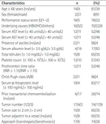
Table 1. Baseline Patients and Tumor Characteristics
Characteristics n (%)
Age ≥ 66 years (no/yes) 14/9 61/39
Sex (female/male) 2/21 9/91
Performance status score (0/1–2) 18/5 78/22 Underlying causes (HBV/HCV/others) 16/5/2 70/22/8 Serum AST level (≤ 40 units/L/> 40 units/L) 12/11 52/48 Serum ALT level (≤ 40 units/L/> 40 units/L) 12/11 52/48
Presence of ascites (no/yes) 22/1 96/4
Serum albumin level (< 3.5 g/dL/≥ 3.5 g/dL) 4/19 17/83 Total bilirubin (≤ 1.0 mg/dL/> 1.0 mg/dL) 15/8 65/35 Platelet count (≥ 100 × 109/L/< 100 × 109/L) 13/10 57/43 Prothrombin time ratio
(INR ≤ 1.15/INR > 1.15) 12/11 52/48
Child-Pugh class (A/B) 22/1 96/4
Serum α-fetoprotein level
(≤ 100 ng/mL/> 100 ng/mL) 19/4 83/17
Prior transarterial chemoembolization
(no/yes) 6/17 26/74
Tumor number (1/2/3) 17/4/2 74/17/9
Tumor size (≤ 2 cm /> 2 cm) 15/8 65/35
Tumor adjacent to a vessel (no/yes) 15/8 65/35 Approach (transhepatic/tansthoracic) 17/6 74/26 Note.-ALT = alanine aminotransferase, AST = aspartate aminotransferase, HBV = hepatitis B virus, HCV = hepatitis C virus, INR = International Nor- malized Ratio
to 8 (average time 3.0). The ablation time ranged from 6 to 24 minutes and a roll-off was obtained in all patients.
No residual viable tumors were noted in all patients after one or two initial sessions of CT fluoroscopy guided RF ablation.
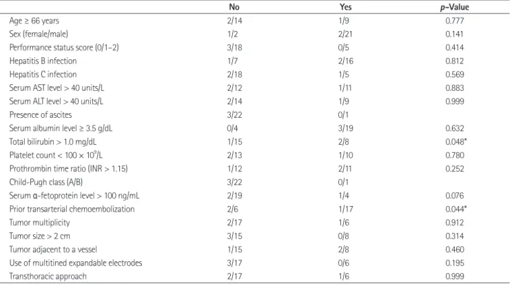
One session of CT fluoroscopy guided RF ablation was per- formed in 21 patients, and in two patients were performed two sessions (Fig. 1). The technique effectiveness rate was 91%. Lo- cal tumor progression occurred in three of the 23 nodules. An additional TACE or RF ablation was performed in two patients to treat these recurrent tumors. The cumulative local tumor progression rates were 0% after 1 year, 12% after 2 years and 20% after 3 years (Fig. 2). Table 2 shows the results of univari- ate analysis for the prognostic factors for local recurrence.
Only the level of serum total bilirubin level (> 1 mg/dL, p = 0.048) and prior TACE (p = 0.044) were statistically significant.
Fig. 1. A 62-year-old woman with hepatocellular carcinoma located in the right hepatic dome.
A. Arterial phase of the contrast-enhanced computed tomography (CT) scan shows a 1.6 cm sized hypervascular nodule with spotty iodized oil uptake in liver (arrow). Delayed phase CT scan (not shown here) reveals washout of contrast media, confirming the radiologic diagnosis of hepa- tocellular carcinoma (HCC).
B. Intermittent CT fluoroscopic spot images are taken to confirm that the electrode is at the appropriate position and trajectory until the elec- trode tip is positioned in the target.
C. On an unenhanced CT scan obtained during the procedure of radiofrequency (RF) ablation under CT fluoroscopy guided transhepatic approach, the tins of a multitined expandable electrode are deployed within the tumor. The duration of application of RF energy was 13 minutes.
D. Portal phase of the contrast-enhanced CT scan obtained immediately after the RF ablation shows a 2.4 cm diameter ablated zone (arrow). The RF needle was not completely retracted before the operator convinced that the tumor has been sufficiently ablated.
E. Arterial (not shown here) and portal phase of the contrast-enhanced CT scan obtained one month after RF ablation shows no definite evi- dence of residual or recurred HCC nodule (arrow).
F. Arterial phase of the contrast-enhanced CT scan obtained 68 months after RF ablation shows no definite evidence of residual or recurred HCC nodules (arrow). Note that the extent of the ablated area has been much reduced than before.
E B
D A
F C
Fig. 2. A diagram of the cumulative local tumor progression rate of hepatocellular carcinoma after RF ablation under CT fluoroscopy. Note that the cumulative 3-year local tumor progression rate was 20%.
0.0 0.1 0.2 0.3
Local tumor progression
0 10 20 30 40 50 60
Months
2-, 3-, 4-, and 5-year overall survival rates from the performance of RF ablation for the HCC nodule were 83%, 63%, 58%, 42%, and 31%, respectively.
DISCUSSION
To perform an ultrasonographically guided RF ablation for the HCC nodules located in the liver dome is difficult in many cases because of their poor visibility or accessibility (2, 20). These tu- mors may become clearly visualized if an artificial ascites or pleural effusion is induced (20-22). However, inducing an artifi- cial pleural effusion can be very difficult in cases of pleural ad- hesion (21, 22). Even after an artificial ascites or pleural effusion are induced successfully, the local tumor progression rates are reported as 20% or more, because it may still be difficult to lo- calize the tumor accurately (20-23). Furthermore, because of the presence of diffusely scattered benign nodules in the back- ground liver parenchyma many small tumors cannot be clearly discerned (24, 25).
Real-time guidance techniques other than US, such as the real In the univariate analysis no other factors were statistically sig-
nificant. Multivariate analysis revealed no independent signifi- cant prognostic factors.
In three of the 23 patients procedure-related complications oc- curred, including minor pleural effusion in two patients, self-lim- ited pneumothorax in one patient and an intraperitoneal hemato- ma in one patient. The latter was the only major complication and it was successfully treated with conservative management. The overall complication rate per patient was 13.0% (3/23) and the major complication rate was 4.3% (1/23). However, no major complication occurred under transabdominal approaches and three of the four complications occurred among the six patients who underwent a RF ablation with a transpulmonary approach.
The chi-squared test revealed that the access route and the oc- currence of complication were statistically associated (p = 0.040).
Fifteen patients died during the follow-up period and among them 10 patients died with progressive HCC. The 1-, 2-, 3-, 4-, and 5-year overall survival rates from the initial diagnosis of HCC were 96%, 91%, 82%, 68%, and 48%, respectively. The 1-,
Table 2. Univariate Analysis of Prognostic Factors Affecting Local Recurrence of the Hepatocellular Carcinoma Nodules Undergoing CT Fluo- roscopy Guided Radiofrequency Ablation
No Yes p-Value
Age ≥ 66 years 2/14 1/9 0.777
Sex (female/male) 1/2 2/21 0.141
Performance status score (0/1–2) 3/18 0/5 0.414
Hepatitis B infection 1/7 2/16 0.812
Hepatitis C infection 2/18 1/5 0.569
Serum AST level > 40 units/L 2/12 1/11 0.883
Serum ALT level > 40 units/L 2/14 1/9 0.999
Presence of ascites 3/22 0/1
Serum albumin level ≥ 3.5 g/dL 0/4 3/19 0.632
Total bilirubin > 1.0 mg/dL 1/15 2/8 0.048*
Platelet count < 100 × 109/L 2/13 1/10 0.780
Prothrombin time ratio (INR > 1.15) 1/12 2/11 0.252
Child-Pugh class (A/B) 3/22 0/1
Serum α-fetoprotein level > 100 ng/mL 2/19 1/4 0.076
Prior transarterial chemoembolization 2/6 1/17 0.044*
Tumor multiplicity 2/17 1/6 0.912
Tumor size > 2 cm 3/15 0/8 0.314
Tumor adjacent to a vessel 1/15 2/8 0.460
Use of multitined expandable electrodes 3/17 0/6 0.195
Transthoracic approach 2/17 1/6 0.999
Note.-*Statistically significant (p < 0.05).
ALT = alanine aminotransferase, AST = aspartate aminotransferase, INR = International Normalized Ratio
complication rates.
The limitations of the present study are as follows:
First, the study was a single arm retrospective analysis. Further studies by other investigators will be needed for generalization.
Prospective controlled studies will provide a strong conclusion on the clinical effectiveness of the intermittent CT fluoroscopy guided RF ablation.
Second, prior TACE might have a significant confounding ef- fect on the clinical effectiveness of the CT fluoroscopy guided RF ablation. Conversely, the present study strongly suggests a prior treatment with TACE may reduce the local tumor pro- gression significantly (32).
Third, the local tumor progression rate for HCC nodules in liver dome was not reduced even with a higher radiation expo- sure to both the patient and the personnel than suggested by the conventional CT guidance or the biplane fluoroscopy guidance.
Finally, the number of patients was too small to reveal poten- tially significant prognostic factors for local tumor progression in the multivariate analysis.
In conclusion, the intermittent CT fluoroscopy-guided RF ab- lation can be performed safely and effectively for the HCCs lo- cated in the liver dome which are not clearly visible on US.
REFERENCES
1. Bruix J, Sherman M; Practice Guidelines Committee, Ameri- can Association for the Study of Liver Diseases. Manage- ment of hepatocellular carcinoma. Hepatology 2005;42:
1208-1236
2. Rhim H, Lim HK, Kim YS, Choi D, Lee WJ. Radiofrequency ablation of hepatic tumors: lessons learned from 3000 procedures. J Gastroenterol Hepatol 2008;23:1492-1500 3. Tateishi R, Shiina S, Teratani T, Obi S, Sato S, Koike Y, et al.
Percutaneous radiofrequency ablation for hepatocellular carcinoma. An analysis of 1000 cases. Cancer 2005;103:
1201-1209
4. Livraghi T, Meloni F, Di Stasi M, Rolle E, Solbiati L, Tinelli C, et al. Sustained complete response and complications rates after radiofrequency ablation of very early hepato- cellular carcinoma in cirrhosis: Is resection still the treat- ment of choice? Hepatology 2008;47:82-89
5. Yang B, Zhang B, Xu Y, Wang W, Shen Y, Zhang A, et al.
time CT fluoroscopy, biplane fluoroscopy or 3-dimensional (3D) fluoroscopic navigation system using cone beam CT, can be potent guiding techniques also (14, 26, 27). However, biplane fluoroscopy guidance techniques or 3D fluoroscopic navigation system are useful only when the tumors are clearly visible on the fluoroscopy because of the iodized oil retention within the tu- mors (27). In addition, with real time CT fluoroscopy guidance technique the radiation hazard can be substantial to both the patient and the operator (14).
In contrast, an intermittent CT fluoroscopy guided RF abla- tion can be with a minimal risk of mistargeting or radiation haz- ard an effective alternative method for HCC regardless of the io- dized oil retention (16). In addition, an easier localization process is the important advantage of CT fluoroscopy if compared with the conventional CT guidance technique. Also, an interactive process is possible between the operator and the patient (16).
In the present study an initial complete ablation was achieved in all cases, but the cumulative rate of local tumor progression at 20% after 3 years. This is similar to the results from previous studies where conventional CT guidance or biplane fluoroscopy guidance techniques were used (20, 26, 28). Such a suboptimal result may be partially attributed to the difficulties to accurate localize the respiratory movements of patients during the proce- dure. The radiation dose needs to be in acceptable ranges for both the patient and the personnel if the intermittent-mode CT fluoroscopy-guided RF ablation was to be justified. The average CT fluoroscopic times in the present study were similar to those reported for other intra-abdominal procedures such as biopsy or drainage procedures using the intermittent-mode CT fluo- roscopy guidance (17, 18).
In the present study, a transabdominal approach was pre- ferred with an intention to minimize possible pleural and lung parenchymal injuries or a metastatic tumor seeding in the tho- rax (29). A frequent incidence of pneumothorax or pleural effu- sion has been reported for CT guided RF ablation using a trans- pulmonary approach (29-31). Among the 17 patients treated with a transdominal approach only one case of minor pleural ef- fusion was encountered as a procedure-related complication which is in accordance with other study results. In contrast, if the transpulmonary approach was used, the complication rate was much higher. So, in the authors’ opinion, transpulmonary approaches should be rather avoided if possible to reduce the
Vasc Interv Radiol 1994;5:71-84
16. Silverman SG, Tuncali K, Adams DF, Nawfel RD, Zou KH, Judy PF. CT fluoroscopy-guided abdominal interventions:
techniques, results, and radiation exposure. Radiology 1999;212:673-681
17. Paulson EK, Sheafor DH, Enterline DS, McAdams HP, Yo- shizumi TT. CT fluoroscopy--guided interventional proce- dures: techniques and radiation dose to radiologists. Radi- ology 2001;220:161-167
18. Carlson SK, Felmlee JP, Bender CE, Ehman RL, Classic KL, Hoskin TL, et al. CT fluoroscopy-guided biopsy of the lung or upper abdomen with a breath-hold monitoring and feedback system: a prospective randomized controlled clinical trial. Radiology 2005;237:701-708
19. Goldberg SN, Grassi CJ, Cardella JF, Charboneau JW, Dodd GD 3rd, Dupuy DE, et al. Image-guided tumor ablation:
standardization of terminology and reporting criteria. J Vasc Interv Radiol 2009;20(7 Suppl):S377-S390
20. Rhim H, Lim HK, Kim YS, Choi D. Percutaneous radiofre- quency ablation with artificial ascites for hepatocellular carcinoma in the hepatic dome: initial experience. AJR Am J Roentgenol 2008;190:91-98
21. Koda M, Ueki M, Maeda Y, Mimura K, Okamoto K, Matsu- naga Y, et al. Percutaneous sonographically guided radio- frequency ablation with artificial pleural effusion for he- patocellular carcinoma located under the diaphragm. AJR Am J Roentgenol 2004;183:583-588
22. Iwai S, Sakaguchi H, Fujii H, Kobayashi S, Morikawa H, Enomoto M, et al. Benefits of artificially induced pleural effusion and/or ascites for percutaneous radiofrequency ablation of hepatocellular carcinoma located on the liver surface and in the hepatic dome. Hepatogastroenterology 2012;59:546-550
23. Kang TW, Rhim H, Kim EY, Kim YS, Choi D, Lee WJ, et al.
Percutaneous radiofrequency ablation for the hepatocel- lular carcinoma abutting the diaphragm: assessment of safety and therapeutic efficacy. Korean J Radiol 2009;10:
34-42
24. Fukunaga T, Kudo M, Tochio H, Okabe Y, Orino A. Natural course of small nodular lesions with intranodular pre- served portal supply in cirrhotic liver. Oncology 2007;72 Suppl 1:24-29
Prospective study of early detection for primary liver can- cer. J Cancer Res Clin Oncol 1997;123:357-360
6. Sherman M, Peltekian KM, Lee C. Screening for hepatocel- lular carcinoma in chronic carriers of hepatitis B virus: in- cidence and prevalence of hepatocellular carcinoma in a North American urban population. Hepatology 1995;22:
432-438
7. Izzo F, Cremona F, Ruffolo F, Palaia R, Parisi V, Curley SA.
Outcome of 67 patients with hepatocellular cancer detect- ed during screening of 1125 patients with chronic hepatitis.
Ann Surg 1998;227:513-518
8. Toyoda M, Kakizaki S, Horiuchi K, Katakai K, Sohara N, Sato K, et al. Computed tomography-guided transpulmo- nary radiofrequency ablation for hepatocellular carcinoma located in hepatic dome. World J Gastroenterol 2006;12:
608-611
9. Park BJ, Byun JH, Jin YH, Won HJ, Shin YM, Kim KW, et al.
CT-guided radiofrequency ablation for hepatocellular car- cinomas that were undetectable at US: therapeutic effec- tiveness and safety. J Vasc Interv Radiol 2009;20:490-499 10. Yamakado K, Nakatsuka A, Ohmori S, Shiraki K, Nakano T,
Ikoma J, et al. Radiofrequency ablation combined with chemoembolization in hepatocellular carcinoma: treat- ment response based on tumor size and morphology. J Vasc Interv Radiol 2002;13:1225-1232
11. Yamakado K, Nakatsuka A, Takaki H, Yokoi H, Usui M, Sak- urai H, et al. Early-stage hepatocellular carcinoma: radio- frequency ablation combined with chemoembolization versus hepatectomy. Radiology 2008;247:260-266
12. Katada K, Kato R, Anno H, Ogura Y, Koga S, Ida Y, et al.
Guidance with real-time CT fluoroscopy: early clinical ex- perience. Radiology 1996;200:851-856
13. Kato R, Katada K, Anno H, Suzuki S, Ida Y, Koga S. Radia- tion dosimetry at CT fluoroscopy: physician’s hand dose and development of needle holders. Radiology 1996;201:
576-578
14. Nawfel RD, Judy PF, Silverman SG, Hooton S, Tuncali K, Adams DF. Patient and personnel exposure during CT fluo- roscopy-guided interventional procedures. Radiology 2000;216:180-184
15. Wagner LK, Eifel PJ, Geise RA. Potential biological effects following high X-ray dose interventional procedures. J
the hepatic dome. Hepatogastroenterology 2008;55:1164- 1166
30. Shibata T, Shibata T, Maetani Y, Kubo T, Itoh K, Togashi K, et al. Transthoracic percutaneous radiofrequency ablation for liver tumors in the hepatic dome. J Vasc Interv Radiol 2004;15:1323-1327
31. Miura H, Yamagami T, Terayama K, Yoshimatsu R, Matsu- moto T, Nishimura T. Pneumothorax induced by radiofre- quency ablation for hepatocellular carcinoma beneath the diaphragm under real-time computed tomography-fluo- roscopic guidance. Acta Radiol 2010;51:613-618
32. Lee MW, Kim YJ, Park SW, Hwang JH, Jung SI, Jeon HJ, et al. Percutaneous radiofrequency ablation of small hepato- cellular carcinoma invisible on both ultrasonography and unenhanced CT: a preliminary study of combined treat- ment with transarterial chemoembolisation. Br J Radiol 2009;82:908-915
25. Kudo M. Multistep human hepatocarcinogenesis: correla- tion of imaging with pathology. J Gastroenterol 2009;44 Suppl 19:112-118
26. Lee MW, Kim YJ, Park SW, Yu NC, Choe WH, Kwon SY, et al. Biplane fluoroscopy-guided radiofrequency ablation combined with chemoembolisation for hepatocellular car- cinoma: initial experience. Br J Radiol 2011;84:691-697 27. Morimoto M, Numata K, Kondo M, Nozaki A, Hamaguchi
S, Takebayashi S, et al. C-arm cone beam CT for hepatic tumor ablation under real-time 3D imaging. AJR Am J Roentgenol 2010;194:W452-W454
28. Kim YK, Kim CS, Lee JM, Chung GH, Chon SB. Efficacy and safety of radiofrequency ablation of hepatocellular carci- noma in the hepatic dome with the CT-guided extratho- racic transhepatic approach. Eur J Radiol 2006;60:100-107 29. Wang ZY, Sun WB, Li MY, Zhang XX, Ding XM. Percutane- ous extrapulmonary radiofrequency ablation for tumors in
간 Dome에 위치한 간세포암의 간헐적 전산화단층촬영 투시 유도하 고주파 열 치료1
박다린
1,2· 조윤구
1· 조현제
1· 김미영
1목적: 간 dome에 위치한 간세포암 치료시 간헐적 전산화단층촬영 투시법을 이용한 고주파 열 치료의 임상적 유용성을 평 가하고자 하였다.
대상과 방법: 2005년부터 2010년까지 23명의 환자를 대상으로 간 dome에 위치한 간세포암에 대해 간헐적 전산화단층 촬영 투시 유도하에 고주파 열치료를 시행하였다. 본 연구는 본원의 임상시험 심사위원회의 승인을 받았다. 국소 종양 재 발률에 영향을 미치는 예후인자를 평가하기 위해 Cox 비례위험 모형을 이용하여 분석하였다. 또한 접근 경로와 시술 후 합병증과의 연관성을 분석하기 위해 카이 자승 검증을 하였다.
결과: 종양 크기는 1.0 cm에서 2.9 cm로 모든 환자에서 초기 국소 치료에 성공하였다. 평균 환자 추적 기간은 31개월이 었고 합병증 발생률은 4.3%였다. 3년 누적 국소 종양 재발률은 20%였다. 국소 재발에 대한 단일 변수분석 결과 혈청 총 빌리루빈 수치와 간색전술 시행 여부가 통계적으로 의미 있는 요인이었고, 다변량 분석에서는 의미 있는 변수는 없었다. 시 술과 연관된 합병증은 23명의 환자 중 3명에서 생겼다.
결론: 간 dome에 위치한 간세포암에 대해 간헐적 전산화단층촬영 투시를 이용한 고주파 열치료는 안전하면서도 효과적 인 치료 방법이다.
1중앙보훈병원 영상의학과, 2연세대학교 의과대학 세브란스병원 영상의학과