Introduction
There have been many studies on the characteristics and differences between two sexes or races with respect to ontogeny, morphology, and size of teeth in dental anthro- pology [1-5]. Also, digital analysis using advanced digital photography and computer-assisted imaging tools are being used in active studies in a diverse population. It has been widely known that there is a variation in the cusp size of teeth according to the order of development and sex, which has been reported in morphometric studies from several countries and periods [6,7].
In a study in the natives of Australia, it was reported that cusps of the maxillary first molar which form early
show less variations than cusps of the maxillary second molar which form later [8]. In addition, sex differences existed in the maxillary second molars than in the maxil- lary first molars. Other researches also showed that late- developing cusps tend to have more variation and sex dif- ferences because of the differences in production and secre- tion of sex hormones [9]. For instance, maxillary second molars have greater variations in crown size as well as sex differences than maxillary first molars. Kondo et al. also reported that the later the calcification occurs, the greater the size variation in cusps in dental casts obtained from Japanese patients [10]. In addition, protocone, the earliest- developing cusp of maxillary molars exhibited lesser sex difference in cusp size [11]. A previous study also demon- strates a high correlation between the order of cusp calci- fication and variation of cusp size in hominoid primates [12].
Mandibular posterior teeth consist of the mesial part and the distal part in the early developmental stage, and the former is called trigonid, the latter is called talonid.
Variations in the Cusps of Mandibular Molars in Koreans
Hong-Il Yoo, Ji-Hye Kim, Sun-Hun Kim
Department of Oral Anatomy, School of Dentistry, Chonnam National University
(Received 30 July 2014, revised 25 August 2014, accepted 5 September 2014, Published Online 30 September 2014)
Abstract: This study aimed to investigate the cusp size and morphological characteristics of permanent mandibu- lar molars in Koreans with reference to the hypoconulid, and to analyze the differences and correlations between both sexes as well as between first and second mandibular molars. We obtained data from dental casts of 110 adults (78 males and 32 females). Mesiodistal and buccolingual diameters of first and second mandibular molars, the area of five cusps (protoconid, metaconid, hypoconid, entoconid, and hypoconulid), as well as the total cusp area and occlusal table area were measured. Paired t-test was performed to analyze the morphological differences between first and second mandibular molars and the sex differences between both sexes using SPSS program.
Crown diameters and cusp areas of mandibular first molars were larger than those of mandibular second molars in both sexes. The hypoconulid was the most variable in size and morphological pattern among the five cusps, and the first molars showed a higher incidence of hypoconulid than the second molars. Except for the entoconid area of the first molar (p==0.06) and the hypoconulid area of the second molar (p==0.24), all other mean values were larger in males than in females, demonstrating a significant sexual dimorphism. These data suggest that the teeth which develop late in ontogeny tend to be smaller in size and more variable in morphological characteristics.
Keywords : Mandibular molar, Koreans, Hypoconulid, Dimorphism
The author (s) agree to abide by the good publication practice guideline for medical journals.
The author (s) declare that there are no conflicts of interest.
Correspondence to : Sun-Hun Kim (Department of Oral Anatomy, Dental Science Research Institute, School of Dentistry, Chonnam National Univer- sity)
E-mail : [email protected]
http://dx.doi.org/10.11637/kjpa.2014.27.3.155 Original Article
During the later stage of development, trigonid is divided into two cusps, protoconid (mesiobuccal cusp) and meta- conid (mesiolingual cusp), and talonid is divided into three cusps, hypoconid (distobuccal cusp), entoconid (distolin- gual cusp), and hypoconulid (distal cusp), resulting in a total of five cusps. Among the five cusps, protoconid is the first to develop and calcify, whereas hypoconulid is the last to develop and calcify, with greater individual vari- ations [5].
Although there are some morphological studies on decid- uous and permanent molar teeth of Koreans [13,14], not much study was performed to investigate the individual cusp size and morphological characteristics of the occlusal surface with reference to the size of hypoconulid of mandi- bular molars, and to assess sex differences in size and mor- phology of mandibular first and second molars yet.
Materials and Methods
1. Sampling group
The total number of participants in this was 164, consist- ing of 110 males and 54 females who were students at the Chonnam National University School of Dentistry. The average age of participants was 30 years.
2. Selection and exceptions to the sampling group
1) Oral examination of participants
Participants with dental restorations such as amalgam,
gold crown, and inlay in mandibular right first and second molars were excluded from the experiment because it is difficult to distinguish the adjacent cusps. Also, partici- pants with missing molars on both sides were excluded.
2) Test for dental casts
Dental casts with ambiguous cusp tips due to attrition or fracture of the occlusal surface were excluded from the experiment. The experiment was mostly performed using mandibular right molars, but mandibular left molars were also included when there were missing teeth, heavy attri- tion, prosthesis, and anomalies on the right side. After ex- cluding all types of exceptions, the final number of partic- ipants was 110 with 78 males and 32 females.
3. Measurement of the occlusal surface
Dental casts were made with dental plaster (GC co., Tokyo, Japan) after taking impression of participants’ teeth.
Cusp tips and marginal ridges were marked by a pencil to distinguish the circumferential occlusal table area. Canon Powershot A640 (Canon, Tokyo, Japan) was used to take an image of the occlusal surfaces of mandibular first molars and second molars in dental casts (Fig. 1). Mesiodistal (MD) diameter, buccolingual (BL) diameter, individual cusp area, total cusp area, and occlusal table area of man- dibular molars in the photographs were measured by Axio Vision LE Rel 4.4 software (Carl Zeiss, Jena, Germany).
The following data were measured: MD diameter, BL diameter, individual areas of five cusps (protoconid, meta- conid, hypoconid, entoconid, and hypoconulid), total cusp
Fig. 1.Images of the dental cast were taken from the occlusal surfaces of mandibular first molars (Left photograph) and second molars (Right photograph) under the same magnification. Cusp tips and marginal ridges were marked by a pencil to distinguish the occlusal table area.
Buccal
Distal
area, and area of occlusal table of mandibular first and sec- ond molars. When measuring these data, the division of each cusp was based on the developmental groove (mesi- obuccal developmental groove; MBDG, distobuccal devel- opmental groove; DBDG, lingual developmental groove;
LDG, central developmental groove; CDG). Total cusp area was calculated by simply adding up individual five cusp areas, and the area of occlusal table was measured according to the boundary of cusp tips and marginal ridges.
In order to check for measurement errors, 30 dental casts were randomly selected and then different observers took photographs and performed graphic analysis of dental casts, repeatedly. Technical errors, which were the differences between the first and second measurements, were calcu- lated by the formula mentioned below [15]. In the formu- la, d indicates the difference between two measurements, and N indicates the number of dental casts which were ran- domly selected and repeatedly measured.
»d2 Error == mmmm 2N
4. Categorization of the occlusal surface
Morphology of the occlusal surface was classified by using the morphological categories of Gregory [16] and Hellman [17], which are often used to describe the morph- ology of the occlusal surface in an anthropological study.
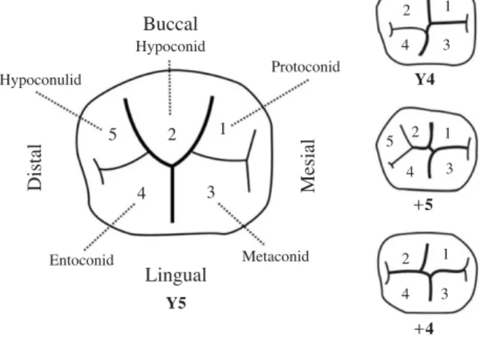
Firstly, occlusal surfaces were divided into the five-cusp
group and the four-cusp group. Then, each group was sub- divided into ‘Y’ pattern if there was contact between the metaconid and hypoconid, and ‘++’ pattern if there was no contact between the metaconid and hypoconid (Fig. 2).
Therefore, there were a total of 4 categories: Y5, ++5, Y4, and ++4 [3].
5. Statistical analysis
Statistical analysis including the distribution of vari- ables was performed with PASW Statistics 18.0 program (IBM-SPSS Corporation, Armonk, NY, USA). Sex differ- ences, differences between mandibular first molars and second molars, and correlation between groups were ana- lyzed by paired t-test. A significance level was determined at p⁄0.05.
Results
1. Crown diameter, total cusp area, and occlusal table ratio of mandibular molars
Table 1 presents the mesiodistal diameter, buccolingual diameter, area of five cusps, total cusp area, and area of occlusal table of mandibular first and second molars. Men showed greater measurements than women in all aspects of the crown in mandibular first and second molars. Also, both men and women showed significantly larger first molars than second molars with respect to the mesiodistal and buccolingual diameters. In addition, first molars had greater total cusp area and area of occlusal table than sec- ond molars.
In both genders, the protoconid had the largest cusp area among five cusps of mandibular first and second molars, whereas the hypoconulid had the smallest cusp area. Men showed a larger area in four cusps except the hypoconulid in the mandibular second molar than the mandibular first molar, whereas women showed a larger area in three cusps except the entoconid and hypoconulid in the mandibular second molar than the mandibular first molar. In terms of the occlusal table ratio, which indicates the ratio of area of the occlusal table to the total cusp area in percentage, men showed an occlusal table ratio of 56.11% for mandi- bular first molars and that of 54.12% for mandibular sec- ond molars. Women showed an occlusal table ratio of 57.69% and 55.73% for mandibular first and second molars, Buccal
Hypoconid
1
1
1
2 1 3 4
2 4 3 5
2 3 4
2 5
4 3
Distal Mesial
Y4
+ +5
+ +4 Lingual
Y5
Protoconid Hypoconulid
Entoconid Metaconid
Fig. 2. Categorization of cusp patterns and developmental grooves of mandibular molars were obtained from Kraus et al. [3] i.e., 5 cusps with Y fissure (Y5), 5 cusps with ++fissure (++5), 4 cusps with Y fissure (Y4), 4 cusps with ++fissure (++4).
respectively. Thus, women had a greater occlusal table ratio than men, and the first molars showed a greater occlu- sal table ratio than the second molars.
2. Categorization of the occlusal surface
Categorization of the occlusal surface based on the morphological category is shown in Table 2. With respect to mandibular first molars, 91.03% of men showed Y fis- sure with five cusps (Y5 pattern), whereas only 8.97% of men showed ++fissure with five cusps (++5 pattern). There was no case with four cusps. Among the women, 68.75%
showed the Y5 pattern, 28.12% showed the ++5 pattern,
3.13% showed the Y4 pattern, and 0% showed the ++4 pattern, respectively. With respect to mandibular second molars, 41.03% of men showed the Y5 pattern, and 26.92%, 24.36%, and 7.69% of men showed the ++5 pattern, Y4 pattern, and ++4 pattern, respectively. Among the women, 37.5% showed the Y5 pattern, and 31.25%, 25.0%, 6.25%
showed the ++5 pattern, Y4 pattern, and ++4 pattern, res- pectively.
3. Sex differences between the mandibular molars
When analyzing the sex differences in measurements of this study, it is clear that the mesiodistal diameter, buccol- Table 1.Crown diameters and areas of mandibular molars
1st mandibular molar
Males Females
N Mean S.D. C.V. N Mean S.D. C.V.
Diameters (mm)
MD 78 10.79 0.55 5.10 32 10.15 0.41 4.04
BL 78 10.02 0.53 5.29 32 9.41 0.40 4.25
Cusp areas (mm2)
Pr 78 22.94 3.05 13.30 32 20.22 2.76 13.65
Me 78 18.04 2.49 13.80 32 16.44 1.79 10.89
Hy 78 18.56 3.18 17.13 32 15.18 2.52 16.60
En 78 17.74 2.57 14.49 32 16.64 3.04 18.27
Hc 78 11.72 3.07 26.19 31 9.42 2.83 30.04
Total cusp areas (mm2) 78 89.00 7.88 8.85 32 77.91 5.10 6.546
Occ. table areas (mm2) 78 49.90 5.96 11.94 32 44.92 4.62 10.28
Occ. table ratio (%) 78 56.11 4.89 8.72 32 57.69 5.09 8.82
2nd mandibular molar
Males Females
N Mean S.D C.V. N Mean S.D C.V.
Diameters (mm)
MD 78 10.48 0.61 5.82 32 9.83 0.38 3.87
BL 78 9.88 0.56 5.67 32 9.29 0.46 4.95
Cusp areas (mm2)
Pr 78 23.40 2.77 11.84 32 20.70 2.96 14.30
Me 78 18.40 2.53 13.75 32 16.74 2.35 14.04
Hy 78 19.84 4.15 20.92 32 16.42 3.78 23.02
En 78 18.28 3.11 17.01 32 15.76 2.27 14.40
Hc 53 6.60 5.00 75.76 22 5.40 4.27 79.07
Total cusp areas (mm2) 78 86.52 8.49 9.81 32 75.03 5.60 7.46
Occ. table areas (mm2) 78 46.73 5.87 12.56 32 41.82 4.83 11.55
Occ. table ratio (%) 78 54.12 5.25 9.70 32 55.73 5.02 9.01
MD; mesiodistal diameter, BL; buccolingual diameter, occ. table ratio (%); occlusal table areas/total cusp areas×100, S.D.; standard deviation, C.V.;
coefficient of variation==S.D / mean×100, Pr; protoconid area, Me; metaconid area, Hy; hypoconid area, En; entoconid area, Hc; hypoconulid area
ingual diameter, area of individual five cusps, total cusp area, and area of occlusal table of the crown of the mandibu- lar first and second molars were greater in men than in women (Table 3). In terms of diameter of the crown, both the mesiodistal diameter and buccolingual diameter showed significant sex differences in mandibular first and second molars, with the mean difference in the range of 0.56~
0.65 mm. Among the areas of five individual cusps, only the entoconid of first molars (p==0.06) and the hypoconulid of second molars (p==0.24) showed no significant sex dif- ferences, and the remaining cusp areas revealed significant sex differences. The mean difference was in the range of 1.10~3.42 mm2. In addition, total cusp area and area of occlusal table showed significant sex differences, and the
respective mean differences in the mandibular first and sec- ond molars were 11.09 mm2, 11.49 mm2, 4.98 mm2, and 4.91 mm2, respectively.
4. Measurement errors
After selecting 30 dental casts randomly, measurement errors were calculated by comparing the first measurement values obtained by taking photographs and performing graphic analysis of dental casts repeatedly. The measure- ment errors calculated for the linear measurement and area were 0.50~0.77 mm and 0.21~0.59 mm2, respectively.
The average reliability obtained by using the 100 (1-Error2/ s2) formula was 99%, demonstrating that there were few Table 3.Sex differences in crown diameters and areas
M1 M2
t-test M.dif t-test M.dif
Diameters (mm)
MD 0.00 0.65 0.00 0.65
BL 0.00 0.61 0.00 0.56
Cusp areas (mm2)
Pr 0.00 2.72 0.00 2.69
Me 0.00 1.60 0.00 1.67
Hy 0.00 3.37 0.00 3.42
En 0.06 1.10 0.00 2.52
Hc 0.00 2.30 0.24 1.20
Total cusp areas (mm2) 0.00 11.09 0.00 11.49
Occ. table areas (mm2) 0.00 4.98 0.00 4.91
Occ. table ratio (%) 0.13 -1.58 0.14 -1.61
Relative cusp ratio (%)
Pr 0.82 -0.13 0.43 -0.47
Me 0.08 -0.88 0.07 -1.02
Hy 0.03 1.31 0.26 1.03
En 0.03 -1.29 0.84 0.13
Hc 0.13 0.99 0.78 0.33
MD; mesiodistal diameter, BL; buccolingual diameter, occ. table ratio (%); occlusal table areas/total cusp areas×100, relative cusp ratio (%); individual cusp area/total cusp area×100, Pr; protoconid area, Me; metaconid area, Hy; hypoconid area, En; entoconid area, Hc; hypoconulid area, M.dif; mean differences Table 2.Morphological categories of mandibular molars
5 cusps 4 cusps
Y fissure (%) ++fissure (%) Y fissure (%) ++fissure (%)
M (N=78) M1 91.03 8.97 0 0
M2 41.03 26.92 24.36 7.69
F (N=32) M1 68.75 28.12 3.13 0
M2 37.5 31.25 25.0 6.25
Y fissure; contact between the metaconid and the hypoconid, ++fissure; no contact between the metaconid and the hypoconid
measurement errors in this study. Since the paired t-test for first and second measurements showed no significant dif- ferences, measurement errors due to photography and graphic analysis seemed to be negligible.
Discussion
The mean age of the sampling group was about 30 when they may have mild to moderate attrition on the occlusal surface in the molar region, so that the cusps with apparent attrition were not used in this study. This study was mostly performed using mandibular right molars, but several left molars were also included when they showed missing teeth, heavy attrition, prosthesis, and anomalies on the right side under the assumption that both sides of mandibular molar teeth have the same morphology. No significantly different results were obtained in the experiment of size and morpho- logical variance in the hypoconulid of mandibular molars in Koreans compared with those in other previous studies in other races. Firstly, men had overall greater measurements than women with respect to the mesiodistal diameter, buc- colingual diameter, individual cusp areas, total cusp area, and area of occlusal table of first and second molars. In addi- tion, both men and women had greater measurements with respect to the total cusp area, area of occlusal table, and occlusal table ratio in the first molars than in the second molars. These results correspond with the results of pre- vious studies, which showed that the faster the cusp devel- ops, the greater the cusp becomes, i.e., late-developing teeth more frequently have reduced overall size than early-devel- oping teeth because the former have more spatial restric- tions [3,5,8,10,18-22].
Women had greater occlusal table ratio than men, and first molars had greater occlusal table ratio than second molars. This result suggests that although women have smaller teeth than men, they may function better during mastication, or in terms of the morphology and men may have teeth with a more remarkable height of contour than women. It can be assumed that the occlusal table ratio of first molars is greater because first molars may play more significant roles in biophysical aspects of mastication than second molars. On observing the difference between man- dibular first molars and second molars based on categories of the occlusal surface, it is obvious that men will have greater total cusp area in first molars than in second molars.
On the other hand, second molars had greater individual areas of four cusps except for the hypoconulid than first molars. This result may indicate that all of the mandibular first molars had five cusps based on the category of the occlusal surface, whereas 67.95% of mandibular second molars had five cusps and 32.05% of mandibular second molars had four cusps in men. In other words, the hypo- conulid appears less frequently in mandibular second molars than in mandibular first molars, causing other four cusps to become relatively larger than those in mandibular first molars. As in other previous reports, our results show- ed that late-developing cusps have greater variations in size and morphology [23].
In case of women, similar to the result in men, mandibu- lar first molars had a greater total cusp area than mandibu- lar second molars. Individual cusp areas of three cusps without the entoconid and hypoconulid were greater in mandibular second molars than in mandibular first molars.
In women also, 96.87% of mandibular first molars had five cusps, and only 3.13% of mandibular first molars had four cusps based on the category of the occlusal surface. In addi- tion, 68.75% of mandibular second molars had five cusps and 31.25% of mandibular second molars had four cusps.
It may be assumed that the areas of other cusps in mandibu- lar second molars become relatively larger, when the hypo- conulid appears less frequently.
With respect to sex differences, men had greater values than women for individual cusp areas, total cusp area, and occlusal table area except for the occlusal table ratio. In the individual cusp area, there was a significant sex differ- ence except for the entoconid of mandibular first molars (p==0.06) and hypoconulid of mandibular second molars (p==0.24). While mandibular first molars showed a signi- ficant sex difference in the size of the latest-developing hypoconulid (p==0.00, mean difference==2.30), mandibu- lar second molars showed no significant sex difference in the size of the hypoconulid (p==0.24, mean difference== 1.20). There may be two explanations for no significant sex difference in the size of the hypoconulid of the mandi- bular second molars. First, the latest-calcifying hypoconu- lid has the greatest variability and the hypoconulid appears less frequently in mandibular second molars because of greater spatial restrictions than mandibular first molars [8,10]. Secondly, it might be assumed that the periods of completion of mandibular first and second molar cusp formation (mandibular first molar: 2.5~3 years of age,
mandibular second molar: 7~8 years of age) are much before adolescence, during which there is production and secretion of sex hormones. That is, sex hormones seem to have little effect on the formation of mandibular first and second molars; instead, it might be due to general sex dif- ferences in body size or genetic differences among indivi- duals [20].
Also, the cusp size is affected by not only the order of formation but also by complex factors such as distance between adjacent cusps, rate of development, formation period, and rate of calcification. Size variations in individ- ual cusps are also affected by the order of ontogeny [21, 24,25]. In conclusion, this study would be meaningful to compare the characteristics of mandibular molars in Ko- reans. A further study focusing on the morphological char- acteristics of anterior and premolar teeth, and systematic comparison and analysis between Koreans and other races is needed.
Acknowledgment
This work was supported by the National Research Foundation of Korea (NRF) grant funded by the Korea government (MSIP) (2011-0030121).
References
1. Jensen E, Kai-Jen Yen P, Moorrees CF, Thomsen SO.
Mesiodistal crown diameters of the deciduous and perma- nent teeth in individuals. J Dent Res. 1957; 36:39-47.
2. Garn SM, Lewis AB, Swindler DR, Kerewsky RS. Genetic control of sexual dimorphism in tooth size. J Dent Res.
1967; 46:963-72.
3. Turner II CG NC, Scott GR. Scoring procedure for key mor- phological traits of the permanent dentition: The arizona state university dental anthropology system. New York:
Wiley-Liss; 1991.
4. Bailey SE, Pilbrow VC, Wood BA. Interobserver error invo- lved in independent attempts to measure cusp base areas of pan m1s. J Anat. 2004; 205:323-31.
5. Hattab FN, al-Khateeb S, Sultan I. Mesiodistal crown diame- ters of permanent teeth in jordanians. Arch Oral Biol. 1996;
41:641-5.
6. Kraus BS, Jordan RE, Abrams L. Dental anatomy and occlu- sion: A study of the masticatory system. Baltimore: The
Williams & Wilkins Company; 1969.
7. Nelson SJ, Ash MM. Wheeler’s dental anatomy, physiolo- gy and occlusion. 8th ed. Elsevier Health Sciences; 2003.
p. 322-3.
8. Takahashi M, Kondo S, Townsend GC, Kanazawa E. Vari- ability in cusp size of human maxillary molars, with partic- ular reference to the hypocone. Arch Oral Biol. 2007; 52:
1146-54.
9. Gingerich PD. Size variability of the teeth in living mam- mals and the diagnosis of closely related sympatric fossil species. J Paleontol. 1974; 48:895-903.
10. Kondo S, Townsend GC, Yamada H. Sexual dimorphism of cusp dimensions in human maxillary molars. Am J Phys Anthropol. 2005; 128:870-7.
11. Macho GA, Moggi-Cecchi J. Reduction of maxillary molars in homo sapiens sapiens: A different perspective. Am J Phys Anthropol. 1992; 87:151-9.
12. Corruccini RS. Molar cusp-size variability in relation to odontogenesis in hominoid primates. Arch Oral Biol. 1979;
24:633-4.
13. Park DE, Kim HK, Lim YS, Nakatsuka M, Kwon HB, Han SH, et al. Different mandibular first molar shapes accord- ing to groove and cusp configuration in relation to suggest- ed bracket position. Eur J Orthod. 2013; 35:730-6.
14. Kim KR, Song JS, Kim SO, Kim SH, Park W, Son HK.
Morphological changes in the crown of mandibular molars with an additional distolingual root. Arch Oral Biol. 2013;
58:248-53.
15. Zar JH. Biostatistical analysis. 5th ed. Pearson: Prentice- Hall; 2010.
16. Gregory WK. Studies on the evolution of the primates-part i the cope-osborn “theory of trituberculy” and the ancestral molar patterns of the primates. Bulletin of the American Museum of Natural History. 1916; 35:239-55.
17. Hellman M. Racial characters of human dentition. Proceed- ings of the American Phylosophical Society. 1928; 67:157- 74.
18. Wood BA, Engleman CA. Analysis of the dental morph- ology of plio-pleistocene hominids. V. Maxillary postca- nine tooth morphology. J Anat. 1988; 161:1-35.
19. Macaluso PJ, Jr. Sex discrimination potential of permanent maxillary molar cusp diameters. J Forensic Odontostom- atol. 2010; 28:22-31.
20. Wells JC. Sexual dimorphism of body composition. Best Pract Res Clin Endocrinol Metab. 2007; 21:415-30.
21. Harris EF, Dinh DP. Intercusp relationships of the perma- nent maxillary first and second molars in american whites.
Am J Phys Anthropol. 2006; 130:514-28.
22. Polychronis G, Christou P, Mavragani M, Halazonetis DJ.
Geometric morphometric 3d shape analysis and covaria- tion of human mandibular and maxillary first molars. Am J Phys Anthropol. 2013; 152:186-96.
23. Townsend G, Richards L, Hughes T. Molar intercuspal dim- ensions: Genetic input to phenotypic variation. J Dent Res.
2003; 82:350-5.
24. Quam R, Bailey S, Wood B. Evolution of m1 crown size and cusp proportions in the genus homo. J Anat. 2009;
214:655-70.
25. Morita W, Yano W, Nagaoka T, Abe M, Nakatsukasa M.
Size and shape variability in human molars during odonto- genesis. J Dent Res. 2014; 93:275-80.
한국인 아래턱큰어금니 교두 변이에 관한 연구
유홍일, 김지혜, 김선헌
전남대학교 치의학전문대학원 구강해부학교실
간추림 : 본 연구 목적은 한국인 아래턱큰어금니 hypoconulid와 관련된 각 교두의 크기 및 형태적 특징 및 성별 차이와 아래턱큰어금니 사이의 크기 및 형태 차이를 알아보기 위함이다.
조사 대상은 전남대학교 치의학전문대학원 남학생 78명, 여학생 32명이었으며, 평균연령은 30세였다. 대상자 의 치아모형을 제작하여 아래턱첫째 및 둘째큰어금니의 최대 안쪽-먼쪽너비, 볼쪽-혀쪽너비, 5개 교두(proto- conid, metaconid, hypoconid, entoconid, hypoconulid)의 개별면적, 총교두면적, 교합상면적을 측정하였다. 성별 차 이와 아래턱첫째 및 둘째큰어금니 간의 차이 및 상관성에 관해서는 paired t-test를 사용하여 분석하였다.
남녀 모두 아래턱큰어금니의 안쪽-먼쪽너비, 볼쪽-혀쪽너비, 총교두면적, 교합상면적 측정값은 첫째큰어금니가 둘째큰어금니보다 크게 나타났다. 아래턱첫째 및 둘째큰어금니 모두 protoconid 면적이 가장 크게 나타났고, hypoconulid 면적이 가장 작게 나타났다. 아래턱큰어금니 5개 교두 중 크기 및 형태 변이가 가장 큰 것은 hypo- conulid였으며, hypoconulid 발현빈도는 아래턱첫째큰어금니가 둘째큰어금니보다 더 높았다. 모든 측정값은 남자 가 여자보다 더 크게 나타났으며, 아래턱첫째큰어금니 entoconid (p==0.06)와 둘째큰어금니 hypoconulid (p==0.24) 면적을 제외한 나머지 측정값들은 모두 남녀간의 유의한 차이가 있는 것으로 나타났다. 한국인은 남녀 모두 다 른 인종에 비해 전반적인 치아 크기(너비, 면적)가 작은 것으로 나타났으며, 남자 치아 크기가 여자보다 큰 것으 로 나타났다. 또한 늦게 발생이 일어나는 치아일수록 치아크기는 작고 변이는 더 큰 경향을 나타내었으며, 제일 늦게 발생하는 hypoconulid 변이 정도가 제일 크게 나타났다. 이상의 결과는 한국인 아래턱큰어금니의 비교치아 형태학 자료를 제공한다.
찾아보기 낱말 : 아래턱큰어금니, hypoconulid, 교두
교신저자 : 김선헌(전남대학교 치의학전문대학원 구강해부학교실) 전자우편 : [email protected]