J Korean Soc Radiol 2018;78(6):389-403 https://doi.org/10.3348/jksr.2018.78.6.389
INTRODUCTION
Pulmonary artery (PA) size is measured where the main PA is bifurcated perpendicular to the vessel wall (1, 2). Truong et al. (3) reported that normative reference values for the main PA were 29 mm in healthy men and 27 mm in healthy women and that the ratio of main PA to ascending aorta diameter was 0.9.
The right and left PA should be approximately equal in size, but the left PA appears to be slightly larger in most subjects.
Many congenital and acquired disorders involve PAs, and their diameters can be affected by these conditions (4). Altera- tions of the PA diameter due to diseases can broadly be catego- rized as involving reductions or enlargements. The advantages of computed tomography (CT) include minimal invasiveness and the ability to produce multi-planar reformatted and three- dimensional images. Additionally, CT can detect PA distal to ob- structions that cannot be seen on an angiogram eg, PA wall
thickening, enhanced PA, and other causes of dyspnea and un- derlying lung, pleural, and mediastinal diseases. Next-genera- tion multi-detector CT (MDCT) instruments that require shorter acquisition times and thinner collimation can be used to evalu- ate PA beyond the segmental level, because of the higher spatial resolution. These images can also be used to lower the indeter- minate CT pulmonary angiography (CTPA) rate because of faster scanning times (4, 5).
In this article, we review the features of various disorders that decrease and increase PA diameters, with an emphasis on their MDCT appearance.
CT PROTOCOls
Optimal contrast PA opacification, with consistent and homog- enous enhancement, is essential given that the causes of inde- terminate CTPA are motion artifacts (74%) and poor contrast
Spectrum of Multi-Detector Computed Tomography Findings that Alter Pulmonary Artery Diameters in Adults
성인에서 폐동맥을 변화시키는 다중검출 전산화단층촬영 소견의 분류
Hye Rim Park, MD, Young Tong Kim, MD*, Sung Shick Jou, MD, Woong Hee Lee, MD
Department of Radiology, Cheonan Hospital, Soonchunhyang University College of Medicine, Cheonan, Korea
Abnormalities in the anatomy of pulmonary arteries may have heritable or environ- mental causes and involve a reduction or enlargement in transverse diameters of the blood vessels eg, congenital and developmental disorders, acquired intrinsic causes, extrinsic compression, and constriction. Pulmonary hypertension, pulmonary artery aneurysm and pseudoaneurysm, and idiopathic dilatation can also increase the diameter of a pulmonary artery. Multi-detector computed tomography (CT) is useful to evaluate each pulmonary artery and to diagnose the conditions that alter the diameter of the pulmonary artery. It is important to be familiar with the CT fea- tures of a variety of disorders to allow for accurate diagnoses and appropriate ther- apeutic management.
Index terms
Multidetector Computed Tomography Pulmonary Artery
Hypertension, Pulmonary
Received July 13, 2017 Revised August 24, 2017 Accepted October 1, 2017
*Corresponding author: Young Tong Kim, MD Department of Radiology, Cheonan Hospital, Soonchunhyang University College of Medicine, 31 Soonchunhyang 6-gil, Dongnam-gu, Cheonan 31151, Korea.
Tel. 82-41-570-3515 Fax. 82-41-570-3516 E-mail: [email protected]
This is an Open Access article distributed under the terms of the Creative Commons Attribution Non-Commercial License (http://creativecommons.org/licenses/by-nc/4.0) which permits unrestricted non-commercial use, distri- bution, and reproduction in any medium, provided the original work is properly cited.
enhancement (40%) (6). Contrast enhancement depends on patient weight, cardiac output, scan duration, and contrast de- livery protocol, but no single injection protocol strategy can be applied universally for CTPA. After a region of interest is placed in the main PA, 80–120 μL/kg or 1.0–1.2 μL/kg of contrast me- dia is injected more than 4 mm/s using an 18-G or 20-G cathe- ter. It is recommended that a biphasic injection protocol is fol- lowed involving contrast injection followed by a saline bolus.
Saline-chasing and flushing reduce streak artifacts that arise from dense concentrations of contrast media in the superior vena cava. Bolus and automated bolus-triggering techniques have been preferred to traditional fixed-delay techniques, be- cause the contrast arrival time for each patient. A caudocranial direction of acquisition is recommended because it reduces re- spiratory motion artifacts in the lower lobe; most emboli are lo- cated in the lower lobes (7), although this is becoming less of a concern with the newest CT scanners and their faster scan times. The pulmonary embolism-specific setting helps to dif- ferentiate between emboli and artifacts. Electrocardiogram (ECG)-gated CTPA reduces cardiac motion and enables diag- nosis of a sub-segmental embolus in the paracardiac PA. How- ever, ECG gating is controversial because of longer scan times and higher radiation doses (8).
DeCReAseD PUlmONARy ARTeRy DIAmeTeR
Disorders that reduce the diameter of PA have been classified into four categories: congenital or developmental disorders, ac- quired intrinsic, extrinsic compression, and constriction (Table 1) (9). Systemic arterial supply to the lungs that includes bron- chial and PA anastomoses with transpleural systemic anasto- moses has been demonstrated in many cases of congenital and acquired diseases such as PA interruption and chronic PA ob- struction (10).
Congenital or Developmental Disorders
To understand congenital and developmental PA disorders, we must know how they develop. The central and peripheral portions of PA develop independently; the proximal portion is derived from the truncus arteriosus, and the peripheral portion is from the lung mesenchyme. The proximal portions of the left
and right sixth aortic arches contribute to both main branches of the PA during the first 16 weeks of intrauterine development.
The distal part of the left sixth arch forms the ductus arteriosus, and there is an involution of the distal part of the right sixth arch (11). An abnormality in this process results in PA inter- ruption or agenesis. The peripheral capillary plexus is formed from the pulmonary mesenchyme by vasculogenesis. By 34 days’ gestation, buds from the sixth arch arteries grow into primi- tive lungs and anastomoses with a capillary network around each prospective main bronchus.
In the pre-acinar region proximal to the alveolar duct, the ar- teries run with airways, and the branching pattern of arteries and airways is complete by mid-term gestation; later, the only Table 1. Classification of Disorders Altering Pulmonary Artery Diameter Decreasing diameter of pulmonary artery
Congenital or developmental disorders Unilateral proximal interruption Congenital stenosis
Hypoplasia
Swyer James syndrome Acquired intrinsic causes
Primary tumor
Chronic pulmonary thromboembolism Vasculitis & rheumatic stenosis Bacterial endocarditis of pulmonary valve Extrinsic compression
Tumors Aneurysm
Constriction of pulmonary arteries Anthracofibrosis
Fibrosing mediastinitis Chronic pericarditis
Increasing diameter of pulmonary artery Pulmonary hypertension
Idiopathic
Lung parenchymal disease
Chronic thromboembolic pulmonary hypertension Miscellaneous causes
Pulmonary artery aneurysm & pseudoaneurysm Infection
Trauma Iatrogenic causes
Idiopathic dilatation of pulmonary artery Pulmonary arteriovenous malformation
pre-acinar arteries change in the dimension and wall thickness.
Lung development can be divided into five stages: embryonic (1–7 weeks gestation), pseudoglandular (5–17 weeks), canalic- ular (16–26 weeks), saccular (24–38 weeks), and alveolar (36 weeks onward). Alveoli multiplied from 36 weeks’ gestation are one-third to one-half of the adult number at birth and increase in number until an age of 2–4 years, and they increase in size until the completion of somatic growth. New arteries develop in the intra-acinar region and multiply as the alveoli develop dur- ing infancy and early childhood. During normal lung develop- ment, the pre-acinar vessels follow the development of the air- way, and the intra-acinar vessels support the development of the alveoli. Abnormalities in fetal lung development also affect both airways and blood vessels (12), and abnormalities in airway or alveoli growth will also affect the number of blood vessels and
structures. Thus, hypoplasia of the pulmonary vessel is always as- sociated with congenital or acquired underdevelopment of the lung or airway.
Pulmonary Artery Interruption
When regression of the distal segment of the sixth arch extends into the proximal segment, a discontinuity occurs between main and distal PA at the lung hilum. This condition is called unilat- eral absence or interruption of PA (13). The term “interruption”
is preferred over “absence” because PA continues to develop in- dependently and the intrapulmonary vascular network remains intact. The lung is supplied through systemic collateral vessels, including the bronchial, intercostal, internal thoracic, subclavi- an, and innominate arteries (Fig. 1B, C). The mediastinal por- tion of the affected PA is entirely absent or terminates within 1
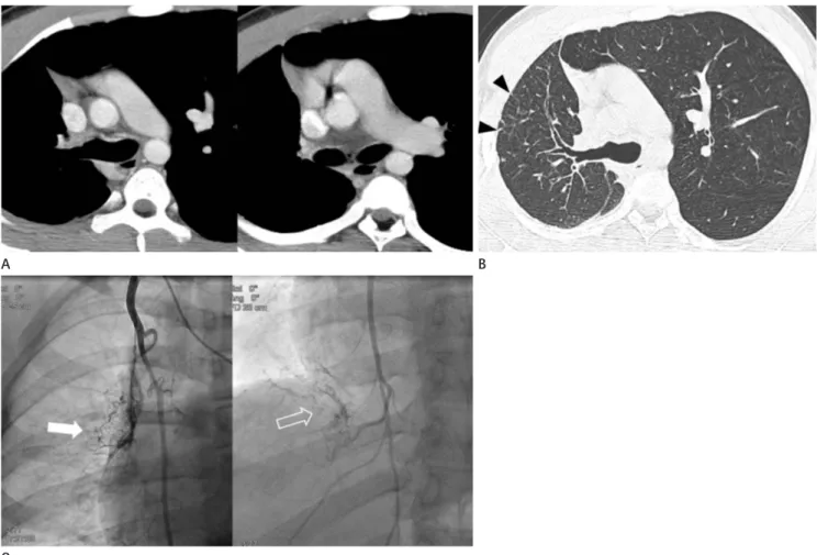
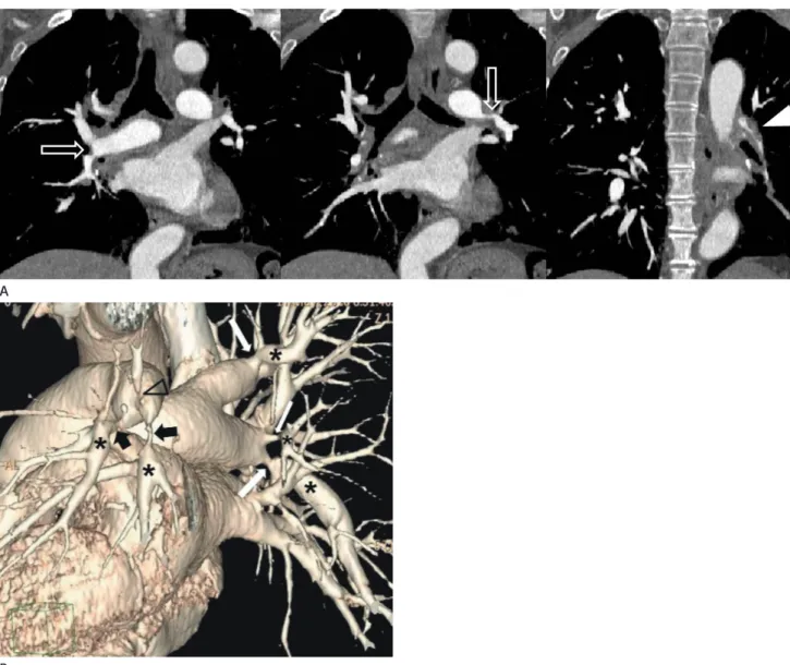
Fig. 1. Interruption of the right pulmonary artery in an 18-year-old man.
A. Axial images show a complete absence of the right main pulmonary artery, a small right lung, and mediastinal shifting to the right side.
B. Axial image with lung window settings shows a small right lung with irregular linear opacities (arrowheads) in the lung periphery.
C. On angiography of the right internal thoracic artery, left (arrow) and right (open arrow) portions of the right lung are supplied by the right in- ternal thoracic artery. These systemic collaterals are shown as reticular opacities in the periphery of the affected lung on computed tomography.
A
C
B
cm of its origin. Although an interruption in the right PA is more common than the left PA, this anomaly is still an isolated finding. A left PA interruption is associated with a high inci- dence of the associated right aortic arch and congenital cardio- vascular anomalies. CT findings include the complete absence of the mediastinal portion of the right or left main PA, enlarged collateral vessels, pleural thickening, and peripheral fine reticu- lar opacities (Fig. 1) (14). Clinical presentation of a PA inter- ruption can be differentiated from other diseases such as simple pulmonary hypoplasia, hypogenetic lung syndrome, Swyer- James syndrome, and tuberculosis sequelae. An angiogram test showing a failure to fill the PA with a contrast agent does not always indicate the absence of a PA anomaly. At surgery or on postmortem examination, a patently small PA has been ob- served, even when the PA did not appear to be filled with a contrast agent during angiography. CT and magnetic resonance have been used to detect small under-perfused PA (15, 16).
Congenital Stenosis of the Pulmonary Artery
Congenital PA stenosis is thought to be a developmental anomaly and may be single or multiple, unilateral or bilateral, and peripheral or central. Franch and Gay (17) reviewed 90 cases of congenital PA stenosis and suggested the following four classifications: I. single central stenosis involving the pulmo-
nary trunk or its point of bifurcation into main PA (Fig. 2), II.
bifurcation stenosis, III. multiple peripheral stenoses, and IV.
combined central and intermediate or peripheral stenoses (17).
Among 90 cases, 34 and 22 patients could be typed as type I and type IV, respectively. Among the 34 type I cases, right PA steno- sis (Fig. 2) was more common (n = 26) than in the main (n = 6) and left (n = 2) PA cases. Associated cardiovascular defects are more common in the main PA than in peripheral branch ste- nosis (18).
Hypoplasia of Pulmonary Artery
Isolated congenital unilateral pulmonary hypoplasia is a rare condition in adulthood (19). Hypoplasia of the PA may be asso- ciated with congenital or acquired underdevelopment of the lung lobe; because the blood vessels follow airway development em- bryologically (20), hypoplasia of the pulmonary vessel is always associated with underdevelopment of the corresponding lung lobe and can be manifested as a parenchymal change (21). Thus, lobar agenesis of the lung is combined with the absence of a corresponding lobar PA, and the ipsilateral PA is hypoplastic (Fig. 3). Unilateral pulmonary vein atresia, which is thought to result from the failure of the common pulmonary vein to in- corporate into the left atrium, is a rare congenital anomaly (Fig.
4). In unilateral pulmonary vein atresia, ipsilateral PA impairs
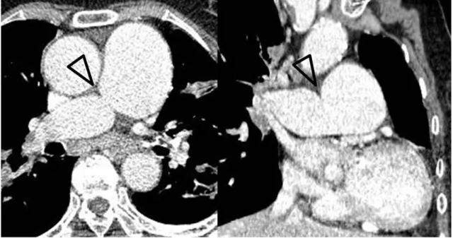
Fig. 2. Constriction of the right pulmonary artery in an 85-year-old woman. Axial and coronal images show focal narrowing of the upper wall of the proximal right pulmonary artery (open arrowheads). The cause of pulmonary artery stenosis is thought to be idiopathic because there is no evidence of arterial wall thickening or surrounding soft tissue mass.
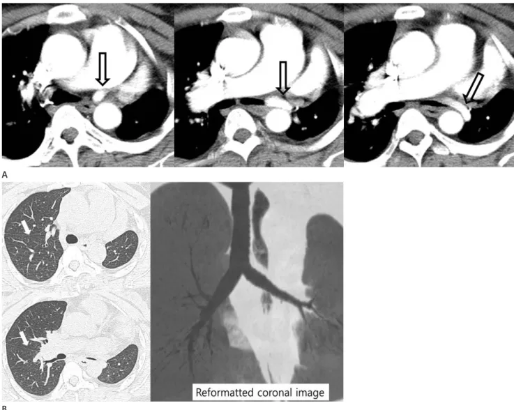
Fig. 3. Hypoplastic left pulmonary artery associated with lobar agenesis of the lung in a 40-year-old woman. Serial axial images with a mediasti- nal setting (A) show an anomalous hypoplastic left pulmonary artery. On the axial image with a lung window setting and coronal reformatted image (B), the normal positioned right upper lobe bronchus is absent, and the right major fissure is displaced anteriorly (arrows). Also, the bifur- cation of the left main bronchus is not seen, and the hypoplastic left lung consists of a single lobe.
A
B
Fig. 4. Hypoplastic right pulmonary artery associated with right pulmonary vein atresia in a 14-year-old teenage girl. Serial axial images show a smooth left atrial margin (black open arrowhead) and absent right superior and inferior pulmonary veins. The adjacent soft tissue opacity (arrow- heads) around the left atrium suggests collaterals. The right pulmonary artery (*) is diffusely narrowed.
growth and results in hypoplastic PA because of preferential perfusion of the contralateral PA (22). Swyer-James syndrome is a manifestation of post-infectious obliterative bronchiolitis that occurs in infancy or childhood (23); the affected lung does not grow normally, resulting PA hypoplasia. CT findings for Swyer-James syndrome include a small ipsilateral PA and a hy- perlucent small or normal-sized hemithorax on the affected side, with air trapping (Fig. 5).
Acquired Intrinsic Disorders
Chronic Pulmonary Thromboembolism
Most acute emboli undergo complete resolution with treat- ment. However, in some patients, the thromboemboli do not resolve completely and may end in endothelialized fibrotic ob- struction of the PA (24). Most cases result in vascular stenosis, but rarely does this translate into complete PA occlusion. CT
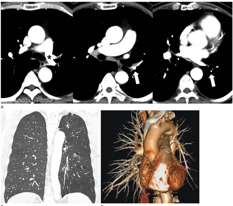
Fig. 5. Hypoplastic left pulmonary artery in Swyer-James syndrome in a 61-year-old man.
A. Serial axial images showed a relatively small left pulmonary artery (*) and decreased interlobar and segmental arteries (arrows) in the left lower lobe.
B. Coronal image with lung window settings shows diffuse air trapping in the left lung with multifocal bronchiectasis and an irregularly shaped nodule in the left upper lobe. The volume of the left lung is not decreased.
C. Anterior volume rendering image shows decreased pulmonary vascularity of the left lung.
A
B C
findings for chronic pulmonary thromboembolism include complete or partial obstruction, eccentric thrombus, calcified thrombus, bands, webs, and post-stenotic dilatation (Fig. 6).
Chronic thrombotic occlusion of one main PA mimics PA in- terruption (Fig. 7) (25). Unlike a chronic pulmonary embolism, a PA interruption is characterized by smooth, abrupt tapering of the artery without an intraluminal change. Multiple bilateral PA abnormalities are a helpful diagnostic clue in a chronic pul- monary thromboembolism (Fig. 6).
Vasculitis of the Pulmonary Artery
Primary large vessel vasculitis (Takayasu arteritis and giant cell arteritis) may involve PA. CT findings are stenosis or occlu- sion of the segmental and sub-segmental arteries with wall thick- ening (26).
Extrinsic Compression
Extrinsic PA compression is caused by tumors or an aortic an- eurysm (Fig. 8) (9). Anterior mediastinal teratoma, Hodgkin’s disease, thymic tumors, bronchial carcinoma, and mesothelio-
Fig. 6. Multiple stenoses and post-stenotic dilatation of the bilateral pulmonary arteries by chronic pulmonary thromboembolism in a 56-year- old man.
A. Coronal images show narrowing bilateral pulmonary arteries at the bifurcation site (open arrows) and irregularly shaped intraluminal thrombi (arrowhead) in the inferior inter-lobar artery of the left lower lobe.
B. The volume rendering three-dimensional image in left posterolateral projection showed multiple narrowing of the right (white arrows) and left (black arrows) pulmonary arteries with post-stenotic dilatation (*). Also, note the discontinuity of the pulmonary artery (open black arrowhead) on the left upper lobe.
A
B
ma of the pericardium may encase or surround the proximal right or left PA, and may include the involvement of the more distal parts of both PAs.
Pulmonary Artery Constriction
Mediastinal fibrosis caused by anthracofibrosis or fibrosing mediastinitis may result in stenosis or PA occlusion (Fig. 9). Al- though the cause is idiopathic, it is thought to be an abnormal immunologic response to histoplasmosis and tuberculosis. CT images show an infiltrative soft tissue mass that is frequently calcified and that obliterates normal fat planes and encases or invades adjacent structures (27).
Idiopathic or constrictive pericarditis may result in PA nar- rowing, but these events are rare occurrences (Fig. 10) (9, 28).
PA stenosis is caused by calcified pericardial bands or rings (29).
INCReAseD PUlmONARy ARTeRy DIAmeTeR
The upper limit of the standard diameter of main PA on CT is 29 mm (Fig. 11), and that of the right interlobar PA is 17 mm (Fig. 12) (30), and the PA dilatation is focal or diffuse. Pulmo- nary hypertension (PH) is the most frequent cause of diffuse PA enlargement. An aneurysm and pseudoaneurysm are con- sidered to be focal PA dilatation going beyond the maximal normal range. Focal dilatation of the peripheral PA is caused by pulmonary arteriovenous malformation. Rarely, diffuse PA dil- atation is idiopathic (Table 1) (4).
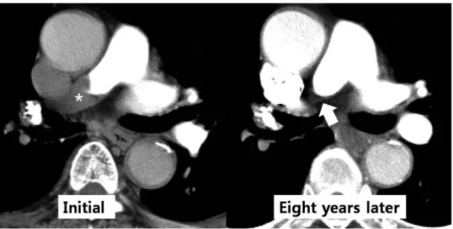
Fig. 7. Complete occlusion of the right pulmonary artery by chronic pulmonary thromboembolism in an 81-year-old man. Initial axial computed tomography image shows thromboembolism (*) of the right pulmonary artery and follow-up axial image obtained eight years later indicates a complete occlusion in the mid-portion of the right pulmonary artery (arrow).
Fig. 8. Extrinsic compression of the right pulmonary artery by an aor- tic aneurysm in a 36-year-old man who was diagnosed with Marfan syndrome. Axial computed tomography image shows extrinsic com- pression of the main and right pulmonary arteries (*) by an aneurysm in the ascending thoracic aorta.
Pulmonary hypertension
The PA is a more compliant vessel than the systemic arterial system, and it is thus more sensitive to changes in pressure and
volume; as a result, an increase in mean PA pressure should correlate with a change in the PA diameter. Many causes of PH can be classified into clinical or hemodynamic categories. Clini-
A
B
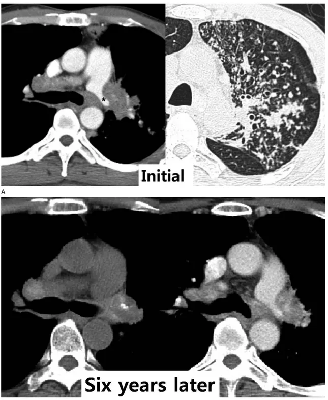
Fig. 9. Constriction of the left pulmonary artery by fibrosing mediastinitis in a 46-year-old woman.
A. Initial axial images show an irregularly shaped mass-like lesion with a narrowing left pulmonary artery (*) and axial image with lung window settings show active pulmonary tuberculosis in the left upper lobe.
B. Axial images after six years show left pulmonary artery narrowing by a soft tissue mass that has focal calcification and diffuses high attenua- tion on unenhanced computed tomography. Despite improved active pulmonary tuberculosis in the left upper lobe, mediastinal fibrosis by tuber- culosis has not changed.
cally, it is organized into five groups: pulmonary arterial hyper- tension including idiopathic or congenital heart diseases (Figs.
11, 12), PH due to left heart disease, PH due to lung diseases and/or hypoxemia, chronic thromboembolic PH, and PH with unclear multifactorial mechanisms (31). Hemodynamically, PH is classified into two categories: precapillary (arterial) and post-
capillary (venous) (32). Precapillary PH is defined as pulmo- nary capillary wedge pressure greater than 25 mm Hg at rest or 30 mm Hg during exercise. The important feature in PH is va- soconstriction, predominantly at sub-segmental levels, which increases vascular resistance and results in dilatation of the cen- tral PA (Figs. 11, 12). PH can be reliably predicted by CT when
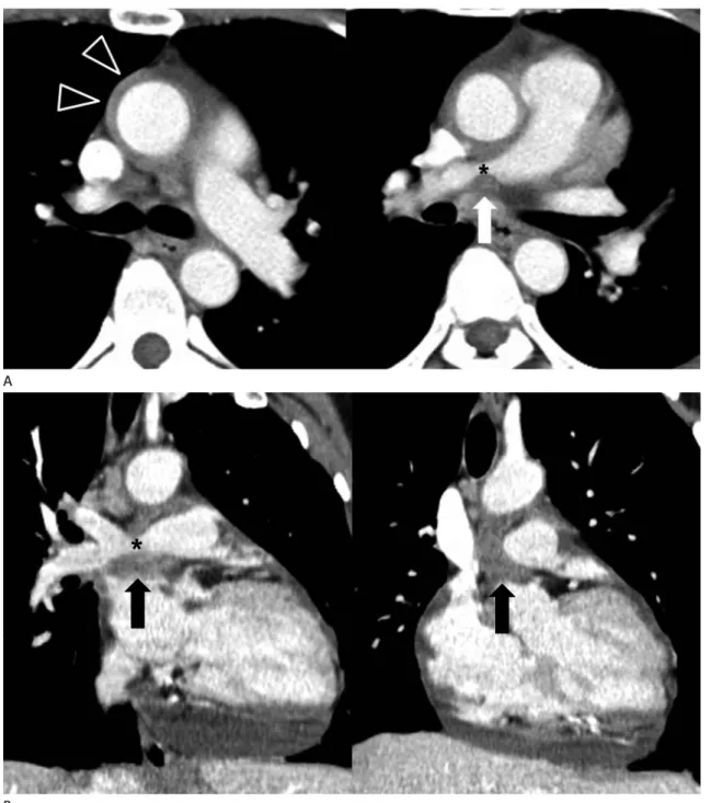
Fig. 10. Constriction of the right pulmonary artery by chronic pericarditis in a 47-year-old woman.
A. Axial images show circumferential soft tissue attenuation (open arrowheads) around the ascending thoracic aorta and focal narrowing of the right pulmonary artery (*) with surrounding soft tissue lesion (arrow).
B. Coronal images show focal narrowing (*) of the right pulmonary artery by a pericardial soft tissue mass (arrows); it was histologically con- firmed as chronic inflammation of the pericardium by pericardiotomy.
B A
the distal main PA is greater than or equal to 29 mm, and the segmental artery-to-bronchus ratio is greater than 1:1 in three of four lung lobes (32, 33). Elevated PA pressure increases right ventricle (RV) pressure, which leads to right ventricular hyper- trophy (RVH). CT findings for PH include central PA dilata- tion, abrupt narrowing or tapering of the peripheral PA, RVH, dilated bronchial arteries, and a mosaic perfusion of the lung (32). The prevalence of chronic thromboembolic PH after acute
pulmonary embolism is suggested to be 0.1–9.1% (34), and CT findings include signs of PH, variable features of thrombi, col- laterals, and mosaic perfusion of the lung. Lung disease is the most common cause of PH, and CT has shown pathologic changes from underlying obstructive or restrictive lung diseases (32). In pulmonary valvular stenosis, blood flow is directed to left PA, resulting in its enlargement (35).
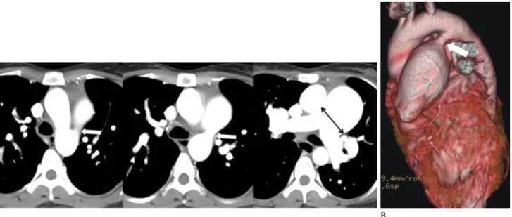
Fig. 11. Pulmonary hypertension secondary to patent ductus arteriosus in a 28-year-old woman.
A, B. Serial axial images (A) show a main pulmonary artery dilated to more than 29 mm. There is direct communication (arrows) between the main pulmonary artery and the descending thoracic aorta on axial (A) and volume rendering images (B). The main pulmonary artery size is typi- cally measured at the level of bifurcation of the main pulmonary artery perpendicular to the vessel wall (double-headed arrow). The upper limit of the normal main pulmonary artery diameter is 29 mm and that of the right interlobar artery is 17 mm on computed tomography.
A B
Fig. 12. Pulmonary hypertension in a 28-year-old woman who has been diagnosed with ASD. Post-contrast axial image shows that the diame- ters of the main and proximal lobar pulmonary arteries are increased secondary to ASD and thromboembolism (*) in the dilated bilateral inter-lo- bar pulmonary arteries; eccentric calcifications (arrows) in the thrombi on pre-contrast image indicates chronic pulmonary thromboembolism.
ASD = atrial septal defect
Pulmonary Artery Aneurysm and Pseudoaneurysm PA an aneurysm can be congenital or acquired, single or mul- tiple, and located centrally or peripherally. Congenital causes
include deficiency of the vessel wall, valvular and post-valvular stenosis, and congenital heart disease. Increased hemodynamic shear stresses and increased flow due to congenital heart dis-
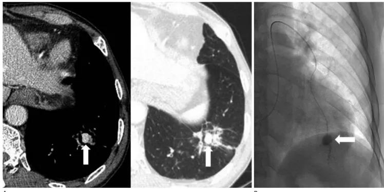
Fig. 13. Pulmonary pseudoaneurysm secondary to fungal infection in a 66-year-old man.
A. Axial images show an aneurysmal sac (*) within the necrotic portion of the diffuse consolidation in the left lung.
B. Pulmonary angiography of the left lung shows an aneurysmal sac (*) in the lingular segment. Bronchoscopic biopsy revealed fungal hyphae and was positive on both periodic acid-Schiff stain and Grocott methenamine silver stain. The sac was diagnosed as pseudoaneurysm by a fungal infection.
A B
Fig. 14. Aneurysm of the peripheral pulmonary artery in a 66-year-old man who was treated 20 days earlier for pneumonia.
A. Axial images show a highly enhanced aneurysm (arrows) and surrounding consolidation in the left lower lobe.
B. Pulmonary angiography shows a small aneurysm (arrow) in the peripheral pulmonary artery in the left lower lobe. It was thought to be a pseudoaneurysm secondary to pneumonia.
A B
ease can result in giant PA aneurysms. Acquired causes include vasculitis, altered connective tissue, and PH. Pseudoaneurysm secondary to pulmonary tuberculosis is known as a Rasmussen an aneurysm and is caused by a weakening of the PA wall from adjacent tuberculosis. Rasmussen aneurysms usually involve the upper lobes and a peripheral PA (30). Infection with pyo- genic bacteria and fungi can cause pseudoaneurysms or, less commonly, aneurysms (Figs. 13, 14) (36). A pseudoaneurysm is usually traumatic or iatrogenic. Moreover, incorrectly posi- tioned Swan-Gans catheters are an increasingly common cause of an iatrogenic PA pseudoaneurysm. Other iatrogenic causes include chest tube insertion, conventional angiography, and a surgical resection or biopsy. Behcet’s disease is the most com- mon cause of an aneurysm of the PA, and the underlying patho- physiology is inflammation of the vasa vasorum of the tunica media, which destroys the elastic fibers of the media and dilates the vessel lumen. Takayasu arteritis is an idiopathic arteritis that leads to stenosis, occlusion, and, occasionally, post-stenotic dil-
atation, and aneurysm formation (Fig. 15) (26). CTPA is useful for evaluating the extent of aneurysms, pseudoaneurysms, and concomitant cardiovascular abnormalities.
Idiopathic Dilatation of the Pulmonary Artery
Idiopathic PA dilatation is a rare congenital anomaly that in- volves abnormal enlargement of the main PA, with or without dilatation of both PAs. To reach this diagnosis, it is necessary to exclude pulmonary and cardiac diseases and to confirm the pres- ence of normal pressure in the RV and PA. The etiology of this disease is unknown, but it is suggested to be caused by congeni- tal weakness of the PA wall. Most patients are asymptomatic, and the disease is usually discovered incidentally on a chest radio- graph (4, 37).
CONClUsION
Many congenital and acquired disorders increase or decrease the diameter of PA, and changes in the PA diameter on CT may be overlooked if the images were not obtained via CTPA. The newest MDCT, with higher spatial resolution and thinner colli- mation, helps to quickly detect a minimal change in PA. Famil- iarity with the CT features of alterations of the PA diameter caused by many congenital and acquired disorders would allow for ac- curate diagnosis and appropriate therapeutic management.
RefeReNCes
1. Edwards PD, Bull RK, Coulden R. CT measurement of main pulmonary artery diameter. Br J Radiol 1998;71:1018-1020 2. Kuriyama K, Gamsu G, Stern RG, Cann CE, Herfkens RJ, Brund- age BH. CT-determined pulmonary artery diameters in pre- dicting pulmonary hypertension. Invest Radiol 1984;19:16- 22
3. Truong QA, Massaro JM, Rogers IS, Mahabadi AA, Kriegel MF, Fox CS, et al. Reference values for normal pulmonary artery dimensions by noncontrast cardiac computed tomog- raphy: the Framingham Heart Study. Circ Cardiovasc Imag- ing 2012;5:147-154
4. Castañer E, Gallardo X, Rimola J, Pallardó Y, Mata JM, Per- endreu J, et al. Congenital and acquired pulmonary artery anomalies in the adult: radiologic overview. Radiographics Fig. 15. Focal dilatation of the peripheral pulmonary arteries in a
54-year-old man who has been diagnosed with Takayasu arteritis. An- terior volume rendering computed tomography image shows focal dilatation of the peripheral pulmonary arteries (arrows) in the left lower lung. Also, note focal narrowing (open arrowhead) and post- stenotic dilatation (*) of the right brachiocephalic artery and stenosis (arrowhead) of the left subclavian artery.
2006;26:349-371
5. Schoepf UJ, Costello P. CT angiography for diagnosis of pul- monary embolism: state of the art. Radiology 2004;230:
329-337
6. Jones SE, Wittram C. The indeterminate CT pulmonary an- giogram: imaging characteristics and patient clinical out- come. Radiology 2005;237:329-337
7. Wittram C. How i do it: CT pulmonary angiography. AJR Am J Roentgenol 2007;188:1255-1261
8. Mayo J, Thakur Y. Pulmonary CT angiography as first-line im- aging for PE: image quality and radiation dose consider- ations. AJR Am J Roentgenol 2013;200:522-528
9. Seymour J, Emanuel R, Pattinson N. Acquired pulmonary ste- nosis. Br Heart J 1968;30:776–785
10. Do KH, Goo JM, Im JG, Kim KW, Chung JW, Park JH. Systemic arterial supply to the lungs in adults: spiral CT findings. Ra- diographics 2001;21:387-402
11. Davies M, Guest PJ. Developmental abnormalities of the great vessels of the thorax and their embryological basis. Br J Ra- diol 2003;76:491-502
12. Hislop A. Developmental biology of the pulmonary circula- tion. Paediatr Respir Rev 2005;6:35-43
13. Anand SH, Jasper A, Mani SE, Joseph E, Mathai J. Proximal interruption of the pulmonary artery: a case series. J Clin Di- agn Res 2015;9:TD04-TD06
14. Ryu DS, Spirn PW, Trotman-Dickenson B, Hunsaker A, Jung SM, Park MS, et al. HRCT findings of proximal interruption of the right pulmonary artery. J Thorac Imaging 2004;19:
171-175
15. Choe YH, Ko JK, Lee HJ, Kang IS, Park PW, Lee YT. MR imag- ing of non-visualized pulmonary arteries at angiography in patients with congenital heart disease. J Korean Med Sci 1998;13:597-602
16. Sondheimer HM, Oliphant M, Schneider B, Kavey RE, Black- man MS, Parker FB Jr. Computerized axial tomography of the chest for visualization of “absent” pulmonary arteries. Cir- culation 1982;65:1020-1025
17. Franch RH, Gay BB Jr. Congenital stenosis of the pulmonary artery branches. a classification, with postmortem findings in two cases. Am J Med 1963;35:512-529
18. Baum D, Khoury GH, Ongley PA, Swan HJ, Kincaid OW. Con- genital stenosis of the pulmonary artery branches. Circula-
tion 1964;29:680-687
19. Isawa T, Taplin GV. Unilateral pulmonary artery agenesis, ste- nosis, and hypoplasia. Radiology 1971;99:605-612 20. Stovin PG. Early lung development. Thorax 1985;40:401-404 21. Park S, Cha YK, Kim JS, Kwon JH, Jeong YJ, Kim SJ. Isolated
unilateral pulmonary artery hypoplasia with accompany- ing pulmonary parenchymal findings on CT: a case report.
J Korean Soc Radiol 2017;76:369-373
22. Dixit R, Kumar J, Chowdhury V, Rajeshwari K, Sethi GR. Case report: isolated unilateral pulmonary vein atresia diagnosed on 128-slice multidetector CT. Indian J Radiol Imaging 2011;
21:253-256
23. Moore AD, Godwin JD, Dietrich PA, Verschakelen JA, Hen- derson WR Jr. Swyer-James syndrome: CT findings in eight patients. AJR Am J Roentgenol 1992;158:1211-1215 24. Castañer E, Gallardo X, Ballesteros E, Andreu M, Pallardó Y,
Mata JM, et al. CT diagnosis of chronic pulmonary throm- boembolism. Radiographics 2009;29:31-50; discussion 50- 53
25. Moser KM, Olson LK, Schlusselberg M, Daily PO, Dembitsky WP. Chronic thromboembolic occlusion in the adult can mimic pulmonary artery agenesis. Chest 1989;95:503-508 26. Castañer E, Alguersuari A, Gallardo X, Andreu M, Pallardó Y,
Mata JM, et al. When to suspect pulmonary vasculitis: ra- diologic and clinical clues. Radiographics 2010;30:33-53 27. Rossi SE, McAdams HP, Rosado-de-Christenson ML, Franks
TJ, Galvin JR. Fibrosing mediastinitis. Radiographics 2001;
21:737-757
28. Friedman MJ, Gabor GE, Fishman MC, Griepp RB. Bilateral pulmonary artery stenosis associated with pericarditis. re- sults of surgery and follow-up by magnetic resonance imag- ing. Chest 1988;93:883-885
29. Hwang YJ, Park CH, Jeon YB, Park KY. Severe pulmonary ar- tery stenosis by a calcified pericardial ring. Eur J Cardiotho- rac Surg 2006;29:619-621
30. Nguyen ET, Silva CI, Seely JM, Chong S, Lee KS, Müller NL.
Pulmonary artery aneurysms and pseudoaneurysms in adults: findings at CT and radiography. AJR Am J Roentgen- ol 2007;188:W126-W134
31. Simonneau G, Gatzoulis MA, Adatia I, Celermajer D, Denton C, Ghofrani A, et al. Updated clinical classification of pul- monary hypertension. J Am Coll Cardiol 2013;62(25
Suppl):D34-D41
32. Grosse C, Grosse A. CT findings in diseases associated with pulmonary hypertension: a current review. Radiographics 2010;30:1753-1777
33. Ng CS, Wells AU, Padley SP. A CT sign of chronic pulmonary arterial hypertension: the ratio of main pulmonary artery to aortic diameter. J Thorac Imaging 1999;14:270-278 34. Lang IM, Madani M. Update on chronic thromboembolic
pulmonary hypertension. Circulation 2014;130:508-518
35. Saremi F, Gera A, Ho SY, Hijazi ZM, Sánchez-Quintana D.
CT and MR imaging of the pulmonary valve. Radiograph- ics 2014;34:51-71
36. Ramachandran L, Dewan S, Kumar V, Wankhade B. Mucor- mycosis causing pulmonary artery aneurysm. Respir Med Case Rep 2015;16:71-73
37. Ugolini P, Mousseaux E, Sadou Y, Sidi D, Mercier LA, Paquet E, et al. Idiopathic dilatation of the pulmonary artery: report of four cases. Magn Reson Imaging 1999;17:933-937
성인에서 폐동맥을 변화시키는 다중검출 전산화단층촬영 소견의 분류
박혜림 · 김영통* · 조성식 · 이웅희
많은 선천적 또는 후천적인 질환들이 폐동맥의 직경을 증가시키거나 또는 감소시킬 수 있다. 선천적 또는 발달장애, 후천 적인 내적요인, 외부의 압력과 폐동맥의 수축 등이 폐동맥의 직경을 감소시킨다. 폐동맥고혈압, 폐동맥류 또는 가성동맥 류와 폐동맥의 특발성 확장은 폐동맥의 직경을 증가시킨다. 전산화단층촬영은 폐동맥을 평가하고 폐동맥의 직경을 변화 시키는 질환을 진단하는 데 있어 유용하다. 이러한 이유로 적절한 진단과 치료를 위해서는 다양한 질환들의 전산화단층촬 영 소견을 아는것이 중요하다.
순천향대학교 천안병원 영상의학과