DOI : 10.3341/jkos.2008.49.4.641
가토에서 트리암시놀론 유리체내 주사가 국소 망막박리에 미치는 영향
김무연1․이영춘2․박영훈2
새빛안과병원1, 가톨릭대학교 의과대학 안과 및 시과학교실2
목적 : 가토에서 실험적 국소 망막박리가 박리된 망막과 박리 주변 유착 망막에 미치는 영향과 유리체내 트리암시놀론 주입이 망막박리, 특히 뮐러세포의 신경아교증(gliosis)에 미치는 영향을 알아보고자 하였다.
대상과 방법 : 유색 가토 12마리를 대상으로 양눈에 유리체절제술을 시행한 후, 망막하로 히알루론산을 주사하여 국소 망막박리를 유발시킨 후 한눈에만 트리암시놀론 5 mg을 유리체내로 주사하고 3일, 7일 그리고 28일째에 각각 4마리 씩 안구 적출하여 박리된 망막과 주변 망막의 조직검사와 전자현미경검사, 그리고 뮐러세포에 대한 면역조직화학염색을 실시하였다.
결과 : 망막박리는 박리 주변 유착 망막에도 변성을 유발하며 특히, 광수용체와 신경절세포의 변성이 관찰되었고, 박리 된 망막의 두께는 감소되었으나 망막내층의 두께는 비교적 유지되었다. 또한 신경아교증이 박리된 망막과 주변 유착 망 막에서도 증가되어 있었다. 하지만 유리체내 트리암시놀론은 이러한 망막변성과 망막두께 그리고 뮐러세포의 신경아교 증에는 영향을 미치지 않았다.
결론 : 국소 망막박리는 박리된 망막과 주변 유착 망막의 조직학적 변성을 유발하며, 이는 망막 재유착술 후 확대된 시 야장애에 대한 원인으로 생각할 수 있다.
<한안지 49(4):641-650, 2008>
<접수일 : 2007년 5월 18일, 심사통과일 : 2007년 10월 25일>
통신저자 : 박 영 훈
경기도 의정부시 금오동 65-1 가톨릭대학교 의정부성모병원 안과 Tel: 031-820-3570, Fax: 031-847-3418 E-mail: [email protected]
열공망막박리는 실명의 주된 원인 중의 하나이며, 유 일한 치료법은 수술로 망막을 재유착 시키는 것이다.
그러나 성공적인 망막박리 수술에도 불구하고 정상 시 력 회복에 실패하는 경우가 있다.1 이는 다양한 종을 대 상으로 한 실험적 망막박리 후 조기에 재유착 시켰을 때 수일에서 수주 안에 손상된 광수용체 외절의 회복이 관찰된다는 점에서 특이한 사실이다.2,3 또한 국소 망막 박리의 재유착 후에 박리된 망막 부위와 일치하지 않는 시야장애를 호소하는 경우가 있다.1 Faude et al4은 가토의 국소 망막박리에서 주변 망막에서도 변성을 관 찰한 것과 연관이 있다고 하였으며, Francke at al5 은 뮐러 세포의 신경아교증(gliosis)이 망막박리 주변 망막의 변성에 관여할 것으로 생각하였다.
실험적 망막박리는 망막 조직의 변형과 재배치를 유 도한다.6 종마다 차이는 존재하지만, 일반적으로 망막
박리 후에 광수용체의 외절은 수시간 안에 손상되고 광 수용체세포의 괴사는 수일 이후에 발생하는 반면에 망 막내층은 수주간 비교적 정상적으로 유지되나 망막내층 의 신경세포의 형태학적, 생화학적 변화가 유도되며 망 막색소상피와 신경아세포의 활성도 관찰된다.4-9 박리 된 망막에서 신경아세포의 활성은 수분 안에 시작되고 신경아교증은 수시간에서 수일 안에 발생한다고 알려져 있다.5-9
유리체내 트리암시놀론의 주사는 당뇨 황반부종, 증 식당뇨망막병증, 만성 포도막염, 낭포황반부종과 삼출망 막박리 등의 질환에서 널리 사용되어 항부종 효과와 항 혈관생성(anti-angiogenic) 효과 등이 인정되고 있 다.10-13 트리암시놀론은 망막의 vascular endothelial growth factor (VEGF) 분비를 감소시켜 혈관망막 장벽에서 누출을 감소시킨다고 추정된다.13,14 그러나 트리암시놀론이 망막박리로 인한 망막의 손상을 예방하 거나 줄이는지에 대해서는 연구가 부족한 실정이다.
이에 저자들은 가토에서 실험적 국소 망막박리를 유 발한 후에 박리된 망막과 주변 유착 망막의 조직학적 변 화와 차이를 관찰하고 신경아세포의 활성과 신경아교증 을 anti-glial fibrillary acidic protein (GFAP)
면역조직화학 염색을 이용하여 확인하고, 유리체내 트 리암시놀론 주사가 이들에 미치는 영향을 알아보고자 하였다.
대상과 방법
1. 가토 국소 망막박리 모델
체중이 2.5 Kg에서 3.0 Kg 사이인 12 마리의 유색 가토(pigmented rabbit)의 양안에 수술하였으며, ketamine hydrochloride (35 mg/Kg)와 xylazine hydrochloride (5 mg/Kg)를 근육 주사하여 전신마 취 하에 시행하였다. 수술 전에 1% tropicamide와 2.5% phenylephrine hydrochloride 안약을 점안 하여 동공을 산동하였다. 수술현미경 하에서 무균적으 로 수술하였으며, 외안각절개와 360도 결막 절개 후에 각막 윤부에서 2.5 mm 떨어진 섬모체 평면부에 MVR blade로 세 군데 공막 천공하여 4 mm infusion canula에 평형염액(balanced salt solution, BSS) 를 연결하고 planoconcave contact lens (DORC, Zuldland, Holland)와 유리체절제기를 이용하여 유 리체절제술을 시행하였다(Ocutome, Alcon, Tokyo, Japan). 33 게이지 subretinal canula (Synergetics, St. Charles, USA)와 41 게이지 injector needle (DORC, Zuldland, Holland)를 이용하여 망막에 근접하여 직각으로 BSS를 주사하여 작은 망막열공과 retinal bleb을 만든 후에 0.25% 히알루산(Biolon, Bio-Technology General, Israel)을 망막하로 주 입하여 국소 망막박리(3-4 유두크기)를 유발하였다.
모든 망막박리는 가토의 medullary ray의 아래쪽에 만들었다. 8-0 vicryl 봉합사로 공막 봉합 후에 결막 봉합하였다. 양안 수술 후에 한쪽 눈에만 트리암시놀론 (triamcinolone acetonide, 40 mg/ml, 동광제약) 5 mg (0.125 ml)을 30 게이지 주사바늘을 이용하여 윤부에서 2.5 mm 떨어진 섬모체평면부로 유리체내 주 사하였다. 수술안에는 하루 2회 항생제 안연고를 점안 하였다.
2. 수술 후 검사
수술 후에 망막은 도상검안경으로 관찰하였으며, 술 후 3일, 7일, 그리고 28일 망막촬영을 하였다. 전체 가 토 12마리/24안 중 2안에서 심한 증식유리체망막병증 이 발생하여 실험에서 제외하였다. 술 후 3일에 가토 4 마리(7안), 7일에 4마리(7안), 그리고 28일에 4마리 (8안)를 각각 과용량 penonobarbital 정맥주사로 죽
인 후에 양안을 적출하였다.
3. 조직검사와 전자현미경 검사 및 면역조직화학염 색검사
적출된 안구는 각막과 수정체를 제거하고 박리된 망 막 부위를 확인 한 후에 조직검사를 위해 10% 포르말 린에 24시간 고정하였다. 국소 망막박리 부위는 박리된 모양과 망막의 색깔(claudinss) 등으로 정상 망막과 구분이 가능하였다.
포르말린에 고정된 안구는 박리된 망막 부위를 4등 분하고 4 µm 두께로 잘라 파라핀 블록을 만들어 Hematoxylin & Eosin 염색을 하였고, 면역조직화 학염색을 위해 조직을 xylene으로 파라핀 제거 후에 100% ethanol로 함수한 후에 antigen retrival하 기 위하여 10 mM citrate buffer (pH 6.0)에 넣어 서 microwave에서 10분간 가열하였다. 실온에서 TBST buffer (TBS‐0.1% tween20)에서 세척 후 H2O2를 10분간 슬라이드 위에 도포하여 조직 내 peroxidase의 활성을 억제시킨 후 증류수로 헹구고 TBS (Tris Buffer Solution)로 세척 후에 결합항체 의 비특이적 결합을 방지하기 위해 normal blocking serum (normal goat serum; Zymed; Invitrogen, Carlsbad, California, USA)과 10분간 반응시켰 다. Normal blocking serum을 버리고 TBST buffer에서 세척 없이 1:100으로 희석한 일차항체 monoclonal mouse anti‐human Glial Fibrillary Acidic Protein (GFAP; Dako, Denmark)를 처 리하여 4℃에서 overnight하였다. 다음날 오전에 TBST buffer에서 세척 후 Biotinylated secondary antibody (Zymed; Invitrogen, Carlsbad, California, USA)와 10분간 반응시켰다. 다시 TBST buffer에서 세척 하여 streptavin‐HRP‐conjugated tertiary antibody (Zymed; Invitrogen, Carlsbad, California, USA)와 10분간 반응 시킨 후 TBST buffer에서 세척하였다. DAB chromagen (Zymed;
Invitrogen, Carlsbad, California, USA)으로 실온 에서 3분간 반응시켜 발색하였고 Meyer’s hematoxylin 으로 대조염색 시행한 후에 Canada balsam으로 봉입 하여 광학현미경(BX-50, Olympus, Tokyo, Japan) 으로 관찰하였다.
전자현미경 관찰을 위해 망막절편을 0.1 M phosphate buffer (pH 7.2)로 완충된 2% paraformaldehyde- 2.5% glutaraldehyde에 4시간 전고정하고, 2%
osmium tetroxide에 1시간 후고정하였다. 이어서 탈수 및 포매과정을 거쳐 조직을 ultramicrotome으
Figure 1. Fundus photographs of a rabbit eye after detachment surgery. (A) After a small retinal hole had been produced by a fluid stream tangential to the retinal surface, an injector needle was introduced through the hole and a solution of 0.25%
sodium hyaluronate was infused between the retina and retinal pigment epithelium to create a localized retinal detachment. Note that a stable local retinal detachment is established at 3 days after surgery. (B) One week after surgery. (C) Four weeks after surgery, the surgically detached retinal area remained in the detached state over the entire period studied (up to four weeks).
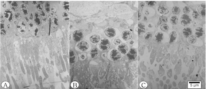
Figure 2. Electron microscopic photographs of rabbit retina. (A) Ultrastructure of a non‐detached area at the superior retina of medullary ray (normal retina). (B) Ultrastructure of the detached area four weeks after surgery, the detached retina underwent considerable cell loss and degeneration, and became rather thin. Note the cystic change of photoreceptor cell nuclei, abnormally swollen inner segments, and severely denatured outer segments. (C) Ultrastructure of a non‐detached neighboring retina of (B), similar (though generally less severe) changes were observed. Note the degenerative changes of photoreceptor nuclei and inner segments.
로 1 µm 두께로 박절편을 작성하고 1% toluidine blue로 염색한 후 광학현미경하에서 확인 후에, 다시 초박절편을 만들어 uranyl acetate 와 lead citrate 로 염색하고 투과전자현미경(JEM-1200EX, JEOL, Japan)으로 관찰하였다.
4. 망막의 두께 측정과 통계분석
망막의 Hematoxylin & Eosin 염색 조직에서 부위 별 망막의 두께를 측정하였다. 격자 모양의 Eyepiece
micrometer (U-OCM SQ 10/10, Olympus, Japan)와 image analysis program (Uthscsa Image tool for windows, version 3)를 이용하여 측정하였으며 망막의 3부위(박리된 망막의 정점, 경계 부위 그리고 주변 정상 망막)를 각각 연속적인 3개의 조직에서 측정하여 각 부위별로 평균두께를 측정 하였다. 통계는 이원분산분석(two-way analysis of variance)과 Wilcoxon rank sum test를 이용하여 분석하였으며 SigmaStat 3.1 프로그램을 사용하 였다.
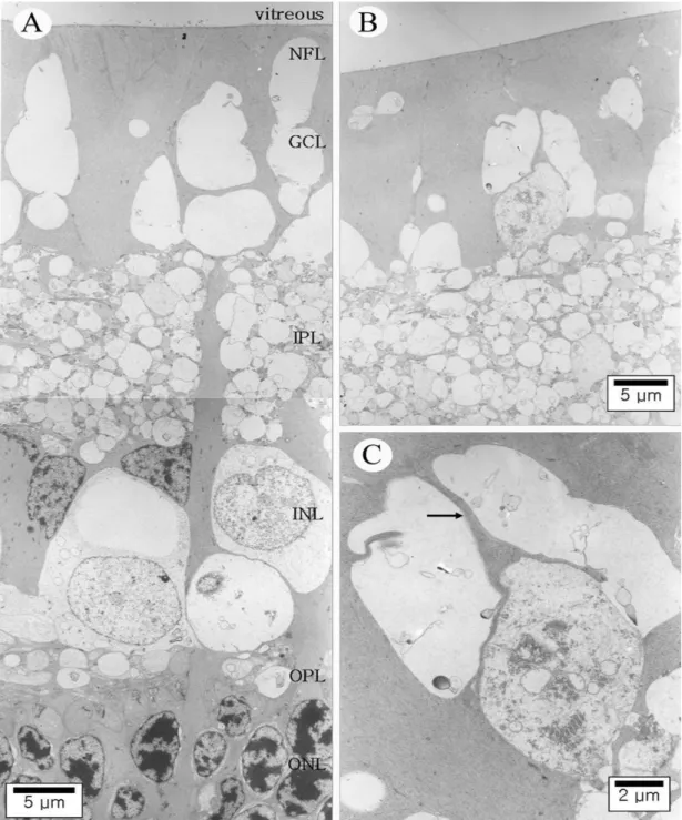
Figure 3. Ultrastructure of the detached rabbit retina four weeks after surgery. (A) The outer nuclear layer (ONL) is severe reduced in thickness. In the nerve fiber layer (NFL) and ganglion cell layer (GCL), empty spaces of various sizes are frequently observed. (B) Large empty spaces are found in the NFL just above a deformed ganglion cell. (C) Higher magnifications of the areas indicated in (B). A degenerating ganglion cell with nucleus consisting of disintegrated chromatin resides in close neighborhood to an intact ganglion cell axon (arrow) and empty spaces indicating sites of axon loss. IPL, inner plexiform layer; INL, inner nuclear layer; OPL, outer plexiform layer.
결 과
모든 대상안(22안/12마리)에서 실험적으로 유도된 국소 망막박리는 실험기간 동안 유지되었다(Fig. 1).
4주간 지속된 박리된 망막에서는 상당한 세포감소와 변
성이 초래되었으며 두께도 감소하였다. 특히 광수용체세 포(photoreceptors)의 변성과 광수용체 외절(outer segment)의 소실이 심하고, 내절의 변성이 심하였다 (Fig. 2B). 외핵층(outer nuclear layer)에 비해 망막내층 들은 비교적 정상 두께를 유지하고 정상적인
Figure 4. Ultrastructure of the detached rabbit retina three days after surgery. (A) Within the vitreous body but in close approximity to the degenerating inner nuclear cell, a macrophage is located. (B) Note the degenerative changes of photoreceptor cell nuclei and denatured inner and outer segments.
신경절세포(ganglion cell)도 관찰되었지만 신경섬유 층(nerve fiber layer)과 신경절세포층에도 심한 변 성은 관찰되었고, 이 두 층에 낭모양의 빈공간들이 다 수 존재하여 정상조직의 소실을 반영하였다(Fig. 3A).
일부 조직에서는 변성 중인 신경절세포를 관찰할 수 있 었는데 빈 공간은 신경절세포의 손실을 의미하고 변형 된 시신경 축삭도 관찰할 수 있었다(Fig. 3B, C). 또 한 대식세포도 변성된 망막 근처의 유리체내에서 관찰 되었다(Fig. 4A). 박리된 망막 부위로부터 일정부분 (수 mm) 떨어진 주변 유착 망막에서도 비록 경하지만 비슷한 망막 변성이 관찰되었고 특히, 광수용체세포층 은 비교적 유지되었으나 일부에서는 변성도 관찰되었다 (Fig. 2C).
박리된 망막이나 주변의 유착 망막 모두에서 외망상 층(outer plexiform layer)과 외핵층의 변형이 관찰 되었고 경계 부분에서도 비전형적인 광수용체 세포의 변성이 관찰되었다. 망막박리 유발 후 3일부터 광수용 체층의 변성이 관찰되었고(Fig. 4B) 시간 경과에 따라 진행하여 2주에는 4주 못지않은 광수용체의 변성이 관 찰되었다. 특히, 박리된 망막 주변의 정상 망막의 광수 용체세포 핵 주변의 세포질과 광수용체의 내절에는 다 양한 정도의 변성과 부종이 특징이었지만 외절은 비교
적 유지되었다(Fig. 2C). 변성된 내절 내부에는 변성 되고 부은 미토콘드리아도 관찰되었다. 이러한 변화들 은 망막박리를 유발하지 않는 정상망막(medullary ray 상층 망막)에서 같은 방법으로 동시에 고정하고 관 찰한 조직(Fig. 1A)에서는 정상 망막소견이 관찰되었 으므로 고정실수(fixation artifacts)의 가능성은 배 제할 수 있을 것 같다. 또한 이러한 박리된 망막과 주변 유착 망막의 전자현미경적인 미세 소견은 트리암시놀 론 주입안에서도 관찰되었지만 정도의 차이는 보이지 않았다.
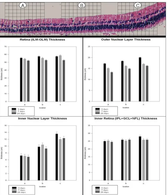
박리된 망막의 신경과 아교세포의 손상의 정도를 확 인하기 위해 박리된 망막의 중심부위(bubble)와 주변 의 박리되지 않은 유착 망막부위의 두께를 측정하였다 (Fig. 5). 국소 망막박리가 28일간 유지된 조직의 경 우, 전체 망막(internal limiting membrane에서 external limiting membrane)의 두께가 bubble 정점(Fig. 5A)에서는 평균 52.03±0.969 um 이나 주변 의 박리되지 않은 망막(Fig. 5C)의 두께는 52.64±2.21 um이었고, 망막내층(inner plexiform layer+ganglion cell layer+nerve fiber layer)과 속핵층은 각각 망 막박리 중심에서 24.505±1.377 um, 6.845±0.837 um이었고 주변의 박리되지 않은 유착 망막에서는 각각
Figure 5. Light micrograph and graphs of the detached (regions A, B) and adhering non‐detached (region C) rabbit retina after detachment surgery. (Upper) Hematoxylin‐Eosin stained paraffin section of one rabbit four weeks after surgery, used for measurements of the thickness of individual retinal layers (ONL, outer nuclear layer; INL, inner nuclear layer), inner retina (IPL, inner plexiform layer; + GCL, ganglion cell layer; + NFL, nerve fiber layer), and total retinal thickness (between ILM, inner limiting membrane, and OLM, outer limiting membrane). (Bottom) Graphs of two‐way analysis of variance (retinal area and duration of detachment). The total retinal thickness gradually decreased in the detached areas, with the most severe degeneration occurring in the center of detachment (region A), while the thickness of the inner retina and INL remained virtually unchanged over several weeks, and ONL was substantially reduced.
25.895±1.829 um, 12.3±1.043 um이었다. 외핵층 의 두께도 망막박리 정점에서는 13.303±0.76 um이고 주변 망막의 두께는 16.208±0.489 um이었다. 트리 암시놀론 주입안과 비주입안에서 각각의 망막두께는 큰 차이를 보이지 않았다(Fig. 6). 각각의 수치는 개체 차 이와 망막박리의 지속기간의 차이가 있고 개체수가 적
어 통계적인 차이는 없었으나 전체 망막의 두께가 망막 박리 부위, 특히 bubble 정점(Fig. 5A, 6A)에서 가 장 심한 감소가 있었으며 망막내층과 속핵층은 망막박 리 유발 후 수주간 비교적 두께가 유지되나 외핵층는 망막박리의 기간에 따라 상당한 두께의 감소를 보였다 (Fig. 5).
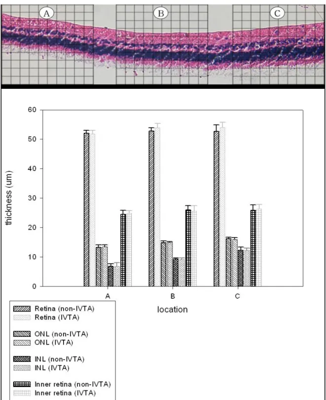
Figure 6. Light micrograph and graph of the detached (regions A, B) and adhering non‐detached (region C) rabbit retina after detachment surgery and intravitreal injection of triamcinolone. (Upper) Hematoxylin‐Eosin stained paraffin section of one rabbit four weeks after retinal detachement surgery and intravitreal injection of triamcinolone (IVTA), used for measurements of the thickness of individual retinal layers (ONL, outer nuclear layer; INL, inner nuclear layer), inner retina (IPL, inner plexiform layer; + GCL, ganglion cell layer; + NFL, nerve fiber layer), and total retinal thickness (between ILM, inner limiting membrane, and OLM, outer limiting membrane). (Bottom) Multiple grouped‐bar graph of Wilcoxon rank sum test (non‐IVTA group and IVTA group). The total retinal thickness, ONL thickness, INL thickness, and inner retinal thickness were not significantly different between non‐IVTA groups and IVTA groups.
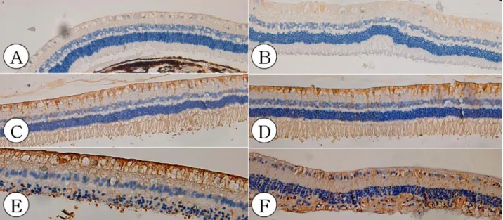
Figure 7. Light micrographs (×200) of the detached and adhering non‐detached rabbit retina. (A, B) Normal rabbit retinas (control eyes) show no glial fibrillary acidic protein (GFAP) immunoreacitivity. (C) Relatively strong GFAP immunoreactivity in adhering non‐detached retina. (D) Relatively strong GFAP immunoreactivity in slice of a rabbit retina which was focally detached for four weeks and was treated with intravitreal triamcinolone injection (IVTA). (E) Strong GFAP immunoreactivity in slice of a rabbit retina which was focally detached for four weeks. (F) Strong GFAP immunoreactivity in slice of a rabbit retina which was focally detached for four weeks and was treated with IVTA. Note that IVTA did not alter these gliotic alterations (GFAP immunoreactivity).
아교세포의 조직면역염색 검사에서는 3일간 박리된 망막에서 신경아교증이 확연하였고 망막박리 주변 유착 망막에서도 신경아교증이 관찰되었다(Fig. 6C, D).
특히 망막박리 3일과 7일 째 조직에서 신경아교증의 정 도가 심함을 확인하였다. 이런 신경아교증은 트리암시 놀론 주입안에서도 관찰되었고 트리암시놀론의 영향을 확인할 수는 없었다(Fig. 6D, F).
고 찰
다른 포유류와 마찬가지로 가토에서도 망막박리 후 에 광수용체세포의 빠른 해체(disorganization)와 소실이 특징이며 박리된 망막의 내층은 비교적 덜 손상 받고 천천히 변성된다고 알려져 있다.4-9 Fuade et al4은 본 연구에서와 같이 광수용체세포의 변성과 신경 절세포(ganglion cell)의 축삭(axon)의 소실이 박리 된 망막뿐만 아니라 박리 주변 유착 망막에서도 관찰되 었다고 보고하였다. 하지만 대부분의 실험적 망막박리 는 주로 망막의 외층의 변화에 국한되어 보고되어왔다.
가토에서는 건강한 망막인 경우에도 신경절세포가 드물 게 분포하며 신경섬유층도 소량의 신경다발이 산재해 있다. 그럼에도 불구하고 실험적으로 신경절세포의 축 삭이 망막박리 후 비교적 초기에 변성됨을 관찰하는 것 은 고무적이다.2-4 이런 신경절세포 축삭의 변성은
retrograde 또는 antegrade하게 박리되지 않은 주 변부의 망막에도 영향을 미치는 것으로 생각된다. 이러 한 결과는 Sasoh et al15이 국소 망막박리 환자들에서 시행한 다초점 망막전위도검사(multifocal ERG) 검 사에서 망막 주변부의 국소 망막박리가 황반부에도 영 향을 미침을 보고한 사실과 일정 부분 일치한다.
본 실험에서 anti‐glial fibrillary acidic protein (GFAP)를 이용한 면역조직화학염색으로 망막박리의 초기에 신경아교증이 심함을 확인할 수 있었고 이는 Uckermann et al8의 보고와 일치한다. 주목할 사실 은 이런 신경아교증이 박리된 망막에 국한되지 않고 박 리 주변 유착 망막에서도 관찰된다는 점이다. 이러한 신경아교증 반응이 박리된 망막에서 주변 망막으로 전 파되는 기전은 알 수 없으나 Uckermann et al8은 fibroblast growth factor 등의 싸이토카인이 박리 된 망막에서 분비되어서 주변 정상 망막으로 확산되어 신경아교증을 유발할 수 있고 medullary ray의 혈관 에서 염증세포의 분비가 원인일 수 있다고 제안하였다.
물론 혈관 분포가 다른 가토 망막박리를 사람과 비교할 수는 없지만 혈관이 풍부한 다른 돼지나 설치류의 망막 박리 모델에서도 유사한 망막변성이 관찰된다고 보고된 바 있다.6,7,9 혈관이 풍부한 실험동물들의 망막박리와 혈관이 제한적인 가토의 망막박리 후의 변성이 비슷 함은 일정 부분 가토 망막박리가 사람 망막박리의
cellular response를 이해하는 데 도움이 될 것으로 생각된다. 특히, 본 실험에서 관찰된 바와 같이 박리된 망막의 주변 유착 망막의 변성은 임상에서 망막박리 범 위보다 확대된 시야장애 내지는 국소망막박리 후 중심 시력의 저하가 생기는 이유를 어느 정도 설명할 수 있 을 것이다.
정상 망막에서 뮐러세포의 주된 기능은 세포외 이온 (extracellular ion)과 수분의 항상성(homeostasis) 의 유지이다. 망막내층의 경우 뮐러세포의 K+clearance 작용에 의해 탈수 상태를 유지한다. 그런데 망막박리 등에 의해 뮐러세포가 신경아교증이 되면 K+clearance 에 장애가 생겨 망막 부종과 낭(cyst) 형성, 광수용체 세포의 변성 등이 유발된다. 망막박리에서 저산소증이 신경세포의 손상과 신경아교증의 주된 원인으로 고려되 며 이런 망막의 저산소증이 blood‐retinal barrier 를 통해 혈청의 누출을 유발한다.5,6,8,9 Yamana et al16도 가토의 망막박리 모델에서 망막열공의 창상 치 유과정에서 뮐러세포의 신경아교증이 중요하다고 anti-GFAP를 이용한 면역화학조직염색을 이용하여 보고하였다.
현재 임상적으로 망막부종의 치료로 유리체내 트리 암시놀론 주사가 활발히 사용되고 있으며, 트리암시놀 론이 저산조증에 동반된 cellular response를 억제한 다는 점에서 저자들은 트리암시놀론이 가토 국소망막박 리 모델에서 glial cell activation을 억제하지 않을 까 가정하였으나 anti‐GFAP를 이용한 면역조직화 학염색에서는 차이를 확인할 수 없었다.
결론적으로 가토를 이용한 망막박리 모델에서 박리 된 망막뿐만 아니라 주변 정상 망막에서도 신경절세포 의 소실과 광수용체세포의 변성이 관찰되었으며 이때 특징적으로 광수용체 내절의 변성이 외절의 변성보다 심한 양상으로 관찰되었다. 또한 뮐러세포의 신경아교 증이 망막박리 초기부터 증가되어 있었으며, 마찬가지 로 박리된 망막의 주변 유착 망막에서도 관찰되었으며 이는 트리암시놀론의 영향을 받지 않았다. 시간이 경과 함에 따라 박리된 망막의 두께는 얇아지며 이는 박리된 망막의 정점부위에 가장 확연하며 특히, 외핵층의 두께 감소가 심하였다. 또한 박리되지 않은 주변 망막에서도 망막두께의 감소가 관찰되었으며 이는 트리암시놀론 주 입안에서 동일하였다.
Francke et al5의 연구와 같이 본 연구에서도 박리 된 망막 주변의 유착 망막의 광수용체세포의 변성이 외 절보다 내절이 심함은 이런 변성이 망막색소상피나 맥 락막에서의 산소나 영양분의 공급 장애라기 보다는 뮐 러세포의 신경아교증에 의한 항상성 유지의 장애라고 생각된다. 만약 이런 가정을 임상에 적용하면 망막박리
의 해부학적 수술 성공에도 불구하고 시력 개선에 한계 가 있거나 주변 망막박리에서도 정상 망막의 기능의 장 애 내지는 시야장애의 확대 등을 일정 부분 설명할 수 있을 것이다. 하지만 이런 가정을 증명하기 위해서는 망막 혈관의 분포가 사람과 유사한 여러 다른 실험동물 에서의 연구가 추가되어야 할 것으로 생각된다.
참고문헌
1) Isashiki M, Ohba N. Recovery of differential light sensitivity following surgery for rhegmatogenous retinal detachment.
Graefes Arch Clin Exp Ophthalmol 1986;224:184-90.
2) Erickson PA, Fisher SK, Anderson DH, et al. Retinal detachment in the cat: The outer nuclear and outer plexiform layers. Invest Ophthalmol Vis Sci 1983;24:927-42.
3) Anderson DH, Guerin CJ, Erickson PA, et al. Morphological recovery in the reattached retina. Invest Ophthalmol Vis Sci 1986;27:168-83.
4) Faude F, Francke M, Makarov F, et al. Experimental retinal detachment causes widespread and multilayered degeneration in rabbit retina. J Neurocytol 2001;30:379-90.
5) Francke M, Faude F, Pannicke T, et al. Glial cell‐mediated spread of retinal degeneration during detachment: A hypothesis based upon studies in rabbits. Vision Res 2005;45:2256-67.
6) Fisher SK, Lewis GP. Cellular effects of detachment and reattachment on the neural retina and the retinal pigment epithelium. In : Ryan SJ, Wilkinson CP, eds. Retina, 4th ed.
St. Louis: Mosby, 2006; v. 3. chap. 115.
7) Jakson TL, Hillenkamp J, Williamson TH, et al. An experimental model of rhegmatogeneous retinal detachment:
Surgical results and glial cell response. Invest Ophthalmol Vis Sci 2003;44:4026-34.
8) Uckermann O, Pannicke T, Wiedemann P, et al. Triamcinolone does not alter glial cell activation in the experimental detached rabbit retina. J Ocul Pharmacol Ther 2005;21:266-74.
9) Fisher SK, Lewis GP, Linberg KA, Verardo MR. Cellular remodeling in mammalian retina: results from studies of experimental retinal detachment. Prog Retin Eye Res 2005;24:395-431.
10) Massin P, Audren F, Haouchine B, et al. Intravitreal triamcinolone acetonide for diabetic diffuse macular edema.
Ophthalmology 2004;111:218-25.
11) Jonas JB. Intravitreal triamcinolone acetonide as treatment for exudative retinal detachment. Br J Ophthalmol 2004;88:587-8.
12) Andrade RE, Muccioli C, Farah ME, et al. Intravitreal triamcinlone in the treatment of serous retinal detachment in Vogt‐Koyanagi‐Harada syndrome. Am J Ophthalmol 2004;137:572-4.
13) Jonas JB, Kreissig I, Degenring R. Intravitreal triamcinolone acetonide for treatment of intraocular proliferative, exudative, and neovascular diseases. Prog Retin Eye Res 2005;24:587-611.
=ABSTRACT=
Structural Alterations of Retinal Detachment After Intravitreal Triamcinolone Injection in Rabbit Eyes
Muyan Kim, M.D.1, Young-Chun Lee, M.D.2, Young-Hoon Park, M.D.2
Saevit Eye Hospital1, Ilsan, Korea
Department of Ophthalmology and Visual Science, College of Medicine, The Catholic University of Korea2, Seoul, Korea
Purpose: To investigate the effect of localized retinal detachment on both the detached and attached regions, and to determine the effect of triamcinolone on Müller cell gliosis.
Methods: Pars plana vitrectomy was performed in both eyes of 12 pigmented rabbits. A dome‐shaped retinal detachment was made by injecting sodium hyaluronate into the subretinal space. Triamcinolone (5 mg) was applied intravitreally to one eye (12 eyes). The detached retinal area and the neighboring attached region were studied by light and electron microscopy 3, 7, and 28 days after surgery. Tissues were prepared in 5 um sections for hematoxylin-eosin staining and immunohistochemistry with antibody to glial fibrillary acidic protein (GFAP).
Results: In addition to the well-known degeneration of photoreceptor cells in the detached retina, an incomplete but severe loss of ganglion cell axons occurs in both the detached and the attached regions. The total retinal thickness gradually decreased in the detached areas, while the thickness of the inner retinal layers remained virtually unchanged over several weeks. Gliotic alterations were apparent in both the detached and non-detached retinal areas, and intravitreal triamcinolone did not alter these gliotic alterations of Müller cells.
Conclusions: It is noteworthy that progressive retinal destruction also occurs in the attached retina after local detachment. This may account for visual impairment in strikingly large areas of the visual field, even after retinal reattachment.
J Korean Ophthalmol Soc 49(4):641-650, 2008
Key Words: Gliosis, Localized retinal detachment, Rabbit, Triamcinolone
Address reprint requests to Young-Hoon Park, M.D.
Department of Ophthalmology, Uijongbu St. Mary’s Hospital, College of Medicine, The Catholic University of Korea
#65-1 Geumo-dong, Uijeongbu-si, Gyeonggi-do 480-717, Korea
Tel: 82-31-820-3570, Fax: 82-31-847-3418, E-mail: [email protected] 14) Sakamoto T, Miyazaki M, Hisatomi T, et al. Triamcinolone-assisted
pars plana vitrectomy improves the surgical procedures and decrease the postoperative blood‐ocular barrier breakdown.
Graefes Arch Clin Exp Ophthalmol 2002;240:423-9.
15) Sasoh M, Yoshida S, Kuze M, Uji Y. The multifocal
electroretinogram in retinal detachment. Doc Ophthalmol 1998;94:239-52.
16) Yammana T, Kita M, Ozaki S, et al. The process of closure of experimental retinal holes in rabbit eyes. Graefes Arch Clin Exp Ophthalmol 2000;238:81-7.