http://www.medicinalcrop.org
http://dx.doi.org/10.7783/KJMCS.2021.29.2.135
다양한 인삼종의 분획물에 대한 C2C12 세포에서의 보호 및 분화 활성 평가
홍준기
1,2
· 홍은영3
· 박준희4
· 이지원5
· 김성수6
· 문지원7
· 황완균8†
Differentiation-promoting and Protective Effects of the Fractions of Various Ginseng Species in C2C12 Cells
Jun Kee Hong
1,2
, Eun Young Hong3
, Jun Hee Park4
, Ji Won Lee5
, Sung Su Kim6
, Ji Won Moon7
and Wan Kyunn Whang8†
INTRODUCTION
Panax ginseng C. A. Meyer, Panax quinquefolium L., and Panax notoginseng Burk. are three major perennial herbal plants in the genus Panax of the Araliaceae family (Lee et al., 2017). These ginsengs have been used as foods or herbal medicines worldwide for thousands of years. Ginseng has
mostly been used as a tonic, and it has been confirmed to improve bodily function, increase vitality, increase resistance to stress and aging, and have immunomodulatory activities (Kim, 2012).
P. ginseng is a essential herbal medicine resource in East Asia owing to its pharmacological properties and physiological activities. P. quinquefolium (American ginseng), which is ABSTRACT
Received: 2020 November 17 1st Revised: 2020 January 19 2nd Revised: 2021 February 15 3rd Revised: 2021 March 3 Accepted: 2021 March 3 This is an open access article distributed under the terms of the Creative Commons Attribution Non-Commercial License (http://
creativecommons.org/licenses/
by-nc/3.0/) which permits unrestricted non-commercial use, distribution, and reproduction in any medium, provided the original work is properly cited.
Background: The effects of Panax ginseng C. A. Meyer in protecting and promoting the differen- tiation of muscle cells have been reported. However, the influences of various ginseng species and various fractions were not investigated.
Methods and Results: In this study, these effects on muscle cells were confirmed using butanol and water fractions of various ginseng species. The fractions of P. ginseng were confirmed to have a cytoprotective effect on C2C12 cells. The myogenin expression level was significantly higher by 1.5 times or more, in the P. ginseng fraction groups than in the other groups, confirming that the P.
ginseng fractions promoted cell differentiation. Both fractions of P. ginseng significantly reduced the rate of production of reactive oxygen species when compared with that of the control group. In the mechanism study, P. ginseng fractions tended to decrease muscle RING-finger protein-1 (MuRF-1), AMP-activated protein kinase (AMPK), forkhead box O3α (Foxo3α), and the expres- sion of the BCL2-associated agonist of cell death (BAD). In particular, AMPK was significantly reduced in the P. ginseng water fraction group when compared to that in the butanol fraction and control groups.
Conclusions: The results of this study confirmed that P. ginseng has superior cell differentiation- promoting and protective effects on muscle cells when compared with the effects of Panax quin- quefolium and Panax notoginseng.
Key Words: Panax Ginseng C. A. Meyer, Panax quinquefolium, Panax notoginseng, Saponin, Non-saponin, C2C12 cell
†
Corresponding author: (Phone) +82-02-820-5611 (E-mail) [email protected]
1
중앙대학교 글로벌혁신신약학과 연구원 / Researcher, Department of Global Innovative Drug, Chung-Ang University. Seoul 06974 Korea.
2
㈜파미니티 연구원 / Researcher, Famenity Co., Ltd., Uiwang 16006, Korea.
3
중앙대학교 글로벌혁신신약학과 연구원 / Researcher, Department of Global Innovative Drug, Chung-Ang University. Seoul 06974 Korea.
4
중앙대학교 글로벌혁신신약학과 연구원 / Researcher, Department of Global Innovative Drug, Chung-Ang University. Seoul 06974 Korea.
5
㈜파미니티 연구원 / Researcher, Famenity Co., Ltd., Uiwang 16006, Korea.
6
㈜파미니티 연구원 / Researcher, Famenity Co., Ltd., Uiwang 16006, Korea.
7
국립원예특작과학원 인삼특작부 인삼과 연구사 / Researcher, Department of Herbal Crop Research, NIHHS, RDA, Eumseong 27709, Korea.
8
중앙대학교 글로벌혁신신약학과 교수 / Professor, Department of Global Innovative Drug, Chung-Ang University. Seoul 06974 Korea.
distributed in North America, is another important ginseng species. P. notoginseng (Sanchi) is available in Japan and the southern area of China (Jee et al., 2014).
Ginseng-related effects include antioxidant (Lee et al., 2004;
Doh et al., 2010; Moon et al., 2019), anticancer (Zhang et al., 2012), antidiabetic (Jung and Kang, 2013; Seo et al., 2016), antihypertensive (Choi et al., 2006), anti-stress (Rai et al., 2003), immunity-promoting (Ha et al., 2009), and memory- enhancing (Kim et al., 2018; Sohn et al., 2008; Lee et al., 2020) effects. Lau et al. (2009) evaluated the antithrombotic efficacy of three ginseng species and found that P. notoginseng showed the highest antithrombotic effect; moreover, it was confirmed that the efficacy was increased when the ginseng was steamed.
Recent studies on the functionality of ginseng have focused on its efficacy in protecting and promoting the production of muscle cells. In rat models of muscle injury due to eccentric exercise, rats treated with dammarane steroids showed decreased muscle cell necrosis and significantly increased distribution of CD68
+M1 macrophages and 3-nitrotyrosine (Yu et al., 2014). The administration of red ginseng extract resulted in the relaxation of rabbit corpus cavernosal smooth muscle following contraction induced by phenylephrine (Choi et al., 1998).
In addition, rats that ingested P. ginseng extract showed significant improvement in exercise time in treadmill test, and the P. ginseng intake group showed a significant increase in serum IGF-1 levels (Sohn et al., 2012). Furthermore, ingestion of ginseng extract in rats significantly protected the muscles from oxidative stress damage caused by exercise (Voces et al., 2004).
G115, a standardized ginseng extract, was also confirmed to decrease lipid peroxidation and inflammatory markers in the muscle (Oliveira et al., 2001). Similar results were confirmed in a clinical trial. The group that ingested red ginseng extract showed significantly reduced exercise-induced production of inflammatory markers, such as IL-6, in the muscles (Jung et al., 2011). Studies on ginseng at the component level confirmed that ginsenoside Rg1, a major constituent of ginseng, improves glycogen supplementation after exercise and decreases the mRNA levels of inflammatory factors, such as TNF-α and IL-10 (Hou et al., 2015).
In this study, to identify the components or fractions responsible for the protective and regeneration-promoting effects of ginseng, the efficacy of saponin and non-saponin
fractions of ginseng using liquid-liquid extraction was investigated. C2C12 muscle cells, developed in 1977, are myoblasts in the pre-differentiation stage into muscle cells and often used as a control cell line in studies of in vitro muscular dystrophy (Denes et al., 2019). The muscle cell-protective effects of the fractions of various ginseng species, namely P.
ginseng, P. quinquefolium, and P. notoginseng, which are representatives of the Panax genus, were compared. In this study, we aimed to confirm the protective and regeneration- promoting effects of P. ginseng in muscle cells compared with those of other ginseng genera and to provide basic research results to elucidate the active components or fractions.
MATERIALS AND METHODS
1. Samples and preparation
Four-year-old P. ginseng grown in Geumsan, Republic of Korea, was purchased in 2019 at Geumsan Ginseng Market, Republic of Korea. P. quinquefolium grown in the United States was purchased in Seongwan Market, Hong Kong. P.
notoginseng grown in the Changbai Mountain area was purchased in 2019 at Changbai Mountain Market, Jilin, China.
Each of the three ginseng species was verified by experts to be suitable for research.
For each ginseng species, 2 g of ginseng powder and 20 ㎖ of 50% methanol (JT Baker, Center Valley, PA, USA) were added to a 50 ㎖ conical tube. The first extraction was then performed in a shaking water bath at 250 rpm and 60 ℃ for 4 h (MaXturdy 45; DAIHAN Scientific Co., Ltd., Wonju, Korea). After the extraction was completed, only the supernatant was recovered by centrifugation at 4,000 rpm for 10 min using a centrifuge (VARISPIN 4, Novapro Co., Ltd., Bucheon, Korea), and 20 ㎖ of 50% methanol was added to the precipitate, which was then used for the second extraction in the same manner. Similarly, only the supernatant was recovered, mixed with the first extract, and then used for analysis.
The mixed extract was mixed with volatilizing methanol in a
vacuum evaporator (N-1100V, Eyela Co., Ltd., Tokyo, Japan)
to obtain a solution of approximately 60 ㎖, and then the same
volume of n-butanol (Daejung Chemicals and Metals Co., Ltd.,
Siheung, Korea) was added to separate the layers. As a result,
the solution was separated into approximately 30 ㎖ of aqueous
layer and approximately 90 ㎖ of saturated butanol layer. The
water-saturated butanol layer was separated using a solvent
through a vacuum evaporator, yielding 596 ㎎ of P. ginseng C butanol fraction [G(B)], 610 ㎎ of P. notoginseng butanol fraction [N(B)], and 650 ㎎ of P. quinquefolium butanol fraction [Q(B)] powders.
The water layer of each ginseng extract was sublimated with water using a freeze dryer (FDS8508, ilShinBioBase, Dongducheon, Korea), yielding 1,921 ㎎ of P. ginseng water fraction [G(W)], 2,330 ㎎ of P. notoginseng water fraction [N(W)], and 2,569 ㎎ of P. quinquefolium water fraction [Q(W)], which were used in the subsequent tests.
2. Cell culture and differentiation methods
Mice-derived C2C12 cells were obtained from the American Type Culture Collection (CRL-1772, ATCC, Manassas, VA, USA) and cultured in a 5% CO
2incubator at 37 ℃. The cells were incubated in Dulbecco's modified Eagle's medium (DMEM) containing 10% fetal bovine serum (FBS) in the cell proliferation phase before differentiation, and the 10% FBS was changed to 2% horse serum to induce differentiation. To eliminate the over-density phenomenon caused by cell proliferation, the appropriate number of cells was maintained by subculturing every 48 h.
During differentiation, C2C12 myoblast cells were seeded at a concentration of 1 × 10
5cells/ ㎖ in a six-well plate and cultured until 70% - 80% confluence. The differentiation medium was collected once a day to confirm whether the cells had differentiated into a myotube shape under a microscope.
3. Confirmation of the cytotoxic and cell-protective effects of ginseng extract
To investigate the cytotoxicity of the ginseng extracts, the viability of C2C12 cells was evaluated using Cell Counting Kit-8 (CK04-20, MedChemExpress, Monmouth Junction, NJ, USA) according to the manufacturer's instruction. C2C12 cells were seeded in a 96-well plate at a concentration of 1 × 10
4cells/ ㎖ day before the experiment.
Subsequently, the cells were treated with 12.5, 25.0, 50.0, or 100.0 ㎍/㎖ of ginseng extract and incubated for 24 h. After that, CCK-8 solution was dispensed at 10 ㎕, which corresponded to 10% of the total volume, and the cells were incubated for 2 h. Absorbance was measured at 450 ㎚ using a microplate reader (Infinite M200 PRO NanoQuant, TECAN Ltd., Zurich, Switzerland).
To confirm the cytoprotective effect of the ginseng extracts, C2C12 cells were divided into a control group (treated with
600 µM H
2O
2only) and ginseng fraction groups [treated with G(B), G(W), Q(B), Q(W), N(B), and N(W) at concentrations of 3.125, 6.250, and 12.500 ㎍/㎖, followed by 600 µM H
2O
2).
To confirm the cell-protective efficacy of the ginseng extracts, C2C12 cells were seeded in a 96-well plate at a concentration of 1 × 10
4cells/ ㎖ day before the experiment.
Next, the ginseng-treated groups were treated with each concentration of the sample. After 1 h of incubation, all groups except the control group were treated with 600 µM H
2O
2. After that, the cells were further incubated for 24 h. Next, 10
㎕ of CCK-8 solution was added to the cells, which were then incubated for another 2 h. Absorbance was measured at 460 ㎚ using a microplate reader (Infinite M200 PRO NanoQuant;
TECAN Ltd., Zurich, Switzerland).
4. Protein separation and western blotting analysis Western blotting analysis was performed to confirm the differentiation of myoblasts into myotubes and confirm changes in the factors indicating the cytoprotective effect of each fraction. After appropriate cell culture and treatment in 100 ㎜ dishes, intracellular protein was isolated using 1 mM phenylmethylsulfonyl fluoride (MedChemExpress, Mnmouth Junction, NJ, USA), 1% protease inhibitor cocktail, and NP40 cell lysis buffer (Invitrogen, Carlsbad, CA, USA).
The separated protein was quantified using a Pierce
™BCA Protein Assay Kit (Invitrogen, Calesbad, CA, USA). Next, 30 µg of protein was electrophoresed using Bolt
™4% - 12% Bis- Tris Plus Gels, and the protein was then transferred to a dry iBlot
®Transfer Stack (Invitrogen), a nitrocellulose membrane of a gel substrate, using an iBlot Gel Transfer Device (Invitrogen, Calesbad, CA, USA).
Each membrane was blocked at room temperature for 1 h
using 5% skim milk and subsequently washed three times with
0.1% Tris-buffered saline in 0.1% Tween 20 buffer. The
membrane was incubated with primary antibodies, including
anti-myogenin [F5D] (ab1835) (1 : 250, Abcam, Cambridge,
England), anti-MyoD1 [5.2F] (ab16148) (1 : 1000, Abcam,
Cambridge, England), anti-MuRF1 (ab96857) (1 : 1000,
Abcam, Cambridge, England), anti-Foxo3α (phospho S253)
(ab47285) (1 : 1000, Abcam, Cambridge, England), and anti-
Foxo3α ab121620 (1 : 2500, Abcam, Cambridge, England),
overnight in a refrigerator at 4 ℃. The membrane was then
incubated with HRP-conjugated IgG secondary antibody (1 :
30000, Abcam, Cambridge, England) for 1 h at room
temperature.
After washing the membrane three times, the developed protein bands were analyzed using a C-DiGit
®Blot Scanner (LI-COR Inc., Lincoln, NE, USA) to determine the protein expression levels. The band was quantified using the Image J Program (National Institutes of Health, Bethesda, MD, USA), and protein expression level was expressed as a fold change relative to the value of the control group.
5. ROS generation measurement
Intracellular ROS levels were measured using the DCF-DA method. C2C12 myoblasts were dispensed into a 6-well plate at 1 × 10
5cells/ ㎖ and cultured for 24 h, and then the ginseng extract sample [G(B), G(W), Q(B), Q(W), N(B), N(W)] was added to serum-free DMEM at a concentration of 12.5 ㎍/㎖
for 24 h. After 1 h, the cells were washed with PBS, and treated with 600 μM H
2O
2for 24 h. After that, it was washed with PBS, and 10 μM DCF-DA was dispensed into each well and cultured in an incubator at 37 ℃ in a 5% CO
2atmosphere for 30 min.
After 30 min, it was washed with PBS, and 1 ㎖ of PBS was dispensed into each well, and fluorescence was measured at excitation 485/20 and emission 528/20 using a microplate reader (Infinite M200 PRO NanoQuant, TECAN Ltd., Zurich, Switzerland).
6. Statistical analysis
Statistical analysis was performed using analysis of variance in the SPSS program (PASW Statics 18, IBM Co., Armonk, NY, USA). Significant differences in the parameters, such as ginsenoside content, were analyzed by Duncan's Multiple Range Test (DMRT) at a significance level of 5% (p < 0.05).
RESULTS AND DISCUSSION
1. Cytotoxicity of ginseng extract in C2C12 cells
In the butanol fraction of all ginseng species, as the concentration increased, cytotoxicity increased. On the contrary, there was no significant change in cell viability in the water fraction group. Therefore, the water fractions of the three ginseng species did not appear to cause cytotoxicity, showing similar cell viability to that of the control group (Fig. 1).
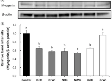
2. Muscle cell differentiation-promoting effect of ginseng extract
To determine whether the ginseng extracts affect myoblast
differentiation into myotubes, C2C12 myoblasts were induced to differentiate into myotubes. After differentiation induction, the myoblasts were confirmed to have differentiated into myotubes morphologically. After H
2O
2was added to the differentiated cells, the length of the tube was shortened, and the cells were killed (data not shown). Each extract sample was used to induce differentiation, and the expression of myogenin, a marker gene that induces skeletal muscle
Fig. 1. C2C12 cell viabilities of Butanol or water fraction of various Panax species. All data are shown as the means± SD (n = 3). Panax ginseng (B); Butanol fraction of Panax ginseng C. A. Meyer, Panax ginseng (W); Water fraction of Panax ginseng C. A. Meyer, Panax quinquefolium (B); Butanol fraction of Panax quinquefolium, Panax quinquefolium (W); Water fraction of Panax quinquefolium, Panax notoginseng (B); Butanol fraction of Panax noto- ginseng, Panax notoginseng (W); Water fraction of Panax notoginseng. *Different superscript letters show significant differences at 5% by Duncan’s Multiple Range Test (DMRT, p < 0.05).
differentiation, was confirmed by western blotting analysis.
Our results showed that the butanol fraction and water fraction of each ginseng species, excluding the water fraction of P. ginseng C, showed similar levels of myogenin expression as Q(B); 0.64 ± 0.05, Q(W); 0.57 ± 0.03, N(B); 0.58 ± 0.07, N(W); 0.54 ± 0.02, and G(B); 0.65 ± 0.01.
Moreover, in the group treated with the non-fraction of P.
ginseng C, myogenin expression level was 0.96 ± 0.06, which was a significantly increased (p < 0.05) compared with in the groups treated with other test materials, further confirming that the water fraction of P. ginseng promoted the differentiation of muscle cells (Fig. 2).
3. Cytoprotective effects of ginseng extract against oxidative stress
Oxidative cell damage in C2C12 cells was induced by H
2O
2treatment, and the protective effect of ginseng extract was confirmed. Following treatment with H
2O
2at various concentrations, the cell viability was approximately 50% after treatment with 600 µM H
2O
2. Thus, this concentration of H
2O
2was used in this experiment.
The cells were pretreated with each ginseng extract for 1 h
and then treated with H
2O
2, a toxic substance, for 24 h.
Subsequently, cell viability was examined. The results showed
Fig. 3. Protective effects of fractions of Panax ginseng on the viability of H2O2-treated C2C12 cells. All data are shown as the means ± SD (n = 3). G(B); butanol fraction of P. ginseng, G(W); water fraction of P. ginseng, Q(B);
butanol fraction of Panax quinquefolium, Q(W); water fraction of P. quinquefolium, N(B); butanol fraction of Panax notoginseng, N(W); water fraction of P. notoginseng.
*Different superscript letters show significant differences at 5% as analyzed by Duncan’s Multiple Range Test (DMRT, p < 0.05).
Fig. 2. Myogenin Expression of Butanol or water fraction of various Panax species in C2C12 cells. (A) Expression of myogenin in C2C12 cell, (B) Graph presents relative intensity compared with the beta-actin band intensity. All data are shown as the means ± SD (n = 3). Q(B);
Butanol fraction of Panax quinquefolium, Q(W); Water fraction of Panax quinquefolium, N(B); Butanol fraction of Panax notoginseng, N(W); Water fraction of Panax notoginseng, G(B); Butanol fraction of Panax ginseng C. A.
Meyer, G(W); Water fraction of Panax ginseng C. A.
Meyer. *Different superscript letters show significant differences at 5% by Duncan’s Multiple Range Test (DMRT, p < 0.05).
that there was no significant difference in cell viability in the groups treated with extracts of P. quinquefolium and P.
notoginseng compared with that in the group treated with H
2O
2alone. However, cell viability was significantly higher (p <
0.05) in the groups treated with P. ginseng extracts. Thus, the ginseng extracts were confirmed to have cell-protective effect against oxidative stress caused by H
2O
2.
In particular, the water fraction of P. ginseng significantly protected cells from oxidative damage, compared with the butanol fraction, in a concentration-dependent manner (p <
0.05, Fig. 3).
4. Effect of each ginseng extract on ROS production and oxidative stress
Cell damage caused by H
2O
2is related to impaired mitochondrial function due to abnormal ROS production in the mitochondria. The protective effect of the ginseng extracts against ROS production was thus assessed.
Intracellular ROS levels were determined using the DCF-DA method. Fluorescence analysis showed that there was no significant difference in fluorescence between the extract groups and the H
2O
2group (169.17 ± 6.50%). However, ROS levels were 140.11 ± 4.30% in the P. ginseng butanol fraction group and 124.98% ± 3.73% in the P. ginseng water fraction group, showing a significant reduction compared with that in the group treated with only H
2O
2(p < 0.05).
These findings confirmed that among the ginseng extracts, P.
ginseng extract had protective effect against oxidative stress. In particular, the water fraction of P. ginseng effectively reduced ROS levels compared with the other extracts (Fig. 4)
5. Mechanism of the cytoprotective effect of ginseng extract against oxidative stress in C2C12 cells
The expression of muscle damage indicator AMP-activated protein kinase (AMPK) and proteolytic signaling transducers muscle RING-finger-1 (MuRF1) and forkhead box O3α (Foxo3α), which are activated by an increase in AMP level due to ATP depletion, was confirmed using western blotting.
The efficacy of the extracts in inhibiting apoptosis due to oxidative stress-induced cell damage was examined.
The results of protein expression levels showed that the P.
ginseng fraction had protective effect on cell viability.
Compared with those in the control group, the expression levels of the protein degradation markers AMPK and MuRF1 in the group treated with H
2O
2were increased, confirming the presence of muscle damage.
The MuRF-1 expression level in the P. ginseng butanol fraction group was 0.85 ± 0.06, showing a decreasing trend compared with that in the H
2O
2group (1.16 ± 0.09), but the decrease was not significant. The MuRF-1 expression in the P.
ginseng water fraction group was 0.67 ± 0.08, showing a significant decrease compared with that in the H
2O
2group (p <
0.05, Fig. 5B).
AMPK expression level showed a decreasing tendency in the H
2O
2alone group (1.17 ± 0.06). The expression level of AMPK was 1.13 ± 0.05 and 0.84 ± 0.07 in the P. ginseng butanol and water fraction groups, respectively, showing significant decreases compared with that in the H
2O
2alone group (p < 0.05, Fig. 5C).
BAD expression in the P. ginseng butanol and water fraction groups were 0.53 ± 0.04 and 0.43 ± 0.02, respectively, showing significant decreases compared with that in the H
2O
2group (p < 0.05, Fig. 5D).
Foxo3α expression in the P. ginseng butanol fraction group was 0.79 ± 0.04, which showed a decreasing trend compared with that in the H
2O
2alone group (1.02 ± 0.07), but the decrease was not significant. Foxo3α expression in the P.
ginseng water fraction group was 0.66 ± 0.03, which was significantly decreased compared with that in the group treated with H
2O
2alone (p < 0.05, Fig. 5E).
Myogenin, which belongs to the MyoD family, plays an important role in the regulation of the differentiation of single nucleated myoblasts into multinucleated myofibers (Lee et al., 2017). Scavenging of ROS from muscle cells is a possible approach to prevent muscle cell damage (Choi et al., 2017).
Abnormally high levels of ROS cause dysfunction of muscle
Fig. 4. Effect of fractions of various Panax species onintracellular ROS levels in C2C12 cells as analyzed by DCF-DA assay. All data are shown as the means ± SD (n
= 3). G(B); Butanol fraction of Panax ginseng, G(W);
water fraction of P. ginseng. *Different superscript letters show significant differences at 5% as analyzed by Duncan’s Multiple Range Test (DMRT, p < 0.05).
cells and damage to intracellular macromolecules such as proteins, lipids, and nucleic acids in muscle cells; they act as a causative factor for cell death (Choi, 2015). Abnormal ROS production by H
2O
2impairs mitochondrial function. The production of BAD, the pro-apoptotic factor, is one of the intrinsic mechanisms of action involved in apoptosis; BAD plays a role in aggregating several mitochondrial signals involved in cell death and blood glucose regulation (Sung et
al., 2006).
AMPK is an important regulator of intracellular energy
balance. When AMPK is activated, the ubiquitin-proteasome
pathway is activated through increases in muscle-specific
ubiquitin ligases, muscle atrophy F-box (MAFbx)/atrogin-1,
and MuRF1 through the transcription factor Foxo3α, resulting
in muscle atrophy due to muscle protein degradation (Choi et
al., 2017; Yeo et al., 2019; Chen et al., 2020; Yoshikawa et
Fig. 5. Effects of Panax ginseng fractions on MuRF-1, AMPK, Foxo3a and BAD expression in H2O2-treated C2C12 cells as analyzed by western blotting. (A) Expression of MuRF-1, AMPK, Foxo3a and BAD. (B) The graph presents the relative intensity of MuRF- 1 compared with that of beta-actin. (C) The graph presents the relative intensity of AMPK compared with that of beta-actin. (D) The graph presents the relative intensity of Foxo3a compared with that of beta-actin. (E) The graph presents the relative intensity of BAD compared with that of beta-actin. All data are shown as the means ± SD (n = 3). G(B); Butanol fraction of P. ginseng, G(W); water fraction of P. ginseng. *Different superscript letters show significant differences at 5% as analyzed by Duncan’s Multiple Range Test (DMRT, p < 0.05).al., 2020).
This study was aimed to evaluate the efficacy of each ginseng and provide basic research results to track the active components or fractions in the ginseng genus for differentiation- promoting and protective effects on muscle cells. There was an evaluation report on each ginseng genus, but not reported about comparison of fractions using each ginseng genus.
Butanol fractions and water fractions of various ginseng species were prepared, and the cytoprotective effect of these fractions against H
2O
2-induced oxidative stress in C2C12 cells was evaluated by measuring myogenin expression and ROS levels. The expression levels of MuRF-1, AMPK, Foxo3α, and BAD were evaluated in the mechanism study.
The P. ginseng fraction showed the highest cytoprotective effect in C2C12 cells subjected to oxidative damage caused by H
2O
2. Myogenin activity, a marker of muscle cell differentiation, was confirmed to increase by 1.5 times or higher after treatment with the water fraction of P. ginseng compared with that after treatment with the butanol fraction of P. ginseng and other ginseng fractions. This trend was also observed in ROS production, but no significant difference was found between the P. ginseng fractions.
These results were thought to be due to the suppression of the expression of MuRF-1, AMPK, Foxo3α, and BAD. In particular, the expression level of AMPK was significantly suppressed by the water fraction of P. ginseng, compared with its butanol fraction.
The findings of this study confirmed that P. ginseng has superior muscle cell-protective effect to that of P. quinquefolium and P. notoginseng. Furthermore, the water fraction of P.
ginseng exhibited the most potent muscle cell-protective effect.
ACKNOWLEDGEMENT
This work was carried out with the support of “Cooperative Research Program for Agriculture Science and Technology Development (PJ0136232020)” Rural Development Admini- stration, Republic of Korea.
REFERENCES
Chen C, Yang J, Lu C, Chiu Y, Chen H, Chung M, Wu Y and Chen F. (2020). Effect of quercetin on dexamethasone-Induced C2C12 skeletal muscle cell injury. Molecules. 25:3267. https://
www.mdpi.com/1420-3049/25/14/3267 (cited by 2020 Nov 10).
Choi DJ, Cho KH, Jung WS, Park SU, Han CH and Lee WC.
(2006). Clinical effects of Korean ginseng, Korean red ginseng, Chinese ginseng, and American ginseng on blood pressure in mild hypertensive subjects. Korean Journal of Oriental Medicine.
27:198-208.
Choi EO, Hwang Bo H, Kim MY, Son DH, Jeong JW, Park C, Hong SH, Kim MJ, Lee JY, Shin SJ and Choi YH. (2017).
Protective effects of ethanol extract mixtures of Sophora flavescens, Glycyrrhiza uralensis and Dictamnus dasycarpus against oxidative stress-induced damage in C2C12 murine myoblasts. Herbal Formula Science. 25:179-191.
Choi YD, Xin ZC and Choi HK. (1998). Effect of Korean red ginseng on the rabbit corpus cavernosal smooth muscle.
International Journal of Impotence Research. 10:37-43.
Choi YH. (2015). Protective effects of isorhamnetin against hydrogen peroxide-induced apoptosis in C2C12 murine myoblasts.
Journal of Korean Medicine for Obesity Research. 15:93-103.
Denes LT, Riley LA, Mijares JR, Arboleda JD, McKee K, Esser KA and Wang ET. (2019). Culturing C2C12 myotubes on micromolded gelatin hydrogels accelerates myotube maturation. Skeletal Muscle. 9:17. https://skeletalmusclejournal.
biomedcentral.com/articles/10.1186/s13395-019-0203-4 (cited by 2020 Nov 1).
Doh ES, Chang JP, Lee KH and Seong NS. (2010). Ginsenoside change and antioxidation activity of fermented ginseng. Korean Journal of Medicinal Crop Science. 18:255-265.
Ha JH, Jeong HS, Oh SH, Kim SS, Jeong MH, Jeong HS, Jung JH, Yu KW and Lee HY. (2009). Comparision of immuno activities of fresh ginseng cultured Phelinus linteus and Hericium erinaceum mycelium associated with ultrasonification extraction. Korean Journal of Medicinal Crop Science. 17:311- 320.
Hou C, Lee S, Kao C, Cheng I, Lin Y, Chuang S, Chen C, Ivy J, Huang C and Kuo C. (2015). Improved Inflammatory balance of human skeletal muscle during exercise after supplementations of the ginseng-based steroid Rg1. PLoS ONE.
10:e0116387. https://journals.plos.org/plosone/article?id=10.1371 /journal.pone.0116387 (cited by 2020 Nov 2).
Jee HS, Chang KH, Park SH, Kim KT and Paik HD. (2014).
Morphological characterization, chemical components, and biofunctional activities of Panax ginseng, Panax quinquefolium, and Panax notoginseng roots: A comparative study. Food Reviews International. 30:91-111.
Jung HL and Kang HY. (2013). Effects of Korean red ginseng supplementation on muscle glucose uptake in high-fat fed rats.
Chinese Journal of Natural Medicines. 11:494-499.
Jung HL, Kwak HE, Kim SS, Kim YC, Lee CD, Byurn HK and Kang HY. (2011). Effects of Panax ginseng supplementa- tion on muscle damage and inflammation after uphill treadmill running in humans. The American Journal of Chinese Medicine.
39:441-450.
Kim DH. (2012). Chemical diversity of Panax ginseng, Panax quinquifolium, and Panax notoginseng. Journal of Ginseng Research. 36:1-15.
Kim KH, Lee DH, Lee HL, Kim CE, Jung KW and Kang KS.
(2018). Beneficial effects of Panax ginseng for the treatment and prevention of neurodegenerative diseases: Past findings and future directions. Journal of Ginseng Research. 42:239-247.
Lau A, Toh D, Chua T, Pang Y, Woo S and Koh H. (2009).
Antiplatelet and anticoagulant effects of Panax notoginseng:
Comparison of raw and steamed Panax notoginseng with Panax ginseng and Panax quinquefolium. Journal of Ethnopharmacology.
125:380-386.
Lee MK, Kim YM, Kim IH, Choi YH and Nam TJ. (2017).
Pyropia yezoensis peptide PYP1-5 protects against dexa- methasone-induced muscle atrophy through the downregulation of atrogin1/MAFbx and MuRF1 in mouse C2C12 myotubes.
Molecular Medicine Reports. 15:3507-3514.
Lee SE, Kim JU, Jeong HS, Choi JH, Ji YJ, Kim HD, Jang GY, Hyun DY and Kim DW. (2020). N-methyl-D-aspartate (NMDA) receptor antagonistic effect and ginsenoside content of Panax ginseng C. A. Meyer cultivar root extracts. Korean Journal of Medicinal Crop Science. 28:9-20.
Lee SE, Lee SW, Bang JK, Yu YJ and Seong NS. (2004).
Antioxidant activities of leaf, stem and root of Panax ginseng C. A. Meyer. Korean Journal of Medicinal Crop Science.
12:237-242.
Moon JW, Jang IB, Yu J, Jang IB, Seo SJ and Lee SW. (2019).
Changes in growth characteristics, biological activity and active compound contents in ginseng of different ages. Korean Journal of Medicinal Crop Science. 27:383-389.
Oliveira ACC, Perez AC, Merino G, Prieto JG and Alvarez AI.
(2001). Protective effects of Panax ginseng on muscle injury and inflammation after eccentric exercise. Comparative Biochemistry and Physiology Part C: Toxicology and Pharmacology. 130:
369-377.
Rai D, Bhatia G, Sen T and Palit G. (2003). Anti-stress effects of Ginkgo biloba and Panax ginseng: A comparative study.
Journal of Pharmacological Sciences. 93:458-464.
Seo YS, Shon MY, Kong R, Kang OH, Zhou T, Kim DY and Kwon DY. (2016). Black ginseng extract exerts anti- hyperglycemic effect via modulation of glucose metabolism in liver and muscle. Journal of Ethnopharmacology. 190:231-240.
Sohn EH, Do H, Kang NS, Jang SA, Park SA, Lee HR, Rhee DK and Pyo SK. (2008). Effects of non-saponin red ginseng components on the function of brain cells. Journal of Ginseng Research. 32:62-66.
Sohn EH, Yang YJ, Koo HJ, Park DW, Kim YJ, Jang KH, Namkoong S and Kang SC. (2012). Effects of Korean ginseng and wild simulated cultivation ginseng for muscle strength and endurance. Korean Journal of Plant Research.
25:657-663.
Sung JH, Jeong MY, Lee ER, Jeong HS and Cho SG. (2006).
Mitochondrial proteins in apoptosis and neuronal degeneration.
Cancer Prevention Research. 11:256-263.
Voces J, Oliveira ACC, Prieto JG, Vila L, Perez AC, Duarte IDG and Alvarez AI. (2004). Ginseng administration protects skeletal muscle from oxidative stress induced by acute exercise in rats. Brazilian Journal of Medical and Biological Research.
37:1863-1871.
Yeo DW, Kang CH, Zhang T and Ji L. (2019). Avenanthramides attenuate inflammation and atrophy in muscle cells. Journal of Sport and Health Science. 8:189-195.
Yoshikawa M, Hosokawa M, Miyashita K, Fujita T, Nishino H and Hashimoto T. (2020). Fucoxanthinol attenuates oxidative stress-induced atrophy and loss in myotubes and reduces the triacylglycerol content in mature adipocytes. Molecular Biology Reports. 47:2703-2711.
Yu SH, Huang CY, Lee SD, Hsu MF, Wang RY, Kao CL and Kuo CH. (2014). Decreased eccentric exercise-induced macrophage infiltration in skeletal muscle after supplementation with a class of ginseng-derived steroids. PLoS ONE. 9:e114649.
https://journals.plos.org/plosone/article?id=10.1371/
journal.pone.0114649 (cited by 2020 Nov 10).
Zhang C, Liu L, Yu Y, Chen B, Tang C and Li X. (2012).
Antitumor effects of ginsenoside Rg3 on human hepatocellular carcinoma cells. Molecular Medicine Reports. 5:1295-1298.