간에 발생한 혈관근육지방종의 압착도말 세포 소견 -1
6
0
0
전체 글
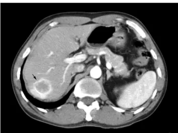
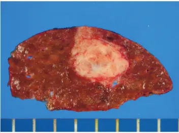
(2) AR Kim et al. : Imprint Cytology of Hepatic Angiomyolipoma. Naked nuclei were noted (Fig. 2B), and intranuclear cytoplasmic pseudoinclusions were occasionally found. Other fields revealed epithelioid tumor cells that had central, round or vesicular nuclei with fine chromatin and small nucleoli. These cells were arranged in solid nests or a trabecular pattern (Fig. 2C). There was no obvious cytologic atypia. Endothelial cells were occasionally found at the edges of the nests or trabeculae. Any mature fat cells were not observed. Immunohistochemical staining was performed on the smears. The tumor cells were positive for smooth musFig. 1. Computed tomography of the liver. Computed tomography shows a well demarcated tumor with peripheral contrast enhancement (arrow).. B twenty years ago. The radioimmunoassay for hepati-. cle actin and HMB-45, and they were negative for hepatocyte common antigen and alpha-fetoprotein (Fig. 2D). Gross and Histologic Findings. tis B virus was positive for both hepatitis B surface antigen and hepatitis B e antigen in the serum. The serum. The segmentectomy specimen measured 9.0×7.0×. level of alpha-fetoprotein was 3.61 ㎍/mL. The comput-. 4.0 cm in size. The tumor measured 4.0×3.0×3.0 cm. ed tomographic scan revealed a well defined mass in. in size (Fig. 3). The cut surface was yellow gray with a. the eighth segment of the liver (Fig. 1). The mass. well-defined margin. Histologic examination showed. showed peripheral enhancement. The radiologic diag-. spindle and epithelioid tumor cells arranged in sheets. nosis was made as "suggestive of inflammatory pseudo-. or a trabecular pattern(Fig. 4A, B). The spindle tumor. tumor". Segmentectomy with a mass resection was per-. cells had eosinophilic, fibrillary cytoplasm. The epithe-. formed. The excised specimen was sent for intraopera-. lioid cells had round vesicular nuclei with pale or clear. tive consultation. The smears imprinted from the hepat-. cytoplasm. Thick-walled blood vessels were noted (Fig.. ic mass were obtained and frozen sectioning then fol-. 4C), but any mature fat cells were not found, and tumor. lowed.. necrosis was not present. Immunohistochemically, both the spindle and epithelioid tumor cells were positive for. Cytologic Findings. smooth muscle actin, HMB-45 and vimentin, and they were negative for hepatocyte common antigen, CD34,. The smears prepared from the touch imprints were. S-100 protein and cytokeratin (AE1/AE3) (Fig. 4B). The. fixed in 95% ethanol and they were stained with hema-. non-neoplastic liver showed chronic hepatitis with sep-. toxylin-eosin and Papanicolaou stains. The smears. tal fibrosis. The patient has survived sixteen months. were highly cellular. Red blood cells were seen in the. after surgery, and there is no evidence of recurrence. background. Spindle-shaped tumor cells were arranged. and metastasis.. in clusters, irregular bundles or single cells (Fig. 2A). The nuclei were elongated or cigar-shaped, with finely granular chromatin and small nucleoli. There was a mild nuclear pleomorphism. The cytoplasm was granular or fibrillary. The cytoplasmic border was indistinct.. The Korean Journal of Cytopathology Vol 19 No 2 / 2008 189.
(3) 김애리 외 : 간의 혈관근육지방종의 세포소견. A. B. C. D. Fig. 2. Cytologic findings. (A) Spindle tumor cells have elongated, cigar-shaped nuclei with eosinophilic fibrillary cytoplasm and indistinct cell border (H&E). (B) Spindle tumor cells show naked nuclei with fine chromatin and small nucleoli (H&E). (C) Epithelioid tumor cells are arranged in solid nests (Papanicolaou stain). (D) Tumor cells are positive for smooth muscle actin (Immuohistochemical stain).. DISCUSSION Angiomyolipoma is a rare benign mesenchymal tumor that's found in the liver. It occurs equally in males and females; the age range of these patients is from 10 to 72 years with a mean age of 50 years.8 Angiomyolipomas can vary considerably in size from less than a centimeter to 36 cm. Most of the tumors are solitary, but a case of multiple lesions has been reported.7 Grossly, it usually presents as a well circumscribed tumor, but it is not encapsulated. The color and consisFig. 3. Gross finding. Tumor shows a well circumscribed graywhite solid cut surface.. 190 대한세포병리학회지 제19권 제2호 / 2008. tency depend on the different proportions of fat and smooth muscle. Hepatic angiomyolipoma is histologi-.
(4) AR Kim et al. : Imprint Cytology of Hepatic Angiomyolipoma. A. B. C. D. Fig. 4. Histologic findings. (A) Spindle tumor cells have elongated, cigar-shaped nuclei with eosinophilic cytoplasm (H&E). (B) Epithelioid tumor cells have round vesicular nuclei with pale or clear cytoplasm with trabecular arrangement (H&E). (C) Thickwalled blood vessels are present (H&E). (D) Epithelioid tumor cells show cytoplasmic positivity for HMB-45 (Immunohistochemical stain).. cally subclassified into the mixed, lipomatous, myomatous and angiomatous. types.9. Much of the adipose tissue generally dissolves during. The tumor of the present. fixation and staining. The presence of nuclear. reported case was composed of a predominantly. pseudoinclusions should not be regarded as a specific. smooth muscle component and it is considered to be. criterion for the diagnosis of angiomyolipoma, but they. the myomatous type.. may be considered as an additional nonspecific cyto-. The cytological features of hepatic angiomyolipoma. logical feature in hepatic angiomyolipoma. 4. appear to be similar to the histologic features.. Intranuclear pseudoinclusions were occasionally found. Extramedullary hematopoiesis elements may be pres-. in the present case.. ent. Mature adipocytes and thick-walled blood vessels. Angiomyolipoma simultaneously expresses smooth. angiomyolipoma.5. muscle and melanocytic markers. These findings sug-. Diagnostic difficulty may arise when the fatty compo-. gest that this tumor has a capacity for muscular and. can be rarely observed in epithelioid. In the. melanocytic differentiation. In the present case, the. present case, any mature fat cells were not identified.. tumor cells were positive for smooth muscle actin and. nent is scant or focal, and it is not. sampled.10. The Korean Journal of Cytopathology Vol 19 No 2 / 2008 191.
(5) 김애리 외 : 간의 혈관근육지방종의 세포소견. HMB-45. Although mature fat cells were not found in. All tumors that show spindle cells, epithelioid cells or. the present case, the cytologic features and the positivi-. mature fat cells should be immunostained for smooth. ty for smooth muscle actin and HMB-45 support the. muscle markers and melanocytic markers to reach a. diagnosis of angiomyolipoma. On cytogenetic study,. correct diagnosis. The biological behavior of the angiomyolipoma is. neither LOH nor MSI appears to play an important role in the pathogenesis of hepatic. angiomyolipoma.11. Hepatic angiomyolipoma should be differentiated. benign, and surgical excision is curative,14 yet malignant hepatic angiomyolipoma has been documented.15-. from smooth muscle tumor, vascular tumor, hepatocel-. 17. lular carcinoma, malignant melanoma and metastatic. surgery, with no evidence of recurrence and metastasis.. carcinoma. Hepatic angiomyolipomas that are predomi-. In conclusion, hepatic angiomyolipoma shows the. nantly composed of spindle cells with cigar-shaped. morphologic spectrum ranging from spindle cells to. nuclei may be mistaken for smooth muscle tumors.. epithelioid tumor cells. Hepatic angiomyolipoma can. Hepatic leiomyosarcomas show hyperchromatic, elon-. be diagnosed by cytologic examination if pathologists. gated, blunt-ended nuclei, and they often show coarse. are aware of its variable cytomorphologic features.. nuclear chromatin and occasionally a perinuclear halo.. Immunohistochemical stains for smooth muscle mark-. Immunohistochemical staining can be helpful for con-. ers and melanocytic markers are useful for making the. firming the diagnosis because smooth muscle tumors. correct diagnosis of this hepatic mass.. In our case, the patient has survived 16 months after. are negative for the melanocytic markers such as HMB45 and melan-A.12 The cytologic features of hemangioma demonstrate the presence of blood in the aspi-. REFERENCES. rate, bland endothelial cells in clusters of various sizes, and longitudinal nuclear grooves in the endothelial cells.13 Tumors with a large quantity of fat may be misdiagnosed as lipomatous or focal fatty change of hepatocytes. True hepatic lipoma is extremely rare. Epithelioid tumor cells with a trabecular pattern and nuclear pleomorphism may resemble hepatocellular carcinoma. Hepatocellular carcinomas show hyperchromatic atypical nuclei, a high nuclear cytoplasmic ratio and prominent nucleoli, and they sometimes produce bile. In order to avoid overdiagnosing angiomyolipoma as hepatocellular carcinoma, a careful search must be done to look for such components as blood vessels, smooth muscles and adipocytes.5 Some angiomyolipomas may resemble malignant melanoma. Immunohistochemically, malignant melanoma is negative for smooth muscle actin. The presence of solid epithelioid areas in angiomyolipoma can mimic metastatic carcinoma and this can create a diagnostic pitfall. The metastatic carcinoma is positive for cytokeratin and it is negative for smooth muscle markers and melanocytic markers.. 192 대한세포병리학회지 제19권 제2호 / 2008. 1. Nguyen GK, Catzavelos C. Solitary angiomyolipoma of the liver. Report of a case initially examined by fine needle aspiration biopsy. Acta Cytol 1990;34:201-4. 2. Ma TK, Tse MK, Tsui WM, Yuen KT. Fine needle aspiration diagnosis of angiomyolipoma of the liver using a cell block with immunohistochemical study. A case report. Acta Cytol 1994;38:257-60. 3. Blasco A, Vargas J, de Agustín P, López-Carreira M. Solitary angiomyolipoma of the liver. Report of a case with diagnosis by fine needle aspiration biopsy. Acta Cytol 1995;39:813-6. 4. Villari D, Grosso M, Vitarelli E, Tuccari G, Barresi G. Nuclear pseudoinclusions in fine-needle aspiration cytology of hepatic angiomyolipoma: case report. Diagn Cytopathol 2000;22:390-3. 5. Khalbuss WE, Fischer G, Bazooband A. Imprint cytology of epithelioid hepatic angiomyolipoma: mimicry of hepatocellular carcinoma. Acta Cytol 2007;51:670-2. 6. Lin KJ, Eng HL, Lu SN, Chiu KW, Kuo FY. Hepatic angiomyolipoma: report of two cases with emphasis on smear cytomorphology and the use of cell block with immunohistochemical stains. Diagn Cytopathol 2004;31:2636. 7. Yim HL, Park KW, Lee KB. Multiple angiomyolipoma of the liver: report of a case with diagnosis by fine needle aspiration cytology. Korean J Cytopathol 1998;9: 79-84..
(6) AR Kim et al. : Imprint Cytology of Hepatic Angiomyolipoma. 8. Ishak KG, Anthony PP, Niederau C, Nakanuma Y. In : Hamilton SR, Aaltonen LA. World Health Organization classification of tumours: pathology and genetics of digestive system. Lyon: IARCPress, 2000;193. 9. Tsui WM, Colombari R, Portmann BC, et al. Hepatic angiomyolipoma: a clinicopathologic study of 30 cases and delineation of unusual morphologic variants. Am J Surg Pathol 1999;23:34-48. 10. Wang SN, Tsai KB, Lee KT. Hepatic angiomyolipoma with trace amounts of fat: a case report and literature review. J Clin Pathol 2006;59:1196-9. 11. Xu AM, Zhang SH, Zheng JM, Zheng WQ, Wu MC. Pathological and molecular analysis of sporadic hepatic angiomyolipoma. Hum Pathol 2006;37:735-41. 12. Atkinson BF. Atlas of diagnostic cytopathology. 2nd ed. Philadelphia: Saunders, 2004; 554-5.. 13. Guy CD, Yuan S, Ballo MS. Spindle-cell lesions of the liver: diagnosis by fine-needle aspiration biopsy. Diagn Cytopathol 2001;25:94-100. 14. Ishak KG, Goodman ZD, Stocker JT. Atlas of tumor pathology. Tumors of the liver and intrahepatic bile ducts. 3rd series Fascicle 31. Washington, D.C.: Armed Forces Institute of Pathology, 2001;99-108. 15. Dalle I, Sciot R, de Vos R, et al. Malignant angiomyolipoma of the liver: a hitherto unreported variant. Histopathology 2000;36:443-50. 16. Parfitt JR, Bella AJ, Izawa JI, Wehrli BM. Malignant neoplasm of perivascular epithelioid cells of the liver. Arch Pathol Lab Med 2006;130:1219-22. 17. Nguyen TT, Gorman B, Shields D, Goodman Z. Malignant hepatic angiomyolipoma: report of a case and review of literature. Am J Surg Pathol 2008;32:793-8.. The Korean Journal of Cytopathology Vol 19 No 2 / 2008 193.
(7)
수치

관련 문서
Injury is a burden on athletes, but the rehabilitation exercise of this study suggests that the improvement of knee muscle function and knee function
The G-kSP placement algorithm proposed here aims to work around the complexity of the formulated ILP. Here, we assume that a network supports a constant number of network
_____ culture appears to be attractive (도시의) to the
이하선의 실질 속에서 하악경의 후내측에서 나와 하악지의 내측면을 따라 앞으로 간다. (귀밑샘 부위에서 갈라져 나와
The 2019 report of The Lancet Countdown on health and climate change: ensuring that the health of a child born today is not defined by a changing
It considers the energy use of the different components that are involved in the distribution and viewing of video content: data centres and content delivery networks
After first field tests, we expect electric passenger drones or eVTOL aircraft (short for electric vertical take-off and landing) to start providing commercial mobility
1 John Owen, Justification by Faith Alone, in The Works of John Owen, ed. John Bolt, trans. Scott Clark, "Do This and Live: Christ's Active Obedience as the
