논문접수일: 2011. 3. 15, 심사완료일: 2011. 6. 20, 게재승인일: 2011. 6. 22 교신저자: 김기석
주소: 제주특별자치도 제주시 대학로 102 Tel: 064) 717-1481, Fax: 064) 757-8276 E-mail: [email protected]
본 연구는 2007년도 대한고혈압학회 연구비 지원에 의하여 수행되었음.
This study was supported by the Research Fund of Korean Society of Hypertension (2007).
고혈압-고지혈증 백서 모델에서 Lectin Like Oxidized Low Density Lipoprotein Receptor-1의 발현
김 기 석1, 김 송 이1, 박 노 관2, 김 동 운2, 주 승 재1, 조 명 찬2
1제주대학교 의학전문대학원 내과학교실, 2충북대학교 의과대학 내과학교실
Expression of Lectin like Oxidized Low Density Lipoprotein Receptor-1 in the Spontaneous Hypertensive Rat with High Cholesterol Diet
Ki-Seok Kim, MD1, No Kwan Park, PhD2, Song-Yi Kim, MD1, Dong-Woon Kim, MD2, Seung-Jae Joo, MD1, Myeong-Chan Cho, MD2
1Department of Internal Medicine, Jeju National University School of Medicine, Jeju;
2Department of Internal Medicine, Chungbuk National University School of Medicine, Cheongju, Korea
❙ABSTRACT❙
Background: Lectin-like, oxidized, low-density lipoprotein receptorreceptors (LOX-1) recognizes recognize vascular oxidized low-density lipoprotein (LDL), which may play an important role in the pathogenesis of atherosclerosis. We investigated the ex- pressions expression of LOX-1 and redox-regulating thyoredoxinthioredoxin systems in a hypertension and hypercholesterolemia rat model. Methods: Spontaneously hypertensive rats (SHR) and Wistar-Kyoto rat (WKY) rats were fed with a normal cholesterol diet (NC) and a high cholesterol diet (HC) for 4 weeks. Plasma LDL cholesterol levels and blood pressure were measured at 1 and 4 weeks. Histological changes of atherosclerosis in the vessel was evaluated by hematoxylin and eosin staining and immunocytochemistry. The expressions expression of LOX-1 and thyoredoixnthioredoxin were measured by Western western blot analysis. Results: In the SHR groupsgroup, blood pressure after 4 weeks was significantly higher than initial levels. LDL-cholesterol levels in the SHR-HC group were increased at 4 weeks (15.3 ± 2.6 mg/dL vs. 20.2 ± 2.6 mg/dL, p < 0.01) compared with the SHR-NC group. In oxyblot analysis, the degree of oxidative stress of in the SHR-HC group was significantly higher than in the SHR-NC group (p < 0.05). The expressions expression of LOX-1 and Trx were was significantly increased in the SHR-HC group compared with the SHR-NC group (p < 0.05) on western blot analysis. Focal overexpressions overexpression of LOX-1 were was observed at the intima layer of the thoracic aorta, and was which wereonly observed in the SHR-HC group. Conclusions: The expressions expression of LOX-1 and oxidative stress were was significantly increased in the “hypertension with hypercholesterol” rat model. These findings suggested suggest that LOX-1 and redox systems may play a certain role in development and progression of atherosclerosis.
(J Korean Soc Hypertens 2011;17(2):57-64) Key words: Atherosclerosis; Hypertension; Oxidative stress
서 론
고혈압과 고지혈증은 심혈관 질환의 주요한 위험인자 중 하나이며, 이들 질환과 연관된 표적 장기의 손상은 심
혈관 질환의 발생과 진행에 있어 중요한 역할을 한다. 표 적장기 손상을 일으키는 주된 원인으로는 고혈압, 고지혈 증, 당뇨 등이 알려져 있으며, 최근에는 산화스트레스 (oxidative stress)의 불균형이 중요한 역할을 하는 것으로 알려지고 있다.1)
산화저밀도지단백질(oxidized-low density lipoprotein, ox-LDL)은 동맥경화 및 심혈관 질환의 발생에 중요한 역 할을 하는 물질로 알려져 있다.2) Lectin like–oxidizes LDL receptor 1 (LOX-1)은 산화저밀도 지단백질 수용체로서 그 간 알려진 SR-AI/II, CD36, SERC 등의 ox-LDL 수용 체와 전혀 다른 구조를 가지는 50 kDa의 type II mem- brane protein으로서 알려지지 않은 protease에 의해 분해 되어 30 kDa의 soluble form으로 혈중에 존재하여 생물학 적 작용에 관여하는 것으로 알려져 있다.3,4) 또한, LOX-1 의 발현은 세포 내의 산환-환원상태와 밀접하게 연관되어 있으며, 과다한 산화스트레스에 의하여 발현이 증가하는 것으로 알려지고 있다.2) 하지만, 동물모델을 이용하여 산 화스트레스와 LOX-1의 연관성에 관한 연구는 아직 국내 에서 미미한 상태이다.
본 연구에서는, 고혈압-고지혈증 동물모델을 이용하여 혈압 및 혈중 저밀도 지단백질의 변화에 따른 혈관조직에 서의 LOX-1과 산화스트레스의 발현을 알아보고, 동맥경 화의 발생과 진행의 과정에서 이들 사이의 연관성을 분석 하고자 하였다.
대상 및 방법
1. 실험동물
고혈압 동물모델로 8주령의 자발성 고혈압쥐(spontane- ous hypertensive rat, SHR; n=10)와 대조군인 Wistar Kyoto rat (WKY, n=10)을 이용하여 1주간의 환경 적응과 안정 기간을 거친 후 실험을 진행하였다. 대조군과 고혈압군을 각각 정상식이군(normal cholesterol diet, NC)과 고지혈 식이군(high cholesterol diet, HC)으로 나누어 4주간 실험 을 진행하였고, 고지혈식이군은 2% cholesterol diet를 시 행하였다. 각 군마다 각각 5 마리의 실험동물을 배정하였다.
좌심실벽의 비후 정도는 각 군의 실험동물을 실험시작 후
1주와 4주 후에 m-mode 초음파를 시행하여 측정하였다.
2. 혈압 및 생화학적 인자의 측정
실험동물의 혈압은 tail-cuff method를 이용하여 측정하 여 측정하였다. 혈압의 측정은 실험동물을 마취하지 않 고, 37℃가 유지되는 chamber 내 설치된 혈압 측정 장치 를 이용하며 실험동물이 충분히 환경에 적응하도록 한 후 5회의 혈압을 측정하고 평균값을 취하여 실험시작 후 1 주, 4주에 각각 혈압을 측정하였다. 혈액의 생화학적 인 자의 분석은 실험동물의 꼬리정맥을 통하여 혈액을 채취 한 후 혈중 LDL-cholesterol치를 실험시작 후 1주, 4주에 측정하여 비교하였다.
3. Western blot analysis
적출한 장기를 각각 homogenized 한 후 4 mL의 solubi- lizing solution (0.5% NP-40 10 mM, Tris HCl [pH 7.2]
150 mM, NaCl 1 mM, PMSF 0.111 unit/mL)으로 30분간 용해시킨다. 동일한 양의 단백질(100 ug)을 10% SDS-pol- yacrylamide gel에 올려놓고 poly vinylidene difluoride membrane (Millipore, Billerica, MA, USA)을 이용하여 전기 영동과 western blot을 시행하였다. 5% non-fat milk로 blocking 한 후, 1차 항체로 thioredoxin (Lab Frontier, Seoul, Korea), LOX-1 (Santa Cruz Biotechnology Inc., Santa Cruz, CA, USA)을 반응시켰다. PBS containing 0.05% Tween-20 버퍼를 이용하여 세척한 후, 4°C에서 2 차 항체인 peroxidase-linked rabbit antibody (Amersham Pharmacia, Piscataway, NJ, USA)를 반응 시킨 후, ECL Western blot detection kit (Amersham Pharmacia)로 LOX-1과 thioredoxin의 발현을 측정하였다.
4. Oxyblot analysis
동일한 양의 단백질(20 ug)을 1% SDS-polyacrylamide gel에 올려놓고 전기영동을 시행한다. Blocking과 dilution 버퍼(Oxyblot kit, Millipore)를 이용하여 1시간 동안 반응시 킨다. 상온에서 1차 항체와 2차 항체(Oxyblot kit)와 1시간 동안 반응 시킨 후 발현을 측정하였다.
Study WKY SHR
period NC HC NC HC
Body Weight (g) 1 wk 134.2 ± 4.3 136.7 ± 3.8 134.6 ± 4.5 137.3 ± 6.2
4 wk 283.9 ± 7.2 276.9 ± 10.2 257.3 ± 9.8 281.2 ± 7.5
SBP (mm Hg) 1 wk 112.7 ± 13.2 107.1 ± 4.7 172.5 ± 16.1† 173.6 ± 8.1†
4 wk 148.7 ± 8.1 136.6 ± 10.1 191.9 ± 16.6† 195.3 ± 6.2†
Wall thickness (mm) 1 wk 1.2 ± 0.2 1.2 ± 0.1 1.3 ± 0.1 1.3 ± 0.1
4 wk 1.6 ± 0.3 1.7 ± 0.1 2.2 ± 0.2* 2.1 ± 0.1*
LDL (mg/dL) 1 wk 6.8 ± 1.5 7.6 ± 0.6 7.5 ± 0.6 5.4 ± 0.9
4 wk 10.2 ± 2.8 15.3 ± 4.5* 15.3 ± 2.6* 20.2 ± 2.6*,†
n = 5 in each group.
WKY, Wistar Kyoto rat; SHR, spontaneous hypertensive rat; NC, normal cholesterol diet; HC, high cholesterol diet; LDL, low density lipoprotein;
SBP, spontaneous hypertensive rat, WKY-NC, WKY-HC vs. SHR-NC, SHR-HC, p<0.01; Wall thickness, WKY-NC, WKY-HC vs. SHR-NC, SHR-HC, p < 0.05; LDL, WKY-NC vs. WKY-HC, SHR-NC, p < 0.05, WKY-NC, WKY-HC, SHR-NC vs. SHR-HC.
*p < 0.05, †p < 0.01.
Table 1. Baseline characteristics of blood pressure and left ventricle wall thickness 5. 조직학적 검사
실험동물에서 전신마취 후 대동맥을 분리하여 적출하였 다. 분리한 장기는 생리 식염수를 혈관내부를 세척하여 혈 액을 제거하고, 주위의 근육과 신경조직을 제거한 후 즉시 액체질소에 냉동시켰다. 샘플절편의 두께가 4 um 이하가 되도록 냉동절편(frozen section) 제작하여 aceton으로 고 정한 후 영하 70℃로 보관한 후 염색에 사용하였다. 냉동절 편은 hematoxylin and eosin (H&E) 염색을 시행하였다.
6. 면역조직화학검사
면역조직화학검사는 immunoperoxidase 기법을 이용하 였다. 적출한 혈관을 파라핀 section을 시행한 후 10 mmol/L의 citrate buffer (PH 6.0)을 이용하여 10분간 가압 멸균을 시행 한 후 3% H2O2를 첨가하여 10분간 반응 시켰 다. 1차 항체로 LOX-1 (Santa Cruz Biotechnology Inc.)을 사용하여 반응하였다. 2차 항체로 goat-IgG를 30분간 반 응 시킨 후, 0.1% 3, 3-di-aminobenzidin을 가하여 5분간 반응하였다. 그 후 LOX-1의 발현을 형광현미경으로 측정 하였다.
7. 통계적 분석
통계적인 분석은 Prism ver. 5.0 (Graph Pad Software Inc., San Diego, CA, US A) 프로그램을 이용하여 분석하고,
모든 수치는 평균± 표준오차로 표시한다. 비교 가능한 변 수는 연속변수인 경우 비모수 검증법인 Mann-Whitney U test를 시행하여 분석한다. 분산분석은 One way ANOVA 검증으로 시행하였으며 Newman-Keuls 방법으로 사후검 정을 시행하였다. 모든 결과는 p-value < 0.05인 경우를 통 계적으로 유의한 결과로 해석하였다.
결 과
1. 혈압 변화 및 심비후
실험시작 후 1주 후 측정한 대조군(WKY)과 고혈압군 (SHR)에서 각 군 간의 몸무게의 차이는 없었으며, 4주 후 의 몸무게도 각 군에서 유의한 차이는 없었다(Table 1).
수축기 혈압은 1주 후 고혈압군(SHR-NC 172.5 ± 16.1 mm Hg, SHR-HC 173.6 ± 8.1 mm Hg)에서 대조군(WKY-NC 112.7 ± 13.2 mm Hg, WKY-HC 107.1 ± 4.7 mm Hg; p <
0.01)에 비하여 유의하게 증가된 소견을 보였다. 4주 후 혈압측정 결과 역시 고혈압군에서 대조군과 비교하여 유 의하게 증가되어 있었다(p < 0.01) (Table 1). 심초음파에 서 심실벽의 비후는 실험 1주 후에는 각 군에서 차이가 없 었으나, 4주후 고혈압군(SHR-NC 2.2 ± 0.2 mm, SHR-HC 2.1 ± 0.1 mm)에서 대조군(WKY-NC 1.6 ± 0.3 mm, WKY-HC 1.7 ± 0.1 mm; p < 0.05)에 비하여 유의하게 증가된 심비후 의 소견을 보였다(Table 1, Fig. 1).
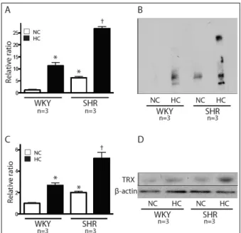
Fig. 1. Change of blood pressure and left ventricular wall thickness.
(A) SBP change at 4 weeks experiment; WKY-NC, WKY-HC vs. SHR-NC, SHR-HC, p < 0.05. (B) Left ventricular wall thick- ness at 4 weeks experiment; WKY-NC, WKY-HC vs. SHR-NC, SHR-HC, p < 0.05. SBP, systolic blood pressure; WKY, Wistar Kyoto rat; SHR, spontaneous hypertensive rat; NC, normal cho- lesterol diet; HC, high cholesterol diet; LDL, low density lipoprotein. n = 5 in each group. *p < 0.05.
Fig. 2. Change of plasma low density lipoprotein levels. Plasma LDL level; WKY-NC vs. WKY-HC, SHR-NC, p < 0.05, WKY-NC, WKY-HC, SHR-NC vs. SHR-HC, p < 0.01, WKY: Wistar Kyoto rat, SHR: spontaneous hypertensive rat, NC: normal cholesterol diet, HC: high cholesterol diet, LDL: low density lipoprotein.
n = 5 in each group. *p < 0.05, †p < 0.01.
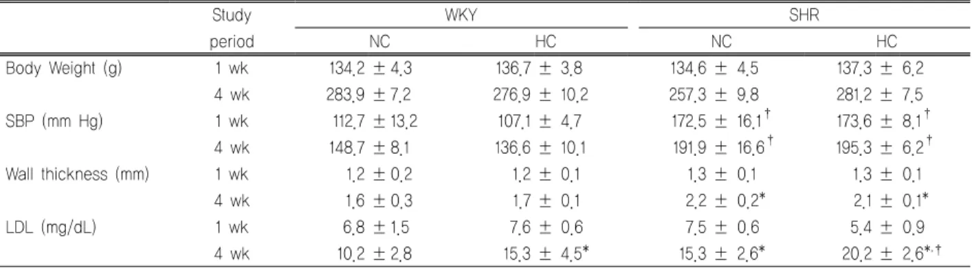
Fig. 3. Expression patterns of oxidative stress in thoracic aorta.
(A) Oxyblot analysis of thoracic aorta, WKY-NC vs. WKY-HC, SHR-NC, p < 0.05, WKY-NC, WKY-HC, SHR-NC vs. SHR-HC, p < 0.01. (B) Representative figure of oxyblot analysis. (C) Expression patterns of TRX in thoracic aorta on western blot analysis, WKY-NC vs. WKY-HC, SHR-NC, p < 0.05, WKY-NC, WKY-HC, SHR-NC vs. SHR-HC, p < 0.01. (D) Representative figure of western blot analysis of TRX. WKY, Wistar Kyoto rat;
SHR, spontaneous hypertensive rat; NC, normal cholesterol diet; HC, high cholesterol diet; TRX, thioredoxin. n = 3 in each group. *p < 0.05, †p < 0.01.
2. 혈중 LDL 농도 변화
실험 시작 1주후에 측정한 혈중 LDL의 농도는 각 군에서 유의한 차이를 보이지 않았다(Table 1). 4주후 측정결과 에서 정상식이를 시행한 대조군에 비하여 고지혈 식이를 시행한 대조군에서 유의하게 높았다(WKY-NC 10.2±2.8 vs. WKY-HC 15.3±4.5, p<0.05). 고지혈 식이의 대조군 (WKY-HC)과 정상식이의 고혈압군(SHR-NC)에서는 혈중 LDL 농도의 유의한 차이가 없었으나, 고지혈식이를 시행 한 고혈압군에서 각 군에 비하여 유의하게 높은 결과를
보였다(WKY-NC 10.2 ± 2.8, WKY-HC 15.3 ± 4.5, SHR-NC 15.3 ± 2.6 vs. SHR-HC 20.2 ± 2.6; p < 0.01) (Table 1, Fig. 2).
3. 산화스트레스의 발현양상
실험 4주후 흉부대동맥을 적출하여 산화스트레스의 발 현양상을 oxyblot 분석을 통하여 측정하였다. 정상식이 대 조군(WKY-NC)에 비하여 고지혈식이 대조군(WKY-HC) 과 정상식이 고혈압군(SHR-NC)에서 산화스트레스의 발 현양상이 유의하게 높았다(p < 0.05) (Fig. 3A, 3B). 또한, 고지혈식이 고혈압군(SHR-HC)에서는 다른 군들과 비교 하여 유의하게 높은 결과를 보였다(p < 0.01) (Fig. 3A, 3B). 산화스트레스의 증가를 반영하는 thirordoxin의 발현 양상 역시 고지혈식이 고혈압군(SHR-HC)에서 다른 군들 에 비해 유의하게 높은 결과를 보였다(p < 0.01) (Fig. 3C, 3D).
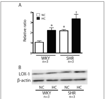
Fig. 4. Expression patterns of lectin-like oxidized low-density lipoprotein receptor (LOX-1) in thoracic aorta. (A) Expression patterns of LOX-1 in thoracic aorta on western blot analysis, WKY-NC vs. WKY-HC, SHR-NC, p < 0.05, WKY-NC, WKY-HC, SHR-NC vs. SHR-HC, p < 0.01. (B) Representative figure of western blot analysis of LOX-1. WKY, Wistar Kyoto rat; SHR, Spontaneous hypertensive rat; NC, normal cholesterol diet;
HC, high cholesterol diet. n=3 in each group. *p< 0.05, †p< 0.01.
Fig. 5. Histological findings of thoracic aorta. (A-D) H&E stain findings of thoracic aorta. (E) Immunohistochemistry finding in SHR-HC group, white arrow tips indicated lectin-like oxidized low-density lipoprotein receptor expressions. WKY, Wistar Kyoto rat; SHR, spontaneous hypertensive rat; NC, normal cholesterol diet; HC, high cholesterol diet.
4. LOX-1의 발현양상
실험 4주 후 적출한 흉부 대동맥을 이용하여 시행한 western blot 분석에서 LOX-1의 발현 양상은 정상식이 대 조군(WKY-NC)에 비하여 나머지 군에서 유의하게 높았 으며(p < 0.05) (Fig. 4), 고지혈식이 고혈압군(SHR-HC)에
서는 고지혈식이 대조군(WKY-HC)와 정상식이 고혈압군 (SHR-NC)에 비하여 유의하게 높은 결과를 보였다(p < 0.01) (Fig. 4).
5. 조직학적 변화
4주 후 흉부 대동맥을 이용하여 시행한 조직학적 검사 에서 각 군에서 유의한 동맥경화의 소견은 관찰되지 않았 다(Fig. 5A). 하지만, 면역형광 염색에서 고지혈식이 고혈 압군(SHR-HC)에서 혈관 내막(intima)에 국한된 LOX-1 의 과잉발현이 관찰되었다(Fig. 5B).
고 찰
본 연구는 고혈압과 고지혈증 백서모델에서 고혈압과 고지혈증이 혈관에서 산화스트레스와 LOX-1의 발현을 증가시키는 것을 확인하였지만, 동맥경화반의 형성을 확 인하지는 못하였다.
동맥경화의 발생과 진행에는 다양한 인자가 관련되어 있지만, 고혈압과 고지혈증은 주요 위험인자 중 하나이다.
고혈압과 고지혈증은 각각의 인자 자체로 체내에서 과도 한 산화스트레스(oxidative stress)를 유발하며, 복합적으 로 작용할 때 과다한 활성산소(reactive oxygen species)의 발현이 일어나 산화-환원계의 심각한 불균형을 초래하게 된다.5) 혈관에서의 LOX-1의 과잉발현은 산화스트레스의 증가와 연관되어 있다고 알려지고 있으며, 동맥경화의 발 생과 진행에 관여하는 것으로 알려져 있다.6) LOX-1은 ox-LDL에 대한 수용체로서의 역할을 할 뿐 아니라, 혈관 긴장도의 변화, 염증세포의 이동 등에 관여하여 혈관의 염증반응과 연관성이 알려져 있으며, 그 자체로 신호전달 물질로의 역할을 하여 각종 활성물질의 분비와 생산과 연 관되어 있으며, 세포고사 과정에서 신호전달 물질의 역할 을 한다고 알려져 있다.7-9) LOX-1의 과잉발현을 유발하는 요인으로는, TNF-α, TGF-β, endothelin-1, angiotensin II 등 의 과잉생산과2) ox-LDL, homocystein, superoxide anion, advanced glycogen end products 등의 산화환원계의 불균 형을 초래하는 물질과 연관성이 알려져 있다.10,11)
LOX-1은 처음에는 단순히 ox-LDL의 수용체로 알려졌
으나, 그 외에 혈관내피세포의 기능 장애를 일으키며, 백 혈구, 단핵구 및 활성화된 혈소판과 반응하여 거품세포 (foam cell) 형성과 연관성이 있는 것으로 알려지고 있 다.6) 혈관평활근세포의 증식과 이동에도 관여하는 것으 로 알려져 있다. LOX-1은 수용체가 활성화된 후 그 자체 가 신호전달체계로의 역할도 수행하여 세포증식과 비후 에 연관된 인자인 ERK1/2, NF-kB 등의 발현을 증가시키 며, 세포고사와 연관된 pro-apoptotic 신호인 Bax의 발현 을 증가시키고, anti-apoptotic 신호계인 Bcl-2의 발현을 감소시켜 세포고사를 일으키는 것으로 알려져 있다.4,7,12) 이런 일련의 과정은 LOX-1의 과잉발현이 심장에서는 심 실재형성(cardiac remodeling)을 일으키고,13) 대동맥에서 는 혈관의 수축과 동맥경화반의 형성에 관여하는 것으로 알려져 있다11). 이러한 결과는 세포실험(in vitro) 및 조직 학적 실험을 통하여 보고되었으며, 생체 내 실험(in vivo) 의 결과는 아직 제한적이다.
본 연구에서는 고지질식이를 통한 고혈압-고지혈증 모 델을 확립하였고, 고혈압에 의한 좌심실의 비대를 확인 할 수 있었다(Table 1). 고지질식이를 시행한 정상 혈압군 (WKY-HC)에서 4주 후 혈중 저밀도지단백의 농도가 유 의하게 증가하였다(Table 1, Fig. 1). 또한, 고지질식이 고 혈압군(SHR-HC)에서 혈중 저밀도지단백의 농도가 가장 현저히 증가하였다(Table 1, Fig. 2). 그리고 흉부 대동맥에 서의 LOX-1의 발현 증가를 western blot 분석을 통하여 확인하였으며 고혈압(SHR-NC) 및 고지혈증군(WKY-HC) 에서 대조군(WKY-NC)에 비하여 발현이 현저하게 증가 되었으며, 고혈압과 고지혈증(SHR-HC)을 동반한 군에서 가장 현저히 증가함을 확인하였다(Fig. 4).
고지혈증 및 고혈압에 의하여 산화스트레스의 증가가 발생하게 되면, 산화-환원계의 체내 방어기전 중 하나인 thirordoxin (TRX) 체계의 활성화가 일어나며 조직에서 TRX의 발현이 증가하게 된다.5) TRX는 심근세포는 물론 혈관 내피세포, 혈관 평활근 세포의 증식, 비후 및 고사 (apoptosis) 등에 관여하며, NF-kB, AKT, ERK 등의 다양 한 신호전달체계의 발현에 관여하는 것으로 알려지고 있 다.14) 이러한 신호전달체계의 활성화는 초기에는 심비후
및 혈관비후의 증상을 보이게 되나, 결국 세포고사와 연 관되어 심부전과 동맥경화의 형태로 발전하여 심혈관 질 환의 진행과 관련이 있다15). TRX의 과잉발현은 고혈압, 심부전, 심근경색증16) 등의 심혈관 질환에서 발생한다고 보고되고 있으며,17) 동맥경화의 발생과 진행에 관하여서 는 아직 연구결과는 아직도 제한적인 상태이다.
일부 연구에서 동맥경화가 진행된 대동맥류 환자의 혈 관조직에서 과잉발현을 보고하였고,18) 혈관 내피세포의 허혈성 손상 모델에서 발현이 증가됨을 보고한 연구결과 가 있다.19)
본 연구에서는 산화스트레스의 과잉발현을 oxyblot 분 석을 통하여 확인하였고, 이는 각각의 위험인자가 증가할 수록 발현양상이 증가함을 확인하였다. 특히, 고혈압과 고지혈증을 동반한 군(SHR-HC)에서 현격히 증가함을 확 인하였다(Fig. 3). TRX의 발현 양상 역시 oxyblot 분석결 과와 일치함을 확인하여 산화스트레스의 증가에 의하여 TRX의 발현의 증가가 일어남을 확인 할 수 있었다(Fig.
3). 하지만, 조직학적 검사에서 동맥경화반의 형성은 모 든 군에서 확인할 수 없었으나, 고혈압과 고지혈증을 동 반한 군에서 혈관내막(intima)에서 국소적으로 LOX-1의 발현 증가를 면역형광염색에서 확인하였다.
본 연구 결과를 요약하면, 고혈압과 고지혈증 동물모델 에서 혈중 저밀도 지단백질의 증가와 혈관조직에서 산화 스트레스 발현이 현저히 증가함을 확인하였고, LOX-1의 발현 또한 현저히 증가함을 확인할 수 있었으나, 동맥경 화반의 형성은 확인하지 못하였다.
본 연구의 제한점으로는 혈관조직의 동맥경화반의 형 성을 확인하기 위하여 충분한 기간 동안 고혈압 및 고지 혈증 모델을 유지하지 못한 점이다. 백서에서 고지혈식이 를 통한 흉부대동맥에서 동맥경화반의 형성은 8-12주 후 에 시작된다는 보고가 있어,20) 본 연구에서 이용한 고혈 압 고지혈증 모델에서도 현미경적 동맥경화반의 확인을 위하여, 고지혈식이의 지속이 필요하였다고 생각된다. 또 한, LOX-1과 TRX와 연관된 하위 신호 전달체계인 ERK 1/2, AKT 및 NF-kB의 발현을 비교하지 못한 점이다. 이 를 위한 추가적인 연구가 추후 필요하겠다.
요 약
연구배경: Lectin-like oxidized LDL receptor (LOX-1) 은 동맥경화의 발생과 진행에서 중요한 역할을 하는 것으 로 알려져 있다. 본 연구에서는 고혈압과 고지혈증 동물 모델에서 혈관 조직에서 LOX-1과 산화-환원계의 조절인 자인 thioredoxin (TRX)의 발현양상을 비교하고자 한다.
방법: 정상식이(normal cholesterol diet, NC)와 고지혈 증식이(high cholesterol diet, HC)를 시행한 정상혈압군 (Wistar Kyoto rat, WKY)과 고혈압군(spontaneously hypertensive rats, SHR)에서 혈압과 심비후의 변화를 비 교하고, 산화스트레스의 발현양상 및 혈중 저밀도지단백 질의 증가양상과 대동맥 조직에서 LOX-1과 TRX의 발현 양상을 비교하였다.
결과: 혈압 증가와 심비후의 발생은 실험 4주후 정상식이 및 고지질식이 고혈압군에서 현저히 증가하였고(p<0.05), 혈중 저밀도 지단백질의 증가는 정상식이 대조군(WKY- NC)을 제외한 모든 군에서 현저히 증가되었다(p<0.05).
산화스트레스의 발현 역시 정상식이 대조군(WKY-NC)을 제외한 모든 군에서 현저히 증가되었으며(p<0.05), 대동맥 조직에서 LOX-1과 TRX의 발현 역시 정상식이 대조군 (WKY-NC)을 제외한 모든 군에서 현저히 증가되었다 (p<0.05). 면역형광검사에서 고지질식이 고혈압군에서 혈 관내막에서 LOX-1의 발현 증가를 확인할 수 있었다.
결론: 고혈압과 고지혈증 동물모델에서 혈중 저밀도 지 단백질의 증가와 혈관조직에서 TRX와 LOX-1의 발현의 증가를 확인할 수 있었으나, 동맥경화반의 형성은 확인하 지 못하였다.
References
1. Ogura S, Kakino A, Sato Y, Fujita Y, Iwamoto S, Otsui K, et al. Lox-1: the multifunctional receptor underlying car- diovascular dysfunction. Circ J. 2009;73:1993-9.
2. Kang BY, Hu C, Prayaga S, Khaidakov M, Sawamura T, Seung KB, et al. LOX-1 dependent overexpression of im- munoglobulins in cardiomyocytes in response to angio- tensin II. Biochem Biophys Res Commun. 2009;379:395-9.
3. Navarra T, Del Turco S, Berti S, Basta G. The lectin-like
oxidized low-density lipoprotein receptor-1 and its soluble form: cardiovascular implications. J Atheroscler Thromb.
2010;17:317-31.
4. Kume N, Kita T. Apoptosis of vascular cells by oxidized LDL: involvement of caspases and LOX-1 and its im- plication in atherosclerotic plaque rupture. Circ Res.
2004;94:269-70.
5. Koharyova M, Kolarova M. Oxidative stress and thio- redoxin system. Gen Physiol Biophys. 2008;27:71-84.
6. Hofnagel O, Luechtenborg B, Stolle K, Lorkowski S, Eschert H, Plenz G, et al. Proinflammatory cytokines regu- late LOX-1 expression in vascular smooth muscle cells.
Arterioscler Thromb Vasc Biol. 2004;24:1789-95.
7. Li DY, Chen HJ, Staples ED, Ozaki K, Annex B, Singh BK, et al. Oxidized low-density lipoprotein receptor LOX-1 and apoptosis in human atherosclerotic lesions. J Cardi- ovasc Pharmacol Ther. 2002;7:147-53.
8. Nowicki M, Muller K, Serke H, Kosacka J, Vilser C, Ricken A, et al. Oxidized low-density lipoprotein (oxLDL)-induced cell death in dorsal root ganglion cell cultures depends not on the lectin-like oxLDL receptor-1 but on the toll-like receptor-4. J Neurosci Res. 2010;88:
403-12.
9. Tanigawa H, Miura S, Zhang B, Uehara Y, Matsuo Y, Fujino M, et al. Low-density lipoprotein oxidized to various degrees activates ERK1/2 through Lox-1. Atherosclerosis.
2006;188:245-50.
10. Kanata S, Akagi M, Nishimura S, Hayakawa S, Yoshida K, Sawamura T, et al. Oxidized LDL binding to LOX-1 upre- gulates VEGF expression in cultured bovine chondrocytes through activation of PPAR-gamma. Biochem Biophys Res Commun. 2006;348:1003-10.
11. Fujita Y, Kakino A, Nishimichi N, Yamaguchi S, Sato Y, Machida S, et al. Oxidized LDL receptor LOX-1 binds to C-reactive protein and mediates its vascular effects. Clin Chem. 2009;55:285-94.
12. Li D, Mehta JL. Intracellular signaling of LOX-1 in endo- thelial cell apoptosis. Circ Res. 2009;104:566-8.
13. Takaya T, Wada H, Morimoto T, Sunagawa Y, Kawamura T, Takanabe-Mori R, et al. Left ventricular expression of lectin-like oxidized low-density lipoprotein receptor-1 in failing rat hearts. Circ J. 2010;74:723-9.
14. Chen J, Hui ST, Couto FM, Mungrue IN, Davis DB, Attie AD, et al. Thioredoxin-interacting protein deficiency in-
duces Akt/Bcl-xL signaling and pancreatic beta-cell mass and protects against diabetes. FASEB J. 2008;22:3581-94.
15. Adluri RS, Thirunavukkarasu M, Zhan L, Akita Y, Samuel SM, Otani H, et al. Thioredoxin 1 enhances neo- vascularization and reduces ventricular remodeling during chronic myocardial infarction: a study using thioredoxin 1 transgenic mice. J Mol Cell Cardiol. 2011;50:239-47.
16. Soejima H, Suefuji H, Miyamoto S, Kajiwaram I, Kojima S, Hokamaki J, et al. Increased plasma thioredoxin in pa- tients with acute myocardial infarction. Clin Cardiol. 2003;
26:583-7.
17. Malik G, Gorbounov N, Das S, Gurusamy N, Otani H, Maulik N, et al. Ischemic preconditioning triggers nuclear translocation of thioredoxin and its interaction with Ref-1 potentiating a survival signal through the PI-3-kinase-Akt
pathway. Antioxid Redox Signal. 2006;8:2101-9.
18. Martinez-Pinna R, Lindholt JS, Blanco-Colio LM, Dejouvencel T, Madrigal-Matute J, Ramos-Mozo P, et al.
Increased levels of thioredoxin in patients with abdominal aortic aneurysms (AAAs). A potential link of oxidative stress with AAA evolution. Atherosclerosis. 2010;212:333-8.
19. Park KJ, Kim YJ, Choi EJ, Park NK, Kim GH, Kim SM, et al. Expression pattern of the thioredoxin system in human endothelial progenitor cells and endothelial cells under hy- poxic injury. Korean Circ J. 2010;40:651-8.
20. Chinellato A, Ragazzi E, Petrelli L, Paro M, Mironov A, Aliev G. Effect of cholesterol-supplemented diet in herit- able hyperlipidemic Yoshida rats: functional and morpho- logical characterization of thoracic aorta. Atherosclerosis.
1994;106:51-63.