333
서 론
지방육종은 성인의 연부조직 기원의 악성종양 중에서 약 20%를 점유하는 가장 높은 발생률을 보이는 종양이며,(1) 소아나 청소년에서도 간혹 발생된다.(2,3) 호발 부위는 하지 와 견갑부 그리고 후복막강, 신장 주위, 장간막 부위 등의 복막강이다.(4) 종괴는 육안적으로 주위의 정상 조직과 비 교적 경계가 좋으나 피막에 둘러싸여 있지는 않다. 병리조 직학적으로는 분화성, 점액양, 원형세포형, 다형태성의 4가 지 전통적인 유형과 여기에 재분화성 유형을 더하는 다섯 가지 유형으로 분류하고 있으며,(5,6) 이들 유형이 서로 혼 합되어 있는 경우도 드물지 않게 관찰된다. 병리조직학적 유형은 종괴의 크기와 함께 이 종양의 예후를 결정하는 가 장 중요한 인자다.(7) 종양의 생물학적 특성상 분화성과 점 액양 지방육종은 전이보다는 국소재발이 많으며, 원형세포 와 다형태성 지방육종은 때로 광범위한 전이를 초래한 다.(8) 따라서 5년 생존율은 분화성 점액양 지방육종에서는 약 70%에 이르지만, 원형세포와 다형태성 지방육종에서는 20% 미만을 보인다. 최근의 분자생물학적 연구에 따르면 지방육종은 분화성, 점액양 원형세포성, 다형태성의 3가지 병리학적 유형으로 분류되고 있다.(9)
악성종양은 생물학적으로 세포의 악성변형, 변형된 세포 의 증식, 국소침윤과 전이의 4단계를 거친다. p53 유전자는 유전체의 방어자로서 유전자 변이를 일으킨 세포를 복구하 거나 제거함으로써 세포의 악성변형을 억제한다.(10) 이 유 전자는 돌연변이에 의하여 종양 발생 억제 능력을 상실하 게 되며, 이러한 현상은 상피세포 기원의 암종뿐만 아니라 간엽세포 기원의 육종에서도 흔히 관찰된다.(11) 돌연변이 를 일으킨 p53 유전자 산물에 의해 생성된 p53 단백질은 정 상 p53 유전자에 의해 생성된 단백질에 비하여 반감기가 길 기 때문에 면역조직화학적 염색을 통하여 종양세포에서 관
복막강 지방육종에서 Ki-67과 p53 단백질 발현의 의의
전남대학교 의과대학 외과학교실 오병렬․성진식․정상영․최수진나
The Expression Ki-67 and p53 Protein in Intra- abdominal Liposarcomas
Byung Ryul Oh, M.D., Jin Sik Sung, M.D., Sang Young Chung, M.D. and Soo Jin Na Choi, M.D.
Purpose: The formation of a liposarcoma is Known to be associated with a mutation of the p53 and MDM2 genes, and the histopathological subtypes of a liposarcoma are related to the prognosis of the patient. This study was per- formd to examine the relationship between the histopatho- logical subtypes, the type of p53 mutation, and the pro- liferative rate.
Methods: Immunohistochemistry was used to measure the p53 protein and Ki-67 (Mib-1 labeling index) expression levels in 24 liposarcomas cases in which the liposarcoma developed primarily in the abdominal cavity.
Results: p53 expression was observed in 11.1% of the well- differentiated liposarcoma cases, 27.3% of the myxoid and round cell liposarcoma cases, and 50% of the pleomorphic liposarcoma cases. There were significant differences between the Ki-67 expression level according to the histopathological subtypes. There were significant differences between p53 positive or negative group and the Ki-67 expression level, and there was a quantitative correlation between them.
Conclusion: The p53 protein was expressed in 25% of all liposarcomas, particularly in pleomorphic liposarcomas be- cause it was expressed more frequently than in the other liposarcoma subtypes (in 2 cases out of 4 cases). The survival rate was much higher in the mucinous round cell liposarcomas which had high p53 and Ki-67 expression levels. The p53 expression level might be a prognostic pre- dictor of a liposarcoma. (J Korean Surg Soc 2004;66:
333-337)
책임저자:최수진나, 광주광역시 동구 학1동 8번지 ꂕ 501-757, 전남대학교병원 외과 Tel: 062-220-6456, Fax: 062-227-1635 E-mail: [email protected]
접수일:2003년 10월 31일, 게재승인일:2004년 1월 6일
Key Word: Liposarcoma 중심 단어: 복막강 지방육종 ꠏ
Department of Surgery, Chonnam National University Medi- cal School, Gwangju, Korea
ꠏꠏꠏꠏꠏꠏꠏꠏꠏꠏꠏꠏꠏꠏꠏꠏꠏꠏꠏꠏꠏꠏꠏꠏꠏꠏꠏꠏꠏꠏꠏꠏꠏꠏꠏꠏꠏꠏꠏꠏꠏꠏꠏꠏꠏꠏꠏꠏꠏꠏꠏꠏꠏꠏꠏꠏꠏꠏꠏꠏꠏꠏꠏꠏꠏꠏꠏꠏꠏꠏꠏꠏꠏꠏꠏꠏꠏꠏꠏꠏꠏꠏꠏꠏꠏꠏꠏꠏꠏꠏꠏꠏꠏꠏꠏꠏꠏꠏꠏꠏꠏꠏꠏꠏꠏꠏꠏꠏꠏꠏꠏꠏꠏꠏꠏ 찰할 수 있다. 지방육종의 발생은 p53 유전자와 함께
MDM2 유전자의 변이가 관여한다고 알려져 있다.(12) 악성 변형된 세포의 성숙은 정상세포에 비하여 세포주기가 단축 되기보다는 증식기에 있는 성장세포의 분획에 영향을 받는 다.(13) 일반적으로 높은 증식기 분획을 갖는 종양은 종괴의 성장 속도가 빠르며, 환자의 생존율을 감소시킨다. Mib-1은 각종 악성종양에서 종양세포의 증식기 분획을 관찰하는 데 사용되고 있다.(14) 종양세포의 증식은 Ki-67과 연관되어 있는데, 이 물질은 G1 단계에서 세포분열 단계에 이르는 동 안 축적되어 세포 분열 동안 최고치에 달하다 세포 분열 직후 최소치로 감소하게 된다. 이 핵 단백질 항원은 G1, S, G2 단계와 세포분열기에는 핵에 존재하나 G0 단계의 세포 의 핵에는 존재하지 않는 것으로 알려져 있다. Ki-67 단백질 은 MDM2 항원군에 속하는 것으로 드러났다.
본 연구에서는 전남대학교 병원에서 외과적 종괴적출술 이 시행된 24예의 복막강 지방육종을 대상으로 하여 병리 조직학적 유형에 따라 p53 단백질의 발현과 증식기세포 분 획에 어떤 차이가 있는지를 비교 관찰하였다.
방 법 1) 대상 환자와 병리 조직
1991년 1월부터 2000년 12월까지 전남대학교병원 외과에 서 복막강(후복막, 전복막 포함) 종괴로 수술을 받은 환자 중 병리조직학적 소견상 지방육종으로 확인된 증례들 중에서 파 라핀 포매 조직절편이 보관 상태가 양호한 24예를 대상으로 하였다. 병리조직학적 진단은 병리학적 소견과 분자생물학적 특성으로 분류한 3가지 유형(분화성, 점액양 원형성, 다형태 성)으로 하였다.
2) p53과 Ki-67에 대한 면역조직화학적 염색
10% 중성 완충 포르말린에 고정한 후 제작한 파라핀 포 매괴를 6μm 두께로 박절하여 Probe-On 슬라이드(Fisher Biotech)에 부착시켜 건조시킨 다음 염색에 사용하였다. 염 색은 Probe-On 슬라이드를 맞대어 생기는 capillary gap action의 원리를 이용하여 개발된 Microprobe Immuno/DNA 염색기(Biomeda)를 이용하였다.(15) 파라핀 절편이 부착된 슬라이드는 탈 파라핀과 함수과정을 거쳐 조직항원이 잘 노출될 수 있도록 1x automation buffer (Biomeda)에 3분간 작용시켰다. 1x automation buffer를 제거한 후 p53 유전자 산물에 대한 단세포군 항체(DO-1, Santa Cruz, CA) 또는 Mib-1 (Anti-Ki-67, Oncogene Science, Uniondale, NY)을 각각 1:50과 1:30으로 희석하여 20분간 작용시킨 후 완충액으 로 세척하였다. 이차 항체로 biotin이 부착된 anti-mouse IgG 를 이용하여 10분간 작용시킨 후 완충액으로 세척하고 strept avidin-peroxidase에 10분간 작용시켰다. 3-amino- ethylcarbamazol (AEC)을 사용하여 발색시킨 후 헤마톡실린
으로 대조 염색을 시행하였다. 종양세포의 핵에 국한되어 갈색으로 발색되었을 때 양성으로 판정하였다. 염색의 전 과정에 있어서 온도는 45oC로 하였으며 음성대조군은 일차 항체 대신 항체 희석액을 작용시켜 이용하였다. p53 양성반 응은 종양조직에서 발현부위가 높은 곳을 400배로 관찰하 면서 1,000개 이상의 세포중 양성세포의 백분율을 구하였 으며, 5% 미만이 염색된 경우는 음성으로 판정하였다.
Ki-67 양성률도 이와 같은 방법으로 백분율을 산정하였다.
3) 통계학적 검증
통계학적 검증에는 SPSS 11.0을 이용하였으며. 유의수준 99%로 하였으며, 통계학적 의미는 P<0.05로 하였다. 지방 육종의 병리조직학적 분류에 따른 p53 단백질과 Ki-67의 산 술평균은 비모수적 검정법인 Kruskal-Wallis test를, p53 단백 질 발현의 존재유무에 따른 병리조직학적 유형의 빈도는 Mann-Whitney test를 이용하여 통계학적 검증을 하였다. 또 한 p53 단백질 발현과 Ki-67 발현 간의 관계는 Pearson 상관 관계를 통하여 분석하였다.
결 과
복강 내에 발생한 지방육종 환자의 연령, 성별 분포, 병리 조직학적 진단, p53 단백질 발현 및 KI-67 양성률은 Table 1에 요약하였다. 임상적으로 진단 당시의 연령은 43세부터 74세 사이의 장년과 노년층에 분포하였다. 성별로는 남성 13명, 여성 11명으로서 성별에 따른 발생 빈도의 차이는 없 었다. 발생부위는 후복막강에서 15예(62.5%), 전복막강에서 9예(37.5%)를 보여 후복막강에서 높은 발생빈도를 보였다.
병리조직학적 유형별로는 분화성 지방육종이 9예(37.5%), 점액양 원형성 지방육종이 11예(45.8%), 다형태성 지방육종 이 4예(16.7%)로서 점액양 원형성 지방육종의 발생빈도가 높았다(Fig. 1)(Table 1).
p53 단백질에 대한 면역조직화학적 염색법에서 양성반응 은 총 24예 중 6예(25.0%)에서 관찰되었다. 양성반응을 나 타낸 종양세포의 비율은 8.5%에서 33.7%까지 다양하였다 (Fig. 1). p53 양성반응은 비교적 분화된 종양세포뿐만 아니 라 다핵성 거대세포에서도 관찰되었다. 병리조직학적 유형 별로는 분화성 지방육종에서 9예 중 1예(11.1%), 점액양 원 형성 지방육종에서 11예 중 3예(27.3%), 다형태성 지방육종 에서는 4예 중 2예(50.0%)에서 양성반응을 보였다. 병리조 직학적 유형에 따른 p53 양성률은 통계학적으로(KrUskal- Wallis Test) 차이가 없었다(P= 0.117). 또한 p53 양성 유무에 따른 병리조직학적 유형에도 차이가 없었다(P=0.396).
Ki-67에 대한 항체를 이용하여 측정한 성장세포 분획은 3.8%에서 30.8%까지 분포하였다. 병리조직학적 유형에 따 른 평균 Ki-67 양성세포는 분화성 지방육종에서 6.6%, 점액 양 원형성 지방육종에서 14.7%, 다형태성 지방육종에서
ꠏꠏꠏꠏꠏꠏꠏꠏꠏꠏꠏꠏꠏꠏꠏꠏꠏꠏꠏꠏꠏꠏꠏꠏꠏꠏꠏꠏꠏꠏꠏꠏꠏꠏꠏꠏꠏꠏꠏꠏꠏꠏꠏꠏꠏꠏꠏꠏꠏꠏꠏꠏꠏꠏꠏꠏꠏꠏꠏꠏꠏꠏꠏꠏꠏꠏꠏꠏꠏꠏꠏꠏꠏꠏꠏꠏꠏꠏꠏꠏꠏꠏꠏꠏꠏꠏꠏꠏꠏꠏꠏꠏꠏꠏꠏꠏꠏꠏꠏꠏꠏꠏꠏꠏꠏꠏꠏꠏꠏꠏꠏꠏꠏꠏꠏ
19.5%를 보였다(Fig. 1). 병리조직학적 유형에 따른 Ki-67 양 성률은 통계학적으로 유의한 차이를 보였다(P=0.004).
전체 증례를 대상으로 p53 음성 또는 양성인 그룹과 Ki-67의 평균값에는 유의한 차이가 있었으며(P=0.002), 이 들 상호간에는 양의 상관관계가 있었다(Pearson Correla- tion=0.590).
본 연구에서는 p53 단백질 발현 유무와 Ki-67 발현 사이 에는 양의 상관관계가 있었으며 이는 조직학적 유형과 함 께 지방육종의 예후인자로 사용될 수 있음을 시사하였다.
고 찰
최근에 발달된 분자생물학적 기법은 지방육종에서도 분 자유전학적 특성을 밝혀왔고, 이에 따라서 병리조직학적 분류법도 다소 보완되었다.(9,16) 1994년에 확립된 WHO 분 류법은 Enzinger 등이 제시한 분류법에 기반을 두고 있으며, 분화성, 점액양, 원형세포, 다형태성의 4가지 유형 외에 재 분화성 유형을 추가하여 다섯 가지로 분류하고 있다(Table 2) (5,6). 이러한 분류법은 최근까지 일반적인 병리조직학적
분류법으로 사용되어왔다. 그러나 본 종양들에서 실시한 세포유전학적 연구결과에 의하면 종양세포에서 원형의 표 지염색체(marker chromosome)가 특징적으로 관찰되는 유 형,(17) t (12;16)(q13; p11) 또는 t (12;22)(q13;q11)의 염색체 전위가 관찰되는 유형,(18) 그리고 다양한 염색체 재배열이 관찰되는 유형(19)의 세 가지로 분류할 수 있었다. 이들 병 리조직학적 소견과 연관지으면 각각 분화성, 점액양 원형 성, 다형태성 지방육종에 해당하였다.(9,16) 이에 따라 방추 세포성 지방육종이나 미분화성 세포가 존재하지 않으면서 점액양 기질이 존재하는 지방육종은 분화성 지방육종으로 재분류되었다(Table 3).
본 연구는 복강에서 발생된 지방육종에 대하여 병리조직 학적 유형에 따른 p53 단백질과 Ki-67의 발현을 비교 관찰 하였다. p53 단백질은 전체 지방육종의 25%에서 발현되었 으며, 병리조직학적 유형에 따라 유의한 차이가 없었으나, 다형태성 지방육종에서는 4예 중 2예에서 발현되어 다른 유형에 비하여 높은 빈도로 관찰되었다.
다른 연구자들의 보고에 의하면 지방육종에서 p53 유전 자의 변이는 최고 25%까지의 빈도를 보였으며,(20,21) 이들 Table 1. Expression of p53 protein and growth fraction in intra-abdominal liposarcomas distinguished histopathologic subtypes ꠚꠚꠚꠚꠚꠚꠚꠚꠚꠚꠚꠚꠚꠚꠚꠚꠚꠚꠚꠚꠚꠚꠚꠚꠚꠚꠚꠚꠚꠚꠚꠚꠚꠚꠚꠚꠚꠚꠚꠚꠚꠚꠚꠚꠚꠚꠚꠚꠚꠚꠚꠚꠚꠚꠚꠚꠚꠚꠚꠚꠚꠚꠚꠚꠚꠚꠚꠚꠚꠚꠚꠚꠚꠚꠚꠚꠚꠚꠚꠚꠚꠚꠚꠚꠚꠚꠚꠚꠚꠚꠚꠚꠚꠚꠚꠚꠚꠚꠚꠚꠚꠚꠚꠚꠚꠚꠚꠚꠚꠚꠚꠚꠚꠚꠚ
Number Age Sex Histopathologic subtype Location p53(%) Ki-67(%)
ꠏꠏꠏꠏꠏꠏꠏꠏꠏꠏꠏꠏꠏꠏꠏꠏꠏꠏꠏꠏꠏꠏꠏꠏꠏꠏꠏꠏꠏꠏꠏꠏꠏꠏꠏꠏꠏꠏꠏꠏꠏꠏꠏꠏꠏꠏꠏꠏꠏꠏꠏꠏꠏꠏꠏꠏꠏꠏꠏꠏꠏꠏꠏꠏꠏꠏꠏꠏꠏꠏꠏꠏꠏꠏꠏꠏꠏꠏꠏꠏꠏꠏꠏꠏꠏꠏꠏꠏꠏꠏꠏꠏꠏꠏꠏꠏꠏꠏꠏꠏꠏꠏꠏꠏꠏꠏꠏꠏꠏꠏꠏꠏꠏꠏꠏ
1 72 M WDL Retroperitoneal - 8.3
2 68 M WDL Peritoneal - 6.5
3 63 F WDL Retroperitoneal - 5.7
4 67 M WDL Retroperitoneal - 5.5
5 74 F WDL Peritoneal - 3.9
6 58 F WDL Retroperitoneal - 9.4
7 50 M WDL Retroperitoneal 8.5 11.3
8 57 M WDL Retroperitoneal - 3.8
9 66 M WDL Retroperitoneal - 7.8
10 63 M M&R Retroperitoneal - 7.9
11 70 F M&R Peritoneal 17.4 12.6
12 55 M M&R Retroperitoneal - 8.6
13 64 M M&R Peritoneal - 7.9
14 58 F M&R Retroperitoneal - 17.3
15 60 F M&R Peritoneal - 6.5
16 61 M M&R Retroperitoneal 12.9 18.3
17 46 M M&R Peritoneal - 22.7
18 52 F M&R Peritoneal - 16.6
19 50 F M&R Retroperitoneal - 8.8
20 58 F M&R Peritoneal 8.7 20.3
21 43 M Pleomorphic Peritoneal - 14.7
22 65 F Pleomorphic Retroperitoneal 20.9 19.6
23 61 F Pleomorphic Retroperitoneal - 13.6
24 73 M Pleomorphic Retroperitoneal 33.7 30.8
ꠏꠏꠏꠏꠏꠏꠏꠏꠏꠏꠏꠏꠏꠏꠏꠏꠏꠏꠏꠏꠏꠏꠏꠏꠏꠏꠏꠏꠏꠏꠏꠏꠏꠏꠏꠏꠏꠏꠏꠏꠏꠏꠏꠏꠏꠏꠏꠏꠏꠏꠏꠏꠏꠏꠏꠏꠏꠏꠏꠏꠏꠏꠏꠏꠏꠏꠏꠏꠏꠏꠏꠏꠏꠏꠏꠏꠏꠏꠏꠏꠏꠏꠏꠏꠏꠏꠏꠏꠏꠏꠏꠏꠏꠏꠏꠏꠏꠏꠏꠏꠏꠏꠏꠏꠏꠏꠏꠏꠏꠏꠏꠏꠏꠏꠏ WDL = well differentiated liposarcoma; M&R = myxoid and round cell liposarcoma
ꠏꠏꠏꠏꠏꠏꠏꠏꠏꠏꠏꠏꠏꠏꠏꠏꠏꠏꠏꠏꠏꠏꠏꠏꠏꠏꠏꠏꠏꠏꠏꠏꠏꠏꠏꠏꠏꠏꠏꠏꠏꠏꠏꠏꠏꠏꠏꠏꠏꠏꠏꠏꠏꠏꠏꠏꠏꠏꠏꠏꠏꠏꠏꠏꠏꠏꠏꠏꠏꠏꠏꠏꠏꠏꠏꠏꠏꠏꠏꠏꠏꠏꠏꠏꠏꠏꠏꠏꠏꠏꠏꠏꠏꠏꠏꠏꠏꠏꠏꠏꠏꠏꠏꠏꠏꠏꠏꠏꠏꠏꠏꠏꠏꠏꠏ
의 결과도 병리조직학적 유형과는 서로 상관관계가 없음을 보여주고 있다. p53 유전자 돌연변이는 분화성보다는 다형 태성 지방육종에서 다소 높은 빈도를 보여(22,23) 비록 증 례수가 적지만 본 연구에서 시행한 P53 단백질에 대한 면역 조직화학적 염색 결과와 비슷하였다.
본 연구에서 Ki-67은 지방육종의 병리조직학적 유형에 따른 차이를 보였다. 즉 분화성에서 가장 낮은 증식지수를 보였으며, 다형태성에서 가장 높은 증식지수를 보였다.
Sprogoe- Jakobsen 등은 점액양 평활근육종과 평활근종의 비교에서 Ki- 67은 p53과 함께 평활근 종양의 악성도와 관 련이 있다고 보고(22,23)하고 있으나, 이와는 상반되게
O'Reilley 등은 44예의 자궁외 평활근육종에서 p53이나 Ki-67 양성률이 종양 환자의 생존율에 영향을 미치지 않는 다고 보고하고 있다.(24) Cox regression 모델을 이용한 최초 의 보고(25)에 의하면 p53 돌연변이나 단백질의 발현 및 Ki-67 양성률은 환자의 생존율과 밀접한 관계가 있었다. 이 중에서도 p53 발현과 Ki-67 증식 세포지수가 높은 점액양 원형세포성 지방육종은 생존율이 현저히 저하되었다. 따라 서 지방육종의 예후는 조직학적 유형 외에도 p53 단백질과 Ki-67 발현이 이용될 수 있음을 시사해 주었다.
Fig. 1. Histopathologic subtypes (A, B, C), and expression of p53 protein (D, E, F) and Mib-1 (G, H, I) by immunohistochemistry in liposarcoma. Case 1 revealed well-differentiated liposarcoma (A, H&E, ×200) which showed negative expression of p53 protein (D, ×100) and 8.3% of Ki-67 positive cells (G, ×100). Case 18 disclosed myxoid and round cell liposarcoma (B, H&E, ×200) which had negative expression of p53 (E, ×100) and 16.6% of Ki-67 positive tumor cells (H, ×100). Case 24 revealed pleomorphic liposarcoma (C, H&E, ×200) which showed strong positive for p53 protein (F, ×200) and Ki-67 (I, ×200) in many tumor cells.
Table 2. World health organization (WHO) classification of liposa- rcoma (1994)
ꠚꠚꠚꠚꠚꠚꠚꠚꠚꠚꠚꠚꠚꠚꠚꠚꠚꠚꠚꠚꠚꠚꠚꠚꠚꠚꠚꠚꠚꠚꠚꠚꠚꠚꠚꠚꠚꠚꠚꠚꠚꠚꠚꠚꠚꠚꠚꠚꠚꠚꠚꠚꠚꠚꠚ Well-differentiated liposarcoma
Adipocytic(lipoma-like) Sclerosing
Inflammatory Dedifferentiated liposarcoma Myxoid liposarcoma Round cell liposarcoma Pleomorphic liposarcoma
ꠏꠏꠏꠏꠏꠏꠏꠏꠏꠏꠏꠏꠏꠏꠏꠏꠏꠏꠏꠏꠏꠏꠏꠏꠏꠏꠏꠏꠏꠏꠏꠏꠏꠏꠏꠏꠏꠏꠏꠏꠏꠏꠏꠏꠏꠏꠏꠏꠏꠏꠏꠏꠏꠏꠏ
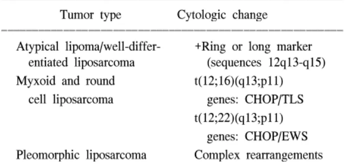
Table 3. Specific (Primary) chromosome changes in liposarcoma ꠚꠚꠚꠚꠚꠚꠚꠚꠚꠚꠚꠚꠚꠚꠚꠚꠚꠚꠚꠚꠚꠚꠚꠚꠚꠚꠚꠚꠚꠚꠚꠚꠚꠚꠚꠚꠚꠚꠚꠚꠚꠚꠚꠚꠚꠚꠚꠚꠚꠚꠚꠚꠚꠚꠚ
Tumor type Cytologic change
ꠏꠏꠏꠏꠏꠏꠏꠏꠏꠏꠏꠏꠏꠏꠏꠏꠏꠏꠏꠏꠏꠏꠏꠏꠏꠏꠏꠏꠏꠏꠏꠏꠏꠏꠏꠏꠏꠏꠏꠏꠏꠏꠏꠏꠏꠏꠏꠏꠏꠏꠏꠏꠏꠏꠏ Atypical lipoma/well-differ- +Ring or long marker entiated liposarcoma (sequences 12q13-q15) Myxoid and round t(12;16)(q13;p11) cell liposarcoma genes: CHOP/TLS
t(12;22)(q13;p11) genes: CHOP/EWS Pleomorphic liposarcoma Complex rearrangements ꠏꠏꠏꠏꠏꠏꠏꠏꠏꠏꠏꠏꠏꠏꠏꠏꠏꠏꠏꠏꠏꠏꠏꠏꠏꠏꠏꠏꠏꠏꠏꠏꠏꠏꠏꠏꠏꠏꠏꠏꠏꠏꠏꠏꠏꠏꠏꠏꠏꠏꠏꠏꠏꠏꠏ E
A B C D
F G H I
ꠏꠏꠏꠏꠏꠏꠏꠏꠏꠏꠏꠏꠏꠏꠏꠏꠏꠏꠏꠏꠏꠏꠏꠏꠏꠏꠏꠏꠏꠏꠏꠏꠏꠏꠏꠏꠏꠏꠏꠏꠏꠏꠏꠏꠏꠏꠏꠏꠏꠏꠏꠏꠏꠏꠏꠏꠏꠏꠏꠏꠏꠏꠏꠏꠏꠏꠏꠏꠏꠏꠏꠏꠏꠏꠏꠏꠏꠏꠏꠏꠏꠏꠏꠏꠏꠏꠏꠏꠏꠏꠏꠏꠏꠏꠏꠏꠏꠏꠏꠏꠏꠏꠏꠏꠏꠏꠏꠏꠏꠏꠏꠏꠏꠏꠏ 결 론
복강에 발생한 지방육종 24예에서 p53 단백질은 전체 증 례의 25%에서 발현되었으며, 병리조직학적 유형에 따라 유 의한 차이가 없었으나, 다형태성 지방육종에서는 4예 중 2 예에서 발현됨으로써 다른 유형에 비하여 높은 빈도로 관 찰되었다.
Ki-67 측정법을 이용한 종양세포의 증식도는 지방육종의 병리조직학적 유형에 따라 유의한 차이가 있었다(다형태 성>점액양 원형성>분화성).
본 연구에서는 p53 단백질 발현 유무에 따른 Ki-67 양성 률 사이에는 유의한 차이가 있었으며, 또한 양의 상관관계 가 있어서 이는 조직학적 유형과 함께 지방육종의 예후인 자로 사용될 수 있음을 시사하였다.
REFERENCES
1) Dei Tos AP. Liposarcoma: New entities and evolving con- cepts. Annals of Diagnostic Pathology 2000;4:4252-66.
2) Dei Tos AP, Pedeutour F. Adipocytic tumors 35-46.
3) Schneider-Stock R, Walter H, Radig K, Rys J, Bosse A, Kuhnen C, et al. MDM2 amplication and loss of hetero- zygosity at Rb and p534 genes: No simultaneous alterations in the oncogenesis of liposarcomas. J Cancer Res Clin Oncol 1998;124:532-40.
4) Reszel PA, Soule EH, Coventry MB. Liposarcoma of extrem- ities and limb girdles. Study of 222 cases. Journal of Bone and Joint Surgery 1996;48:229-44.
5) Enzinger FM, Weiss SW. Soft tissue tumors. Third edition.
Mosby 431-66.
6) Weiss SW. Histologic typing of soft tissue tumors. World health organization histologic classification of tumors berlin.
Germany. Springer. 1994.
7) Reitan JB, Kaalhus O, Brennhovd IO, Sanger EM, Talle K.
Prognostic factors in liposarcoma. Cancer 1985;55:2482-90.
8) Enzinger FM, Winslow DJ. Liposarcoma. A study of 103 cases. Virchows Arch 1962;335:367-88.
9) Paul D. Barrett, Archie J Malcolm. Molecular abnormalities in liposarcoma. Journal of Pathology 1997;181:3-4.
10) Lane DP. Cancer. p53, guardian of the genome. Nature 1992;
358:15-6.
11) Schneider-Stock R, Walter H, Rys J, Radig K, Hoang-Vu C, Roessner A. No correlation of c-myc over expression and p53 mutations in liposarcomas. Virchows Arch 1998;22:315-21.
12) Pilotti S, Della Torre G, Lavarino C, Di Palma S, Sozzi G, Minoletti F, et al. Distinct mdm2/p53 expression patterns in liposarcoma subgroups: Implications for different pathogenic mechanisms. J Pathol 1997;181:14-24.
13) Dei Tos AP, Doglioni C, Piccinin S, Maestro R, Mentzel T, Barbareschi M, et al. Molecular abnormalities of the p53 pathway in differentiated liposarcoma. J Pathol 1997;181:8-13.
14) Schneider-Stock R, Epplen JT, Walter H, Radig K, Rys J, Epplen C, et al. Telomeric lengths and telomerase activity in liposarcomas. Mol Carcinog 1999;24:144-51.
15) Lee MC, Kang MW, Yang BS, Park CS, Juhng SW, Jung S, et al. Proliferating cell nuclear antigen (PCNA) and nuclear organizer regions (NORs) in benign, atypical and malignant meningiomas. Brain Tumor Pathology 1995;12:133-9.
16) Antonescu CR, Tschernyavsky SJ, Decuseara R, Leung DH, Woodruff JM, Brennan MF, et al. Prognostic impact of p53 status, TLS- CHOP fusion transcript structure, and histologic grade in myxoid liposarcoma: A molecular and clinicopatho- logic study of 82 cases. Clin Cancer Res 2001;7: 3977-87.
17) Dal Cin P, Kools P, Sciot R, De Wever I, Van Damme B, Van de Ven W, et al. Cytogenetic and fluorescence in situ hybridization investigation of ring chromosomes charicterizing a specific pathologic subgroup of tissue tumors. Cancer Genet Cytogenet 1993;68:85-90.
18) Knight JC, Renwick PJ, Cin PD, Van den Berghe H, Fletcher CD. Translocation t (12:16)(q13:p11) in myxoid liposarcoma and round cell liposarcoma: Molecular and cytogenic analysis.
Cancer Res 1995;55:24-7.
19) Mentzel T, Bosemberg M, Fletcher CDM. Pleomorphic liposarcoma: Clinicopathologic and prognostic analysis of 31 cases. Modern Pathology 1999;12:13.
20) Florenes VA, Malandsmo GM, Forus A, Andreassen A, Myclebost O, Fodstad O. Mdm2 gene amplification and tran- script levels in human sarcomas: relationship to tp53 gene status. Journal of Natl Cancer Inst 1994;86:1297-302.
21) Leach FS, Tokino T, Meltzner M, Burrell M, Oliner JD, Smith S, Hill De, Sidransky D, Kinzler KW, Vogelstein B. p53 muta- tion and mdm2 amplification in human soft tissue sarcomas.
Cancer Res 1993;53:2231-4.
22) Sprogoe-Jakobsen S, Holund B.. Immunohistochemistry (Ki-67 and p53) as a tool in determining malignancy in smooth mus- cle neoplasms exemplified by a myxoid leiomyosarcoma of the uterus). APMIS 1996;104:705-8.
23) Taubert H, Wurl P, Meye A, Berger D, Thamm B, Neumann K, et al. Molecular and immunohistochemical p53 status in liposarcoma and malignant fibroushistiocytoma: identification of seven new mutations for soft tissue sarcomas. Cancer 1995;76:1187-96.
24) O'Reilley PE, Raab SS, Niemann TH, Rodgers JR, Robinson RA. p53 proliferating cell nuclear antigen and Ki-67 expres- sion in extrauterine leiomyosarcomas. Mod Pathol 1997;10:
91-7.
25) Schneider-Stock R, Ziegeler A, Haeckel C, Franke DS, Rys J, Roessner A. Prognostic relevance of p53 alterations and Mib-1 proliferation index in subgroups of primary liposar- comas. Clin Cancer Res 1999;5:2830-5.