• 검색 결과가 없습니다.

Treadmill Exercise Stress Echocardiography in Patients With No History of Coronary Artery Disease: A Single-Center Experience in Korean Population

N/A
N/A
Protected

Academic year: 2022

Share "Treadmill Exercise Stress Echocardiography in Patients With No History of Coronary Artery Disease: A Single-Center Experience in Korean Population"

Copied!
7
0
0

로드 중.... (전체 텍스트 보기)

전체 글

(1)

528

Open Access

Treadmill Exercise Stress Echocardiography in Patients With No History of Coronary Artery Disease:

A Single-Center Experience in Korean Population

Jeong Yoon Jang, MD, Il Suk Sohn, MD, Jong Nim Kim, RN, Jeong Hwan Park, MD, Chang Bum Park, MD, Eun Sun Jin, MD, Jin Man Cho, MD, Chong Jin Kim, MD, and Jong Hoa Bae, MD

Department of Cardiology, Kyung Hee University Hospital at Gangdong, Seoul, Korea ABSTRACT

Background and Objectives: Treadmill exercise stress echocardiography (TSE) has superior diagnostic accuracy than exer- cise electrocardiography (ECG). The objectives of the study are 1) to define the diagnostic accuracy and safety of TSE in pa- tients without a history of coronary artery disease (CAD), 2) to identify the clinical characteristics that predict positive TSE results and 3) to assess the differential predictive value between TSE and concomitant exercise ECG in a Korean population.

Subjects and Methods: A total of 1,287 patients among 1,500 consecutive patients with no prior history of CAD and who were referred for TSE during a 4-year 3-month period were enrolled. Results: Of the 1,287 patients, 95 (7.4%) showed posi- tive TSE results (newly developed regional wall motion abnormality). Among the 154 patients with coronary angiography, 94 patients (61%) showed significant CAD (30 of 77 patients with negative TSE results and 64 of 77 patients with positive TSE results). The TSE positive population had more cardiovascular risk factors and showed a higher Duke treadmill score and wall motion score index than the TSE negative group. TSE showed relatively good sensitivity (68%), specificity (78%) and posi- tive and negative predictive values (83% and 61%, respectively), and TSE also had higher diagnostic accuracy than concomi- tant exercise ECG (72% vs. 64%, respectively). Conclusion: TSE is safe and offers greater diagnostic power for CAD than ex- ercise ECG in Korean population without a history of CAD. Its prognostic value in this population needs to be confirmed in a larger prospective study. (Korean Circ J 2011;41:528-534)

KEY WORDS: Echocardiography, stress; Electrocardiography; Exercise test; Coronary artery disease.

Received: December 2, 2010

Revision Received: December 21, 2010 Accepted: January 24, 2011

Correspondence: Il Suk Sohn, MD, Department of Cardiology, Kyung Hee University Hospital at Gangdong, 149 Sangil-dong, Gangdong-gu, Seoul 134-727, Korea

Tel: 82-2-440-6108, Fax: 82-2-440-7242 E-mail: [email protected]

• The authors have no financial conflicts of interest.

cc

This is an Open Access article distributed under the terms of the Cre- ative Commons Attribution Non-Commercial License (http://creativecom- mons.org/licenses/by-nc/3.0) which permits unrestricted non-commer- cial use, distribution, and reproduction in any medium, provided the origi- nal work is properly cited.

Introduction

Several diagnostic methods are currently being used in de- ciding clinically to apply coronary angiography (CAG) or for early detection of coronary artery disease (CAD) even in pa-

tients without chest pain. Because the treadmill exercise test can give extra load to the heart and increase the myocardial oxygen consumption, exercise electrocardiography (ECG) is widely used for the initial assessment of patients with chest pain once the diagnosis of acute coronary syndrome has been excluded. Treadmill exercise stress echocardiography (TSE) has superior diagnostic accuracy than exercise ECG, and TSE has been used as a noninvasive method for diagnosis, risk stratification and predicting the prognosis in patients with known or suspected CAD.

1-7)

TSE provides similar diagnostic and prognostic accuracy

as radionuclide stress perfusion imaging, but at a substan-

tially lower cost, and TSE is without environmental impact

and with no biohazards for the patient and the physician.

4)8)

Despite TSE being an ideal physiological stress, TSE is techni-

cally much more demanding than pharma-cologic stress and

TSE is not commonly used in Korea.

(2)

The objectives of the present study are 1) to define the diag- nostic accuracy and safety of TSE for detecting ischemia in patients without a history of CAD, 2) to identify the clinical ch- aracteristics that predict positive TSE results and 3) to assess the differential diagnostic power between TSE and concom- itant exercise ECG in a Korean population.

Subjects and Methods

Study subjects

Consecutive patients who had undergone TSE at our insti- tution from March 2006 to May 2010 were enrolled. Patients were excluded if 1) they had a history of myocardial infarc- tion or ischemic heart disease, had undergone a percutane- ous coronary intervention (PCI) procedure or a coronary ar- tery bypass graft before the test, 2) they had significant valvu- lar heart disease, hypertrophic cardiomyopathy, left ventri- cular dysfunction (an ejection fraction <45%), regional wall motion abnormality on resting echocardiography and 3) they had atrial fibrillation on ECG. We also excluded those pa- tients who could not perform maximal exercise.

Framingham’s risk score (10-year mortality) was calculated for all the patients based on their age, gender, smoking status, the presence of diabetes mellitus, blood pressure, and the cho- lesterol level.

9)

The percentage of risk was then calculated. The clinical variables, including hypertension (defined as systolic blood pressure ≥140 mmHg and diastolic blood pressure ≥90 mmHg or the use of antihypertensive drugs), diabetes or in- sulin requirement or oral hypoglycemic agents, dyslipidemia (defined as fasting plasma total cholesterol level ≥240 mg/

dL, triglyceride level ≥400 mg/dL or the use of lipid-lowering agents) and the smoking status (current smoker, never smoker or former smoker) were assessed from the medical records and by interviewing the patients. Written informed consent was obtained from all subjects, and this study was approved by the Institutional Review Board of our institution.

Treadmill exercise test protocol

The study participants underwent maximal treadmill exer- cise using the Bruce protocol with a 12-lead ECG monitor, and a physician trained in Basic Life Support and Advanced Cardiac Life Support attended the exercise test.

4)

The clinical symptoms and blood pressure were monitored throughout the study. Treadmill exercise was terminated if the target heart rate (85% of the maximal age predicted heart rate) was re- ached, if the patients developed limiting symptoms (signifi- cant chest pain, dyspnea), serious ventricular arrhythmia, se- vere hypertension (systolic >220 mmHg; diastolic >120 mmHg), significant blood pressure depression (<90 mmHg for the sys- tolic blood pressure) or marked ST-segment depression or ele- vation as per the standard recommendations.

2-4)

Before and immediately after exercise, two-dimensional

echocardiography with harmonic imaging was obtained in the left lateral decubitus position from the apical four-, and two-chamber views and from the parasternal long- and short- axis views.

2)4)

The post-exercise echocardiographic images were acquired within 30 to 60 seconds after the termination of peak exercise by an experienced echocardiographer. The images were digitalized, recorded and analyzed by visual in- spection. The early post-exercise images with the best endo- cardial definition were selected for comparison with the rest- ing images.

Interpretation of exercise electrocardiography and echocardiography

Positive exercise ECG was defined as positive in cases of horizontal or downsloping ST-segment depression or eleva- tion ≥1 mm at 80 ms after the J point in two or more contigu- ous leads. Duke treadmill score taking into account the pati- ent’s symptoms, extent of ST-segment shift during exercise and total exercise time was calculated.

10)

The rate-pressure product, which is an indicator of the oxygen requirements of the heart, was calculated as the product of the maximal heart rate and the maximal blood pressure during the treadmill exercise.

11)

The exercise ECG results were confirmed by a cardiologist who was blinded to the echocardiographic results.

The 17-segment model of the left ventricle was used and a 4-point scale was used to assess wall motion after exercise (1, normal; 2, hypokinesis; 3, akinesis; 4, dyskinesis).

12)

The TSE results were interpreted by a cardiologist who was blinded to the ECG results. A positive TSE result was defined as newly developed regional wall motion abnormalities.

1-4)

Coronary angiography and revascularization The attending cardiologist decided to proceed with CAG after considering both the TSE result and the clinical status within 30 days after the test. Standard techniques were used by the physician performing the CAG by utilizing a visual qu- alitative scoring system for image analysis with CAD defined as >50% luminal diameter narrowing. PCI, including balloon angioplasty and stent insertion, was performed based on the decision of the attending cardiologist.

Statistical analysis

Continuous variables are expressed as means±SDs and cate-

gorical variables are expressed as percentages. To compare

between the positive TSE group and negative TSE group, the

continuous variables were analyzed using Student’s t-test and

the categorical variables were analyzed using the Chi-square

test. A p<0.05 was used to reject the null hypothesis. The sta-

tistical analysis was performed using Statistical Package for

the Social Sciences (SPSS) version 13.0 (SPSS Inc., Chicago, IL,

USA). For the calculation of the sensitivity, specificity, the pre-

dictive value and the diagnostic accuracy of the exercise tests,

(3)

the numbers of patients were assessed according to the TSE results or the concomitant exercise ECG and the presence of significant CAD on CAG.

Results

Patient characteristics

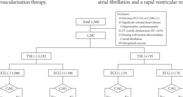
Among the 1,500 participants referred for TSE to our insti- tution during the 4-year 3-month period, 15 patients with a history of previous PCI, 1 patient who underwent coronary bypass surgery before TSE, 43 patients with significant val- vular heart disease with more than a moderate degree of re- gurgitation or stenosis, 5 patients with hypertrophic cardio- myopathy, 25 patients with left ventricular dysfunction (an ejection fraction <45%), 23 patients with wall motion abnor- malities seen on resting echocardiography, 2 patients with atrial fibrillation on the initial ECG and 99 patients with sub- optimal exercise (early stop) (55 with leg discomfort, 44 with dyspnea) were excluded (Fig. 1).

A total of 1,287 participants met the inclusion criteria for this study. Of the 1,287 patients, 95 (7.4%) showed positive TSE results and 154 patients (12%) underwent CAG. Among the 1,192 patients who showed negative TSE results, 77 under- went CAG due to continued chest pain (n=16), positive re- sults for other cardiovascular imaging modalities (45 patients showed positive results on coronary multi-detector computed tomography, 6 patients showed positive results on a nuclear scan) and the patient’s request (n=10). Of the 154 patients with CAG, 94 patients (61%) showed significant CAD (30 of 77 patients with negative TSE results and 64 of 77 patients with positive TSE results) and finally 91 patients (59%) under- went successful revascularization therapy.

Comparison between the patients with positive treadmill exercise stress echocardiography results and negative treadmill exercise stress

echocardiography results

The baseline characteristics of the subjects are described in Table 1. The mean age was 53.2±10.7 years. A total of 523 (41%) of the 1,287 patients were referred for TSE due to typi- cal chest pain. The patients with positive TSE results were more often older in age and were males, and had diabetes and hypertension more often as compared to those with negative TSE results.

The TSE positive group showed a higher Framingham risk score, a lower total cholesterol level and higher blood pres- sure, and a lower high density lipoprotein-cholesterol level than the TSE negative group. There was no significant differ- ence in the incidence of dyslipidemia, current smokers and the reasons for the TSE test.

Treadmill exercise stress echocardiography and exercise electrocardiography findings

The TSE parameters according to the TSE results are present- ed in Table 2. The TSE positive group showed a significantly higher diastolic blood pressure, a higher Duke treadmill score and a higher wall motion score index, but a lower maximal heart rate, exercise time, maximal achieved work load and rate-pressure product than the TSE negative group. The TSE positive group had a higher incidence of exercise limiting chest pain and positive results for concomitant exercise ECG than the TSE negative group. Adverse events, except for chest pain during the test, were reported in 4 patients (0.3%) among the 1,287 patients, and this included 1 patient with paroxysmal atrial fibrillation and a rapid ventricular response, 1 patient

Total 1,500

1,287

Exclusion:

016 Previous PCI (15) or CABG (1) 043 Significant valvular heart disease 005 Hypertrophic cardiomyopathy 025 LV systolic dysfunction (EF <45%) 023 Resting wall motion abnormalities 002 Atrial fibrillation

099 Suboptimal exercise

CAG CAG

Yes No Yes No

54 Stenosis (+) 24 (-) 30

1,032 ECG (-) 1,086

TSE (-) 1,192 TSE (+) 95

ECG (+) 106 ECG (-) 19 ECG (+) 76

23 Stenosis (+) 6 (-) 17

83 13

Stenosis (+) 7 (-) 6

6 64

Stenosis (+) 57 (-) 7

12 CAG

Yes No

CAG

Yes No

Fig. 1. Schematic diagram of the study population according to the exercise test and coronary angiography results. PCI: percutaneous cor-

onary intervention, CABG: coronary artery bypass graft, LV: left ventricular, EF: ejection fraction, ECG: electrocardiography, TSE: treadmill

exercise stress echocardiography, CAG: coronary angiography.

(4)

with paroxysmal supraventricular tachycardia, 1 patients who fainted (near-syncope) due to hypotension and 1 patient with vomiting immediately after peak exercise. All of them recov- ered spontaneously. No fatal ventricular arrhythmias or acute myocardial infarction occurred during the testing.

Comparison between treadmill exercise stress echocardiography and concomitant exercise electrocardiography

We analyzed the patient numbers according to the TSE re- sults or concomitant exercise ECG and the presence of signi- ficant CAD on CAG to compare the sensitivity, specificity, Table 1. Baseline characteristics of the subjects according to the results of treadmill exercise stress echocardiography (TSE)

Variables Total (n=1,287) TSE (-) (n=1,192) TSE (+) (n=95) p

Age (years) 53.2±10.7 52.7±10.7 59.0±8.8 <0.001

Male sex, n (%) 743 (58) 668 (56) 75 (79) <0.001

Body mass index (kg/m

2

) 24.9±3.00 24.9±3.10 25.4±2.9 0.087

Diabetes mellitus, n (%) 159 (12) 133 (11) 26 (27) <0.001

Hypertension, n (%) 473 (37) 424 (36) 49 (52) 0.002

Dyslipidemia, n (%) 144 (11) 133 (11) 11 (17) 0.900

Smoking, n (%) 0.086

Non and former 1,023 (80) 954 (80) 69 (73)

Current 264 (21) 238 (20) 26 (27)

Framingham risk score 8.7±7.1 8.1±6.7 14.7±8.2 <0.001

Reasons for performing the test, n (%) 0.108

Typical chest pain 523 (41) 477 (40) 46 (48)

Atypical chest pain 348 (27) 330 (28) 18 (19)

Others* 416 (32) 385 (32) 31 (33)

Total cholesterol (mg/dL) 192.0±38.20 192.9±38.20 182.2±36.5 0.013

Triglyceride (mg/dL) 146.6±114.1 146.1±115.3 151.4±99.4 0.687

HDL-C (mg/dL) 49.3±11.6 49.7±11.7 044.6±10.2 <0.001

LDL-C (mg/dL) 118.9±31.90 119.1±31.90 117.2±31.7 0.588

LVEF (%) 64.8±4.80 64.9±4.80 64.6±5.1 0.661

Heart rate (beats/min) 67.1±9.70 67.1±9.60 068.0±10.4 0.343

Systolic blood pressure (mmHg) 118.8±16.60 118.1±16.40 127.0±15.9 <0.001

Diastolic blood pressure (mmHg) 71.2±12.0 70. 9±11.8. 074.2±13.0 0.010

The data is expressed as means±SDs or as the numbers (%). *Patients without symptoms, but who were suspicious of having ischemic heart disease on electrocardiography, or coronary calcium and/or stenosis on cardiac multi-detector computed tomography. HDL-C: high density lipoprotein-cholesterol, LDL-C: low density lipoprotein-cholesterol, LVEF: left ventricular ejection fraction, TSE: treadmill exercise stress echocardiography

Table 2. Treadmill exercise parameters according to the results of treadmill exercise stress echocardiography (TSE)

Variables Total (n=1,287) TSE (-) (n=1,192) TSE (+) (n=95) p

Maximal HR (beats/min) 167.9±19.4 169.3±18.5 149.0±20.9 <0.001

Maximal systolic BP (mmHg) 171.9±24.4 171.6±24.5 174.8±22.5 0.228

Maximal diastolic BP (mmHg) 077.6±14.8 077.3±14.8 080.8±13.4 0.028

Exercise time (minutes) 10.5±1.9 10.6±1.8 08.9±2.5 <0.001

Rate-pressure product 28,892.7±5,471.0 29,107.0±5,392.1 26,203.6±5,763.4 <0.001

Work load, METs 12.2±2.1 12.4±2.0 10.5±2.9 <0.001

Limiting chest pain, n (%) 021 (1.6) 00.02 (0.2) 19 (20) <0.001

Duke Treadmill score 08.3±6.3 09.3±4.7 0-3.5±10.2 <0.001

Exercise ECG positive, n (%) 182 (14) 106 (9) 76 (80) <0.001

Wall motion score index 01.0±0.1 01.0±0.0 01.2±0.2 <0.001

Events after exercise,* n (%) 00.4 (0.3) 000.4 (0.3) 0 (0) 0.572

The data are expressed as means±SDs or as the numbers (%). *Events after exercise; 1 PAF, 1 PSVT, 1 Near-syncope, 1 Vomiting. HR: heart rate,

BP: blood pressure, METs: metabolic equivalents, ECG: electrocardiography, PAF: paroxysmal atrial fibrillation, PSVT: paroxysmal supra-

ventricular tachycardia, TSE: treadmill exercise stress echocardiography

(5)

predictive value and diagnostic accuracy of the tests (Table 3).

TSE showed relatively good sensitivity (68%), specificity (78%) and positive and negative predictive value (83 and 61%, respec- tively), and it also had higher diagnostic accuracy than con- comitant exercise ECG (72% vs. 64%, respectively).

Concordance in the coronary artery territory

between induced ischemia during treadmill exercise stress echocardiography and coronary artery disease on coronary angiography

Among the 95 patients who showed positive TSE results, 77 (81%) underwent CAG. According to the coronary artery territory, 46 of 77 patients with positive TSE results showed newly developed wall motion abnormality in the left anterior descending artery territory, 28 patients showed newly devel- oped wall motion abnormality in the right coronary artery territory and 3 patients showed newly developed wall motion abnormality in the left circumflex artery territory (Table 4).

The concordance rate between induced ischemia during TSE and CAD on CAG was 93% (43/46) for the left anterior de- scending artery, 57% (16/28) for the right coronary artery and 67% (2/3) for the left circumflex artery, respectively. The

rate of false positivity for induced ischemia (positive TSE re- sults for a specific coronary artery territory, but without ste- nosis of that coronary artery on CAG) was 7% (3/46) for the left anterior descending artery, 32% (9/28) for the right cor- onary artery and 33% (1/3) for the left circumflex artery, re- spectively.

Discussion

This study demonstrates that TSE is safe and it has higher diagnostic power than exercise ECG for CAD in Korean popul- ation without a history of CAD. To the best of our knowledge, this is the first and largest study on TSE in a Korean population.

Diagnostic accuracy and safety of treadmill exercise stress echocardiography to predict coronary artery disease and comparison with exercise

electrocardiography

In this study, we excluded the patients with a previous his- tory of CAD (PCI or bypass surgery) and silent ischemic wall motion abnormalities seen on resting echocardiography.

TSE showed a relatively good sensitivity, specificity and posi- tive and negative predictive values, and it also had higher di- agnostic accuracy than concomitant exercise ECG (72% vs.

64%, respectively).

Induced myocardial ischemia results in a typical cascade of events that occur in a time sequence; perfusion heteroge- neity is followed by metabolic changes, changes in the regio- nal mechanical function and only at a later stage is global left ventricular dysfunction, ECG changes and pain noted.

13)

The concept of the ischemic cascade is translated clinically into a gradient of sensitivity of the different available clinical mark- ers of ischemia, with chest pain being the least sensitive and re- gional malperfusion being the most sensitive.

4)

This is the con- ceptual basis of the advantages of a perfusion scan or stress echocardiography over exercise ECG.

13)

Wall motion abnor- malities occur earlier than ECG changes and angina in the ischemic cascade, and therefore they provide a particular ad- vantage in patients with submaximal stress test in whom the duration or the severity of ischemia may not be enough to in- duce ECG changes or angina.

2)4)

The diagnostic yield of exercise testing largely depends on the clinical risk profile, and the latter constitutes the pre-test probability of an individual patient.

14)15)

It is well known that the pre-test probability for CAD is mainly related to the pa- tient’s history, age, gender and the characteristics of chest pain.

In the present study, the TSE positive population had more cardiovascular risk factors and a higher Framingham risk score, and showed a higher Duke treadmill score and wall mo- tion score index, and a lower achieved work load, which was mainly due to the higher incidence of exercise limiting chest pain than that in the TSE negative group. Based on the results Table 3. Comparison of the sensitivity, specificity, predictive value

and diagnostic accuracy between treadmill exercise stress echo- cardiography (TSE) and exercise electrocardiography (ECG) to predict significant coronary artery disease

TSE ECG

Sensitivity (%) 68 67

Specificity (%) 78 60

Positive predictive value (%) 83 72

Negative predictive value (%) 61 54

Diagnostic accuracy (%) 72 64

Table 4. Concordance in the coronary artery territory between induced ischemia during treadmill exercise stress echocardiogra- phy (TSE) and significant stenosis noted on coronary angiogra- phy (CAG) in the patients with positive TSE test

Induced ischemia

during TSE Stenosis on CAG Remarks (%) Left anterior

descending artery, 46

Left anterior descending artery, 43

Concordance, 43/46 (93) No stenosis, 3 False-positive,

3/46 (7) Right coronary

artery, 28

Right coronary artery, 16

Concordance, 16/28 (57) Other vessel, 3 Discordance,

3/28 (11) No stenosis, 9 False-positive,

9/28 (32) Left circumflex

artery, 3 Left circumflex, 2 Concordance, 2/3 (67) No stenosis, 1 False-positive,

1/3 (33)

(6)

of the study, it appears that to select TSE or to refer a patient for CAG to detect significant CAD, individualized stratifica- tion using the Framingham risk score and Duke treadmill score is also important in patients with suspected angina even though the treadmill exercise ECG has shown negative results.

As for the issue of safety, exercise is usually safer than phar- macological stress. In the present study, only 4 cases of adverse events occurred among 1,287 patients and these adverse ev- ents resolved spontaneously. TSE is a safe test to evaluate CAD.

The diagnostic role of treadmill exercise stress echocardiography in other cardiovascular diseases

TSE is usually used as a non-invasive diagnostic technique in patients with suspected CAD. In addition, it provides as- sessment of additional cardiac abnormalities that cause chest pain such as hypertrophic cardiomyopathy, significant val- vular disease and silent ischemia even in patients without symp- toms, and is sometimes used for assessing life-threatening con- ditions like aortic dissection, pulmonary embolism and even acute myocardial infarction in patients with vague symptoms, although these can be the contraindications for exercise test- ing. Among the 1,500 patients referred for TSE in the pres- ent study, we found 43 patients with significant valvular heart disease with more than a moderate degree of regurgitation or stenosis, 5 with hypertrophic cardiomyopathy, 25 with left ven- tricular dysfunction (ejection fraction <45%) and 23 with wall motion abnormalities on resting echocardiography.

Technical considerations during performance of stress echocardiography

Echocardiographic scanning during treadmill exercise is not feasible, so immediate post-exercise imaging should be done as soon as possible within 1 minute from cessation of exercise.

To accomplish this, the patient is moved immediately from the treadmill to an imaging bed and placed in the left lateral de- cubitus position, so TSE is technically much more challeng- ing than pharmacological stress or supine bicycle exercise. Fur- thermore, in terms of the concordance of the coronary artery territory between induced ischemia during TSE and CAD on CAG, TSE results might be acceptable for the left anterior descending artery (93%), but not for the right coronary artery (57%) and left circumflex artery (67%). The false positivity for induced ischemia (positive TSE results for a specific coro- nary artery territory, but without stenosis of that coronary ar- tery on CAG) was relatively high for the right coronary artery and left circumflex artery. Localized basal inferior wall ab- normalities, a hypertensive response to exercise and other fac- tors are related to false-positive or false-negative results during stress echocardiography, and patients with these factors sh- ould be carefully managed.

16-18)

Study limitations

There were several limitations to this study that should be noted. This was a single-center, retrospective observational study. Hence, several inherent limitations to this study may be unavoidable. Secondly, CAG was performed in only 154 parti- cipants, and all patients who had abnormal stress echocar- diographic results did not undergo CAG. If all participants, and especially the TSE positive population, were evaluated for CAD, then the sensitivity and specificity might change. Third- ly, the survival status of all participants was not documented and the mortality or major adverse cardiac events were not ob- tained, so a further prospective study is needed. Fourthly, the known factors that may lead to false-positive or false-negative TSE results were not thoroughly assessed, as has been mention- ed earlier.

In conclusion, TSE is safe and it has greater diagnostic po- wer than exercise ECG for CAD in Korean population with- out a history of CAD. The prognostic value of TSE in this popul- ation needs further confirmation in a larger prospective study.

REFERENCES

1) Marwick TH, Nemec JJ, Pashkow FJ, Stewart WJ, Salcedo EE. Accu- racy and limitations of exercise echocardiography in a routine clinical setting. J Am Coll Cardiol 1992;19:74-81.

2) Armstrong WF, Pellikka PA, Ryan T, Crouse L, Zoghbi WA. Stress echocardiography: recommendations for performance and interpret- ation of stress echocardiography. Stress Echocardiography Task Force of the Nomenclature and Standards Committee of the American Society of Echocardiography. J Am Soc Echocardiogr 1998;11:97-104.

3) Cheitlin MD, Armstrong WF, Aurigemma GP, et al. ACC/AHA/ASE 2003 Guideline Update for the Clinical Application of Echocardiogra- phy: summary article. A report of the American College of Cardiology/

American Heart Association Task Force on Practice Guidelines (ACC/

AHA/ASE Committee to Update the 1997 Guidelines for the Clinical Application of Echocardiography). J Am Soc Echocardiogr 2003;16:

1091-110.

4) Sicari R, Nihoyannopoulos P, Evangelista A, et al. Stress echocardiog- raphy expert consensus statement: European Association of Echocar- diography (EAE) (a registered branch of the ESC). Eur J Echocar- diogr 2008;9:415-37.

5) Bae JH. Exercise stress echocardiography. Korean Circ J 1992;22:

517-20.

6) Kim KS, Song YS. The usefulness of exercise stress echocardiography in diagnosis of coronary artery disease. Korean Circ J 1992;22:583-90.

7) Kim KS, Kang HS, Choue CW, Kim MS, Song JS, Bae JH. Valida- tion of treadmill exercise two-dimensional echocardiography in pati- ents with coronary artery disease. Korean Circ J 1992;22:591-8.

8) Kang HS, Choue CW, Kim KS, Kim MS, Song JS, Bae JH. Useful- ness of 2-dimensional echocardiography and technetium-99m MIBI SPECT with high-dose dipyridamole for detection of coronary artery disease. Korean Circ J 1992;22:968-75.

9) Wilson PW, D’Agostino RB, Levy D, Belanger AM, Silbershatz H, Kannel WB. Prediction of coronary heart disease using risk factor ca- tegories. Circulation 1998;97:1837-47.

10) Mark DB, Shaw L, Harrell FE Jr, et al. Prognostic value of a treadmill exercise score in outpatients with suspected coronary artery disease.

N Engl J Med 1991;325:849-53.

11) Gobel FL, Norstrom LA, Nelson RR, Jorgensen CR, Wang Y. The rate-

pressure product as an index of myocardial oxygen consumption dur-

ing exercise in patients with angina pectoris. Circulation 1978;57:

(7)

549-56.

12) Hendel RC, Budoff MJ, Cardella JF, et al. ACC/AHA/ACR/ASE/

ASNC/HRS/NASCI/RSNA/SAIP/SCAI/SCCT/SCMR/SIR 2008 Key Data Elements and Definitions for Cardiac Imaging A Report of the American College of Cardiology/American Heart Association Task Force on Clinical Data Standards (Writing Committee to Develop Cli- nical Data Standards for Cardiac Imaging). J Am Coll Cardiol 2009;

53:91-124.

13) Ross J Jr. Mechanisms of regional ischemia and antianginal drug action during exercise. Prog Cardiovasc Dis 1989;31:455-66.

14) Diamond GA, Forrester JS. Analysis of probability as an aid in the cli- nical diagnosis of coronary-artery disease. N Engl J Med 1979;300:

1350-8.

15) Patterson RE, Horowitz SF. Importance of epidemiology and biosta- tistics in deciding clinical strategies for using diagnostic tests: a sim- plified approach using examples from coronary artery disease. J Am Coll Cardiol 1989;13:1653-65.

16) Ha JW, Juracan EM, Mahoney DW, et al. Hypertensive response to exercise: a potential cause for new wall motion abnormality in the ab- sence of coronary artery disease. J Am Coll Cardiol 2002;39:323-7.

17) Marwick TH. Stress echocardiography. Heart 2003;89:113-8.

18) Shin JH, Shiota T, Kim YJ, et al. False-positive exercise echocardio- grams: impact of sex and blood pressure response. Am Heart J 2003;

146:914-9.

수치

Fig. 1. Schematic diagram of the study population according to the exercise test and coronary angiography results
Table 2. Treadmill exercise parameters according to the results of treadmill exercise stress echocardiography (TSE)
Table 4. Concordance in the coronary artery territory between  induced ischemia during treadmill exercise stress  echocardiogra-phy (TSE) and significant stenosis noted on coronary  angiogra-phy (CAG) in the patients with positive TSE test

참조

관련 문서

This study divided obese women into an exercise group and a control group, and through regular aerobic exercise for 10 weeks, find out whether there is a difference in

The purpose of this study was to investigate how the Da Vinci body board exercise program affects the body balance and physical stress of women with body imbalance.. For

The purpose and necessity of this study was to investigate the effect of the 8-week GX complex exercise program on the stress index, blood lipid and APG

à An element of material near the center of the shaft has a low stress and a small moment arm and thus contributes less to the twisting moment than an

The change in active oxygen was decreased after exercise in the exercise group than in the control group, and there was a statistically significant

• Symmetric beams under the action of arbitrary loadings which are in the plane of symmetry and oriented normal to the center line of the beam. •

The purpose of this study was to investigate how the 8-week aerobic exercise program affects the physical strength and stress of obese women in the

The effect of the stress management program on the stress coping and perceived stress of the patients with finger replantation. Unpublished master's