Korean Circulation Journal
Introduction
Hypertension is associated with alterations in the endothelial function of arterial resistance and is often accompanied by severe complications, e.g., stroke, ischemic heart disease and nephroscler-
Print ISSN 1738-5520 • On-line ISSN 1738-5555
The Number of Endothelial Progenitor Cells is Decreased in Patients With Non-Dipper Hypertension
Seunghwan Kim, MD 1 , Nam-Ho Kim, MD 1,2 , Yong Kwon Kim, MD 1 , Jong Hyun Yoo, MD 1 , Seong Nam Shin, MD 1 , Jum Suk Ko, MD 1,2 , Yun Kyeong Kim, MD 1,2 , Sang Jae Rhee, MD 1,2 , Kyeong Ho Yun, MD 1,2 ,
Eun Mi Lee, MD 1,2 , Nam Jin Yoo, MD 1,2 , Seok Kyu Oh, MD 1,2 , and Jin-Won Jeong, MD 1,2
1
Department of Cardiovascular Center, Regional Cardiocerebrovascular Disease Center, Wonkwang University Hospital,
2Institute of Wonkwang Medical Science, Iksan, Korea
Background and Objectives: Circulating endothelial progenitor cells (EPCs) play a key role in the maintenance of endothelial homeosta- sis and promote vascular repair. A reduced number of EPCs and the functional activity have been associated with several cardiovascular risk factors. However, the relationship between the number of EPCs and circadian rhythm of the blood pressure (BP) remains unclear. The purpose of the present study was to evaluate the relationship between the circadian rhythm of the BP and EPCs in patients with essential hypertension.
Subjects and Methods: A total of 45 patients with essential hypertension who were newly identified by outpatient BP measurements, underwent 24-hour ambulatory BP monitoring. Among the 45 patients with essential hypertension, 20 were classified as dippers (12 men and 8 women; mean age 48±14 years) and 25 as non-dippers (14 men and 11 women; mean age 52±18 years). The EPC count was isolated from the peripheral bloodstream and quantified by flow cytometry.
Results: The baseline clinical characteristics were similar between the dipper and non-dipper hypertensive patients. The circulating EPCs were statistically reduced in the non-dipper patients as compared to the dippers (104±60 vs. 66±47 EPCs per 106 mononuclear cells, p=0.027). The circulating EPC level correlated positively with the circadian changes in the systolic and diastolic BP (r=0.435, p=0.003, and r=0.310, p=0.038, respectively).
Conclusion: The present study demonstrated that the EPC count was reduced in the peripheral bloodstream in non-dipper hypertensive patients. (Korean Circ J 2012;42:329-334)
KEY WORDS: Hypertension; Circadian rhythm; Stem cells.
Received: August 2, 2011
Revision Received: October 1, 2011 Accepted: November 16, 2011
Correspondence: Nam-Ho Kim, MD, Department of Internal Medicine, Wonkwang University Hospital, Institute of Wonkwang Medical Science, 895 Muwang-ro, Iksan 570-711, Korea
Tel: 82-63-859-2523, Fax: 82-63-852-8480 E-mail: [email protected]
• The authors have no financial conflicts of interest.
This is an Open Access article distributed under the terms of the Creative Commons Attribution Non-Commercial License (http://creativecommons.
org/licenses/by-nc/3.0) which permits unrestricted non-commercial use, distribution, and reproduction in any medium, provided the original work is properly cited.
osis, which are associated with vascular damage. The dysfunctional endothelium and resulting structural changes may be responsible for the adverse outcomes of hypertension.
1)Blood pressure (BP) tends to be higher in the early morning hours than at other times of the day, and the nighttime BP is generally 10- 20% lower than the daytime BP. A non-dipper rhythm refers to a circadian change in BP in which a nocturnal decrease in the BP is attenuated or absent. Patients with a non-dipper circadian rhythm of the BP have a greater risk of cerebrovascular and cardiovascular complications than those with a dipper circadian rhythm.
2)Endothelial progenitor cells (EPCs) derived from the bone mar-
row circulate in the peripheral bloodstream and have been impli-
cated in neoangiogenesis after tissue ischemia has occurred, and
are candidates for vascular regeneration.
3)Thus, circulating EPCs
play a key role in the maintenance of endothelial homeostasis and
promote vascular repair. They may also have a predictive value for
cardiovascular events. A reduced EPC count and associated func-
tional activity have been associated with several cardiovascular risk factors, but their relationship to non-dipper hypertension remains unclear.
4-6)Therefore, we conducted the present study to investigate whether the circulating EPC level was altered in patients with non-dipper hy- pertension who were at high risk for cardiovascular disease and to evaluate the relationship between the circadian rhythm of the BP and circulating EPC level in patients with essential hypertension.
Subjects and Methods
Subjects
Between December 2007 and May 2008, 47 patients with essen- tial hypertension who were recently identified by outpatient BP mea- surements and had not previously received antihypertensive ther- apy were included in this study. After 5 minutes of rest in the sitting position, BP was measured by a calibrated mercury sphygmomano- meter, and defined as the average of at least 2 measurements re- corded 3 minutes apart. Two patients were excluded because of their refusal, and blood samples were obtained from the remaining 45 patients. Patients with recent cardiovascular events, concomi- tant malignant diseases or active inflammation were excluded from this study. All individuals were on an unrestricted diet and gave their informed consent according to the protocol approved by the Ethical Committee of our Hospital.
Ambulatory blood pressure monitoring
All patients underwent 24-hour ambulatory BP monitoring (TO- NOPORT V, GE Marquette, Milwaukee, WI, USA). The patients were divided into two groups, dippers and non-dippers, according to their nocturnal decrease in BP. Patients whose nocturnal decrease in sys-
tolic BP was ≥10% of the daytime systolic BP were classified as dip- pers, and those whose nocturnal decrease in systolic BP was <10%
of the daytime systolic BP were classified as non-dippers.
7)Isolation of circulating endothelial progenitor cells
In all participants, the total EPC count was assessed by using an in vitro assay, as described previously.
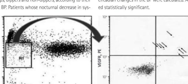
8)In brief, mononuclear cells (MNCs) were obtained from peripheral blood samples (100 μL), and the EPCs were identified by flow cytometry (BD Biosciences, San Jose, CA, USA). Their phenotype was determined by immunohisto- chemistry after staining with 20 μL fluorescein PE-Cy5-conjugated anti-CD45 monoclonal antibody (Dynona, Korea), 20 μL of fluores- cein isothiocyanate (FITC)-conjugated anti-CD34 monoclonal anti- body (Dynona, Korea) and 10 μL of PE-conjugated anti-vascular endothelial growth factor receptor 2 (VEGFR2) monoclonal antibody (R&D, Minneapolis, MN, USA), and further incubated in a dark room for 1 hour. After appropriate gating with low cytoplasmic granularity and a low expression of CD45, the number of CD34
+VEGFR2
+cells was quantified and expressed as the absolute number of cells per 1×10
6peripheral MNCs. The total number of CD45
lowCD34
+VEG- FR2
+cells was then counted (Fig. 1).
Statistical analysis
All statistical analysis was performed using the Statistical Pack- age for the Social Sciences (SPSS Inc., Chicago, IL, USA) for Win- dows 12.0. All data are expressed as means±standard deviations.
The Student’s t-test was used for comparisons of the measured values between the dipper and non-dipper hypertensive patients.
A Pearson’s correlation coefficient between the number of EPSs and circadian changes in the BP were calculated. A p<0.05 was consider- ed statistically significant.
Fig. 1. Quantification of EPCs by flow cytometry. Circulating EPCs were identified by flow cytometry with low cytoplasmic granularity (R1) and with ex- pression of cell surface antigens such as CD45
lowCD34
+VEGFR2
+. Arrows indicate CD45
lowCD34
+VEGFR2
+cells. EPCs: endothelial progenitor cells, VEGFR2:
vascular endothelial growth factor receptor 2, SSC: side scather, FITC: fluorescein isothiocyanate.
10
410
310
210
110
010
410
310
210
110
0SSC-Height CD34 FITC
CD 45 P E cy 5 VE GF R
2P E
0 200 400 600 800 1000 10
010
110
210
310
4Results
Subject characteristics
Among the 45 patients with essential hypertension, 20 were clas- sified as dippers (12 men and 8 women; mean age 48±14 years) and
25 as non-dippers (14 men and 11 women; mean age 52±18 years).
The baseline characteristics of the dipper and non-dipper hyper- tensive patients are shown in Table 1. The clinical characteristics and laboratory parameters were similar between the dipper and non- dippers except for the number of lymphocytes (2.5±0.6 vs. 2.1±
0.7×10
3/uL; p=0.02) and left ventricular ejection fraction (70.8±6.0%
vs. 64.8±11.1%; p=0.04).
Comparison of differences in the endothelial progenitor cell count between the dippers and non-dippers
The difference in the circulating number of EPCs between the dippers and non-dippers is presented in Fig. 2. The absolute num- ber of circulating EPCs was significantly reduced in the non-dipper hypertensive patients as compared to the dippers (104±60 vs.
66±47 EPCs per 10
6MNCs, p=0.027) (Fig. 2).
Relationship between the endothelial progenitor cell count and parameters
The correlation between the EPC count and parameters is shown in Table 2. The EPC count correlated positively with the number of lymphocytes and the glucose level (r=0.625, p<0.001, and r=0.318, p=0.035, respectively).
Relationship between the endothelial progenitor cell count and circadian changes in the blood pressure
The correlation between the EPC count and the circadian changes in the BP is presented in Fig. 3. The number of circulating EPCs cor- related positively with the circadian changes in the systolic blood pressure and diastolic blood pressure (r=0.435, p=0.003, and r=
0.310, p=0.038, respectively) (Fig. 3).
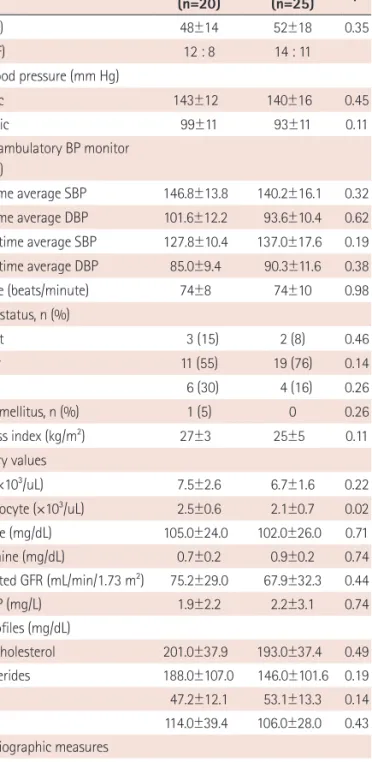
Table 1. Baseline characteristics of the patients evaluated in this study Dipper
(n=20)
Non-dipper
(n=25) p
Age (year) 48±14 52±18 0.35
Sex (M : F) 12 : 8 14 : 11
Office blood pressure (mm Hg)
Systolic 143±12 140±16 0.45
Diastolic 99±11 93±11 0.11
24-hour ambulatory BP monitor (mm Hg)
Day-time average SBP 146.8±13.8 140.2±16.1 0.32 Day-time average DBP 101.6±12.2 93.6±10.4 0.62 Night-time average SBP 127.8±10.4 137.0±17.6 0.19 Night-time average DBP 85.0±9.4 90.3±11.6 0.38
Heart rate (beats/minute) 74±8 74±10 0.98
Smoking status, n (%)
Current 3 (15) 2 (8) 0.46
Former 11 (55) 19 (76) 0.14
Never 6 (30) 4 (16) 0.26
Diabetes mellitus, n (%) 1 (5) 0 0.26
Body mass index (kg/m
2) 27±3 25±5 0.11
Laboratory values
WBC (×10
3/uL) 7.5±2.6 6.7±1.6 0.22
Lymphocyte (×10
3/uL) 2.5±0.6 2.1±0.7 0.02
Glucose (mg/dL) 105.0±24.0 102.0±26.0 0.71
Creatinine (mg/dL) 0.7±0.2 0.9±0.2 0.74
Estimated GFR (mL/min/1.73 m
2) 75.2±29.0 67.9±32.3 0.44
hs-CRP (mg/L) 1.9±2.2 2.2±3.1 0.74
Lipids profiles (mg/dL)
Total-cholesterol 201.0±37.9 193.0±37.4 0.49
Triglycerides 188.0±107.0 146.0±101.6 0.19
HDL-C 47.2±12.1 53.1±13.3 0.14
LDL-C 114.0±39.4 106.0±28.0 0.43
Echocardiographic measures
LV ejection fraction (%) 70.8±6.0 64.8±11.1 0.04 Medication, n (%)
Statin 0 0
Data are expressed as mean±SD or number (%). SBP: systolic blood pres- sure, DBP: diastolic blood pressure, WBC: white blood cell, GFR: glomerular filtration rate, hs-CRP: high sensitivity C-reactive protein, HDL-C: high density lipoprotein-cholesterol, LDL-C: low density lipoprotein-cholesterol, LV: left ventricular, ACE: angiotensin-converting enzyme, ARB: angiotensin receptor blocker, CCB: calcium channel blocker
Fig. 2. Comparison of the difference in the EPC count between the dippers and non-dippers. The absolute number of circulating EPCs was significantly reduced in the non-dipper hypertensive patients as compared to the dippers (104±60 vs. 66±47 EPCs per 10
6mononuclear cells, p=0.027). EPC: endo- thelial progenitor cell, VEGFR2: vascular endothelial growth factor receptor 2, MNCs: mononuclear cells.
150
100
50
low++6