• 검색 결과가 없습니다.

Late Stent Thrombosis After Drug-Eluting Stent Implantation: A Rare Case of Accelerated Neo-Atherosclerosis and Early Manifestation of Neointimal Rupture

N/A
N/A
Protected

Academic year: 2022

Share "Late Stent Thrombosis After Drug-Eluting Stent Implantation: A Rare Case of Accelerated Neo-Atherosclerosis and Early Manifestation of Neointimal Rupture"

Copied!
4
0
0

로드 중.... (전체 텍스트 보기)

전체 글

(1)

409

Print ISSN 1738-5520 / On-line ISSN 1738-5555 Copyright © 2011 The Korean Society of Cardiology CASE REPORT

DOI 10.4070/kcj.2011.41.7.409

Open Access

Late Stent Thrombosis After Drug-Eluting Stent Implantation:

A Rare Case of Accelerated Neo-Atherosclerosis and Early Manifestation of Neointimal Rupture

Young-June Yang, MD

1

, Mihyun Kim, MD

1

, Choongki Kim, MD

1

, Junbeom Park, MD

1

, Jaewon Oh, MD

1

, Hoyoun Won, MD

1

, Byeong-Keuk Kim, MD

1

, and Myeong-Ki Hong, MD

1,2

1

Division of Cardiology, Severance Cardiovascular Hospital,

2

Severance Biomedical Science Institute, Yonsei University College of Medicine, Seoul, Korea

ABSTRACT

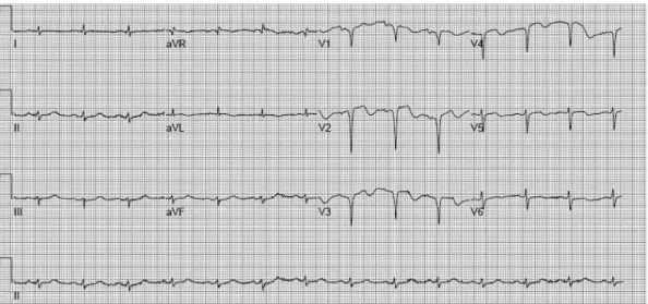
An 80-year old woman suffered from sudden onset of chest pain and dyspnea, and visited the emergency room. She re- ceived stent implantation with a biolimus A9-eluting stent (Nobori

®

3.0×24 mm) at a the mid-portion of the left anterior descending artery 5 months prior to admission. The emergency 5-month follow-up angiogram was performed under the impression of late stent thrombosis. The follow-up angiogram showed subtotal occlusion at the mid-portion of the left ante- rior descending artery, which was the same segment of previous stent implantation 5 months ago. Immediately after throm- bus aspiration with the thrombus aspiration catheter, the optical coherence tomography showed layered appearance of neo- intimal hyperplasia and neointimal rupture within the previously stented segment. Thus, neointimal rupture within accelerated growth of neointimal tissue was observed within a relatively shorter period (i.e., about 5 months) after stent im- plantation. (Korean Circ J 2011;41:409-412)

KEY WORDS: Stents; Thrombosis; Neointima.

Received: March 3, 2011 Accepted: April 11, 2011

Correspondence: Myeong-Ki Hong, MD, Division of Cardiology, Sev- erance Cardiovascular Hospital, Yonsei University College of Medicine, 250 Seongsan-ro, Seodaemun-gu, Seoul 120-752, Korea

Tel: 82-2-2228-8458, Fax: 82-2-2227-7732 E-mail: [email protected]

• The authors have no financial conflicts of interest.

cc

This is an Open Access article distributed under the terms of the Cre- ative Commons Attribution Non-Commercial License (http://creativecom- mons.org/licenses/by-nc/3.0) which permits unrestricted non-commer- cial use, distribution, and reproduction in any medium, provided the origi- nal work is properly cited.

Introduction

Implantation of 1st generation drug-eluting stents (DESs) has significantly reduced the incidence of restenosis and the need for repeat revascularization, compared to previously used bare-metal stents.

1)2)

However, increased use of DESs has rais- ed safety issues regarding the occurrence of stent thrombo- sis.

3)4)

In the 1st generation DES, persistence of polymer even after complete elution of drugs may cause either hypersensi- tivity reaction or inflammation in the stented coronary seg- ments, and both these conditions are associated with increas-

ed risk for stent thrombosis.

5)

To overcome this life-threaten- ing complication, many newly designed DESs have been de- veloped. Compared to previous DESs, the biolimus A9-elut- ing stent (BES, Nobori

®

, Terumo Corporation, Tokyo, Japan) has the characteristics of abluminally applied drug polymer matrix and biodegradable polymer (i.e., polymer degradation over the period of 6 to 9 months).

6)

We experienced a rare case of late stent thrombosis in patients who underwent im- plantation of a BES and follow-up optical coherence tomog- raphy (OCT) for investigation of late stent thrombosis.

Case

An 80-year old woman suffered from sudden onset of ch-

est pain and dyspnea, and visited the emergency room. She

had a history of hypertension, hyperthyroidism and major

depressive disorder. Five months prior to admission, she visit-

ed the cardiology outpatient clinic for the further evaluation

of syncope. The coronary computer tomography and corona-

ry angiography revealed significant stenosis at the mid-por-

tion of the left anterior descending artery (mLAD; B2, 80%,

III, 0) (Fig. 2A) and a BES (Nobori

®

3.0×24 mm) was success-

(2)

410 Early Neointimal Rupture

fully deployed, without residual narrowing (Fig. 2B) at that time.

Pulmonary congestion in both lung fields, suggestive of pul- monary edema, was noticed on the chest X-ray; newly de-

veloped Q wave was visible at leads V1-4 on the electrocardio- gram (Fig. 1); recent regional wall motion abnormality (i.e., akinesia without thinning) was detected in the left anterior de-

Fig. 1. First electrocardiogram at recorded in the emergency room; the Q wave was visible at on leads V1-4.

Fig. 2. Pre-intervention (A) and post-intervention (B) angiographic findings in lesion of the left anterior descending artery (arrow in A) are sh- own 5-months before the onset of late stent thrombosis. At the onset of late stent thrombosis, emergency follow-up angiogram showed sub- total occlusion at the mid-portion of the left anterior descending artery (C). The final angiogram, performed after balloon angioplasty with a drug eluting balloon, showed no residual stenosis at the mid-portion of the left anterior descending artery and a jailed diagonal branch (D).

A

C

B

D

(3)

Young-June Yang, et al. 411

scending artery territory, and the left ventricular ejection frac- tion was reduced by 30% on the echocardiogram at this time.

Cardiac enzyme was slightly elevated (Troponin-T 0.124 ng/

mL). Emergency follow-up angiogram was performed un- der the impression of non-ST elevation myocardial infarction, caused by late stent thrombosis. The follow-up angiogram showed subtotal occlusion at the mid-portion of the left ante- rior descending artery, the same segment of previous stent implantation 5 months ago (B2, 99%, II, 0) (Fig. 2C). Imme- diately after thrombus aspiration with the thrombus aspira- tion catheter (Thrombuster II

®

, Kaneka Corp. Japan), OCT was performed for investigation of both morphologic featur- es and underlying cause of late stent thrombosis. The OCT sh- owed layered appearance of neointmal hyperplasia and neo- intimal rupture within the previously stented segment (Fig. 3A and B). Considering that the stent thrombosis was not due to delay of stent strut endothelialization, balloon angioplasty with a drug eluting balloon (SeQuent please

®

, B. Braun Mel- sungen AG, Germany) was successfully performed at the mid- portion of the left anterior descending artery with no residual stenosis, however jailed diagonal branch (Figs. 2D and 3C).

After the surgical procedure, she was discharged 3 days later, without any significant complications.

Discussion

Occurrence of stent thrombosis has been the major con- cern after implantation of 1st generation DESs. Iakovou et al.

3)

reported 1.3% incidence of stent thrombosis in patients who received DES implantation. In addition, 45% of the patients who experienced stent thrombosis died. Daemen et al.

4)

also reported that stent thrombosis occurring within 30 days after DES implantation increases 0.6% annually. While occurr- ence of acute or subacute stent thrombosis (i.e., onset of stent thrombosis within 30 days after stent implantation) is mainly related to procedural factors, development of late or very late stent thrombosis has different characteristics. Previous pa-

thologic studies showed that delayed stent strut endothelial- ization and potential prothrombotic properties of the DES it- self (i.e., persistence of polymer even after complete elution of drugs) are important factors that may lead to late stent throm- bosis.

7)8)

A recent intravascular ultrasound study by Lee et al.

9)

showed that neointimal rupture within the stent area was observed in 43.5% of 23 patients with very late stent thrombo- sis, after 1st generation DES implantation. A pathologic study reported that DES therapy may accelerate atherosclerosis, while reducing restenosis.

10)

Therefore, new DESs with a bio- degradable polymer have been developed to avoid complic- ations caused by persistence of polymer. Several studies com- pared the outcomes (i.e., cardaic death, myocardial infarc- tion, and stent thrombosis) of BESs with those of 1st gener- ation DESs and concluded that BESs were not inferior to 1st generation DESs and that both suitable alternatives and long term outcomes were neeed.

11)

Despite of these favorable conditions of a BESs compared to 1st generation DESs, accelerated neointimal proliferation, abnormal neointimal morphology (i.e., layered appearance of the neointima) and additional neointimal rupture were ob- served in this patient with late stent thrombosis within a rel- atively shorter period of time (i.e., about 5 months) after im- plantation of a BES. Due to high resolution images of the OCT, the cause of stent thrombosis was more precisely diagnosed than in previous cases using IVUS or other modalities.

REFERENCES

1) Moses JW, Leon MB, Popma JJ, et al. Sirolimus-eluting stents versus standard stents in patients with stenosis in a native coronary artery. N Engl J Med 2003;349:1315-23.

2) Stone GW, Ellis SG, Cox DA, et al. A polymer-based, paclitaxel-elut- ing stent in patients with coronary artery disease. N Engl J Med 2004;

350:221-31.

3) Iakovou I, Schmidt T, Bonizzoni E, et al. Incidence, predictors, and outcome of thrombosis after successful implantation of drug-eluting st- ents. JAMA 2005;293:2126-30.

4) Daemen J, Wenaweser P, Tsuchida K, et al. Early and late coronary stent thrombosis of sirolimus-eluting and paclitaxel-eluting stents in Fig. 3. The optical coherence tomography (OCT) showed layered appearance (arrowhead) of neointmal hyperplasia and neointimal rupture (arrow) within the previously stented segment (A and B). OCT image after balloon angioplasty with a drug eluting balloon (C).

A B C

(4)

412 Early Neointimal Rupture

routine clinical practice: data from a large two-institutional cohort st- udy. Lancet 2007;369:667-78.

5) Cook S, Ladich E, Nakazawa G, et al. Correlation of intravascular ultrasound findings with histopathological analysis of thrombus as- pirates in patients with very late drug-eluting stent thrombosis. Cir- culation 2009;120:391-9.

6) Chevalier B, Silber S, Park SJ, et al. Randomized comparison of the Nobori biolimus A9-eluting coronary stent with the Taxus Liberté pa- clitaxel-eluting coronary stent in patients with stenosis in native cor- onary arteries: the NOBORI 1 trial: phase 2. Circ Cardiovasc Interv 2009;2:188-95.

7) Finn AV, Joner M, Nakazawa G, et al. Pathological correlates of late drug-eluting stent thrombosis: strut coverage as a marker of endoth- elialization. Circulation 2007;115:2435-41.

8) Farb A, Burke AP, Kolodgie FD, Virmani R. Pathological mechani- sms of fatal late coronary stent thrombosis in humans. Circulation 2003;108:1701-6.

9) Lee CW, Kang SJ, Park DW, et al. Intravascular ultrasound findings in patients with very late stent thrombosis after either drug-eluting or bare-metal stent implantation. J Am Coll Cardiol 2010;55:1936-42.

10) Nakazawa G, Vorpahl M, Finn AV, Narula J, Virmani R. One step for- ward and two steps back with drug-eluting-stents: from preventing restenosis to causing late thrombosis and nouveau atherosclerosis.

JACC Cardiovasc Imaging 2009;2:625-8.

11) Garg S, Sarno G, Serruys PW, et al. The twelve-month outcomes of a

biolimus eluting stent with a biodegradable polymer compared with

a sirolimus eluting stent with a durable polymer. EuroIntervention

2010;6:233-9.

수치

Fig. 1. First electrocardiogram at recorded in the emergency room; the Q wave was visible at on leads V1-4

참조

관련 문서

After liberation, it has been suggested that the objective of Korean police activities and duties are maintenance of social order and public interest, but

The corrosion potential(E corr ), corrosion current density(I corr ), and corrosion rate of Ni-Ti files obtained from anodic polarization curves after electrochemical

Thirdly, it appeared that social support had meaningful effect on self-efficacy and the quality of life, and in the case of the difference analysis by

5) After GBR, the membrane was removed i n i ni ti al ti me, the usage of nonabsorbabl e membrane and autogenous bone resul ted i n the mostfavorabl e bone formati

It was summarized as follows that group participating in after-school basketball activities for 12 weeks showed decrease of the body fat(%) and increase

English cleft construction is one of the constructions that have structural properties and semantic properties of themselves.. At first, I observe the

After that, we look forward to the results and expect that it will be used as a tool to determine the direction of brand development and renewal by

 Early advancements using chemical principles (Mass Transfer & Polymer Materials)..  Nasal sprays that deliver finely atomized amounts of a