
Hyaluronidase Inhibitory and Antioxidant Activities of Enzymatic Hydrolysate from Jeju Island Red Sea Cucumber (Stichopus japonicus) for Novel Anti-aging Cosmeceuticals
11
0
0
전체 글
(2) J . Mar . Biosci. Biotechnol. 2018,. p.. 62 - 72. Vol.. healthy [4]. HA synthesized by epidermal keratinocytes and dermal fibroblast is normally found in extracellular matrix (ECM) beneath our skin. HAse is one of dermal enzymatic which able to degrade HA by hydrolyzing the disaccharides at hexosaminidic β (1–4) linkages [5, 6]. HAse pervert skin to lack of water-holding capacity and promote aging-skin problems. Hence, natural compound with anti-HAse potential should be explored for anti-aging active ingredients in cosmeceuticals. In the past decade, the numbers of publication of new bioactive substances derived from marine resources has continuously increased all over the world. Marine organisms with the rich source of biological and chemical diversity displayed great medical benefits. Various compounds of marine organisms demonstrated a highly potential of biological activities and inhibitors of physiological processes. In addition, marine organisms have been discovered to be productive sources which provide great cosmetic activities derived from nature [7, 8]. This is due to the natural active compounds that are safer for humans which are receiving more attention in the research and development of industries than the synthetic compounds [9]. Therefore, marine organisms are popular components used for cosmeceuticals in terms of their productive ingredients because of their safe and effective substances. Sea cucumbers (Stiphopus japonicus), soft-bodied worm-like echinoderms, are well-known as high commercial costly sea food around East Asian countries especially China, Japan and Korea. In addition, sea cucumbers have been used for their benefits in terms of traditional medical treatments for many centuries [10]. S. jaonicus were classified into three groups by their colors which are green, black and red [11]. RSCs are normally found in unique habitat such as gravel beds off shore and observed in different genetic and pigment compositions compared with other color types (blue, green) [12,13]. As a result of their uniqueness, RSCs exhibited various activities and potentials both in pharmaceutical and biomedical utilizations such as anti-cancer, anti-oxidation, anti-inflammation, anti-melanogenesis and anti-bacteria effects [14]. Although. 10,. No.. 2. [Research. Paper]. such results indicate the potential of the RSCs as natural cosmeceutical candidates, RSCs collected from Jeju Island have not been extensively studied in terms of HAse inhibitory and antioxidant activities. Therefore, in this study, RSCs collected from Jeju Island, South Korea were extracted by using various enzymatic hydrolysis protein and investigated for their HAse inhibitory and antioxidant activities aimed to be an alternative for a natural active ingredient of anti-aging substance in cosmeceuticals.. Materials and Methods. Materials The Jeju RSC flesh tissue dried by far infrared radiation dryer was kindly provided by the Jeju International Marine Science Research & Education Center of Korea Institute of Ocean Science & Technology (KIOST). Al, Neu, Pro were purchased from Novo Co. (Novozyme Nordisk, Bagsvaerd, Denmark) and Try, Pep, Chy were purchased from Sigma Chemical Co. (St. Louis, MO, USA). Antibodies to HAS2 and HYAL2 were purchased from Invitrogen Life Technologies (Carlsbad, CA, USA). All other chemicals and reagents used were of analytical grade. Preparation of enzymatic hydrolysate from RSC Enzymatic hydrolysis of the RSC was performed using various enzymes at their optimal conditions described in a previously reported method [15]. The grounded dried RSC powder (1 g) was first homogenized with 100 ml of distilled water, and 10 μl of the enzymes was then added. Enzymatic hydrolysis was conducted for 24 h, after the hydrolysate was boiled for 10 min at 100°C to inactivate the enzyme. The hydrolysate was clarified by centrifugation at 3000×g for 20 min to remove any unhydrolyzed residue. The supernatants of the RSC enzymatic hydrolysates were filtered, adjusted to pH 7.0, and stored for subsequent use in experiments. Total antioxidant capacity measurement methods The methods used to assess Total antioxidant capacity (TAC) were trolox equivalent antioxidant capacity. - 63 -.
(3) J . Mar . Biosci. Biotechnol. 2018,. p.. 62 - 72. Vol.. (TEAC) and oxygen radical absorbance capacity (ORAC). TEAC assay TEAC method was described by Re et al. [16]. The ability of a sample to inhibit the ABTS+radical was compared with an antioxidant standard (Trolox). The ABTS·+ was generated by ABTS solution with potassiumpersulfate (K2S2O8). For this reason, 7.4 mM ABTS solution and 2.6 mM potassium persulfate solution was mixed in equal quantities and kept for 12~16 h in dark at room temperature. This stock solution was stable for 2~3 days when stored in the dark. The working solution was prepared by diluting the stock solution in methanol until its absorbance was 1.1±0.02 units at 734 nm measured by the spectrophotometer. 150 μl of ABTS working solution and 50 μl of each of hydrolysates were added in a 96 well microplate for 2 h in a dark condition and then the absorbance was measured at 734 nm. The concentration of ABTS content in the sample was reported as μM Trolox equivalent (TE)/mg sample. ORAC assay ORAC is based on the decrease of fluorescein fluorescence in presence of the chemical oxidant AAPH. The assay was described by Ou et al. with some modifications [17]. 221 mM AAPH was dissolved in phosphate buffer and kept in an ice bath. Fluorescein (FL) stock solution was made in PBS and kept at 4℃ in dark condition. The stock solution can be stored for several months. A fresh FL working solution was made daily by further diluting the stock solution in PBS. 50 μl FL working solution and 50 μl of each hydrolysate (blank or 20 μM Trolox) was placed in the back 96-well plate for 15 min at 37℃. Then 50 μl of AAPH solution was added using a multichannel pipette. The plate was immediately placed in the reader and measurement were taken every 5 min for 60 min at 37℃. Fluorescence conditions were as follows: excitation at 485 nm and emission at 520 nm. The standard curve was linear between 0 and 20 μM Trolox. Results are expressed as μM TE/mg sample.. 10,. No.. 2. [Research. Paper]. HAse inhibition assay HAse inhibitory activity was measured as previously described with few modifications [18]. Ten μl of HAse (Type-1-S from bovine testes, Sigma-Aldrich Co., USA) dissolved in 0.1 M acetate buffer (pH 3.5) was mixed with 10 μl of each enzymatic hydrolysate, and incubated at 37 °C for 20 min. Twenty μl of 12.5 mM calcium chloride was added to the reaction mixture, and then the mixture was incubated at 37°C for 20 min. This Ca2+activated HAse was treated with 50 μl of sodiumhyaluronate dissolved in 0.1 M acetate buffer (pH3.5), and then incubated at 37 °C for 40 min. Two μl of 0.4 N sodiumhydroxide and 20 μl of 0.4 N potassiumtetraborate were added to the reaction mixture, and then incubated in a boilingwater bath at 100°C for 3 min. After cooling to room temperature, 600 μl of DMAB solution (0.4 g of p-dimethylaminobenzaldehyde dissolved in 35 ml of 100% acetic acid and 5 ml of 10 N hydrochloric acid) was added to the reaction mixture which was then incubated at 37°C for 20 min. Absorbance was measured at the wave length of 585 nm. Cell culture HaCaT were maintained at 37°C in an incubator, under a humidified atmosphere containing 5% CO2. The cells were cultured in Dulbecco’s modified Eagle’s medium (DMEM) containing 10% heat-inactivated fetal bovine serum (FBS) and 1% antibiotics. Measurement of cytotoxicity The cytotoxicity of enzymatic hydrolysate against the HaCaT was determined by colorimetric assays [19]. Cells were seeded into a 96-well plate at a concentration of 1×10⁵ cells/ml. After 16 h, the cells were treated with various concentrations (25, 50, 100, 200 and 400 μg/ml) of enzymatic hydrolysate. The cells were incubated for an additional 24 h at 37°C. There after, 3-(4, 5-dimethyl2-thiazolyl)2,5-diphenyl tetrazoliumbromide (MTT) stock solution (50 μl : 2 mg/ml in PBS) was then added to each well. After incubating for 4 h, the plate was centrifuged at 800×g. - 64 -.
(4) J . Mar . Biosci. Biotechnol. 2018,. p.. 62 - 72. Vol.. for 10 min and the supernatant was aspirated. The formazan crystals in each well were dissolved in DMSO. The amount of purple formazan was determined by measuring the absorbance at 540 nm. Assay of intracellular ROS levels Intracellular ROS levels were measured by the dichlorofluorescein assay [20]. 2′,7′-dichlorodihydrofluorescein diacetate (DCF-DA) can be deacetylated in cells, where it can react quantitatively with intracellular radicals to be converted into its fluorescent product, DCF, which was retained within the cells. Therefore, DCF-DA was used to evaluate the generation of ROS. Cells were seeded in 96-well plates at a concentration of 1.0 × 10⁵ cells/ml. After 16 h, the cells were treated with samples and incubated at 37 ℃ under a humidified atmosphere. After 30 min, AAPH was added at a concentration of 15 mM, and then the cells were incubated for an additional 30 min at 37℃. Finally, DCF-DA was introduced to the cells, and 2′,7′dichloro dihydrofluorescein fluorescence was detected at an excitation wavelength of 485 nm and an emission wavelength of 535 nm, using a fluorescence plate reader. Assay of cell viability Cell viability was then estimated via an MTT assay [19]. The cells were seeded in 96-well plates at a concentration of 1.0 × 10⁵cells/ml. After 16 h, the cells were treated with samples. After 1 h, 15 mM AAPH was added to the cell culture medium and incubated for 24 h at 37℃. MTT stock solution was then applied to each of the wells. After incubating for 4 h, the plate was centrifuged at 800×g for 10 min and the supernatant was aspirated. The formazan crystals in each well were dissolved in DMSO. The amount of purple formazan was determined by measuring the absorbance at 540 nm. HA measurement HaCaT cells were grown to high density in 6-well plates. Immediately before experiments, cells were washed two times with serum-free medium to completely remove HA accumulated during cell growth. Subsequently, HaCaT cells were cultured with or. 10,. No.. 2. [Research. Paper]. without enzymatic hydrolysate in serum-free medium for 24 h. At the indicated time, aliquots of medium were removed, centrifuged at 15,000 × g for 5 min, and supernatants were analyzed for HA using an enzyme-linked immunosorbent assay (ELISA) kit (R&D Systems, Inc. MN, USA). Western blot analysis HaCaT cells were treated with the indicated concentrations of samples and harvested. The cell lysates were prepared with ice-cold lysis buffer (20 mM Tris, 5 mM EDTA, 10 mM Na4P2O7, 100 mM NaF, 2 mM Na3VO4, 1% NP-40, 10 mg/ml aprotinin, 10 mg/ml leupeptin, and 1 mM PMSF). Cell lysates were centrifuged at 12,000×g for 20 min at 4°C and the protein concentrations were determined by using the BCATM protein assay kit. The lysate containing 40 μg of protein was subjected to electrophoresis on a sodiumdodecylsulfate (SDS)-polyacrylamide gel, and the gel was transferred to a nitrocellulose membrane. The membrane was blocked in 5% non fat dry milk in TBST (25mM Tris–HCl, 137 mM NaCl, 2.65 mM KCl, 0.05% Tween 20, pH 7.4) for 2 h. The primary antibodies were use data 1:1000 dilution. The membranes were incubated with the primary antibodies at 4 °C overnight. There after, the membranes were washed with TBST and then incubated with the secondary antibodies used at 1:3000 dilution. Signals were developed using an ECLwestern blotting detection kit and exposed to X-ray films. Statistical analysis The data are presented as means ± standard error (SE). Statistical comparisons of the mean values were performed by analysis of variance (ANOVA), followed by a Duncan’s multiple range test using SPSS software. Statistical significance was considered at p < 0.05. Results. Extraction yield of enzymatic hydrolysates In this study, RSCs collected from Jeju Island, were. - 65 -.
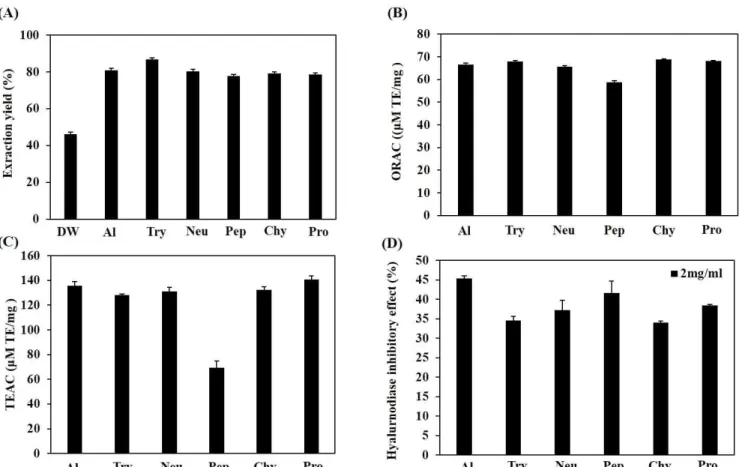
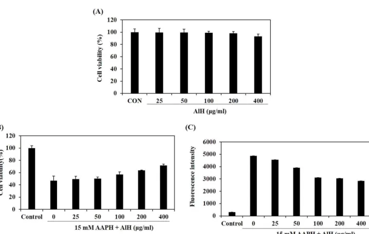
(5) J . Mar . Biosci. Biotechnol. 2018,. p.. 62 - 72. Vol.. investigated and extracted by using enzymatic hydrolysis. Six various proteolytic enzymes which are commercially used for hydrolysis such as Al, Try, Neu, Pep, Chy, and Pro, were chosen and explored for their extraction yields. Each of enzymatic hydrolysates gave extraction yields over than 75%. Among them, Try hydrolysate showed the greatest extraction yield as high as 86.65% as shown in Fig. 1A. Total antioxidant capacity and HAse inhibitory effect of enzymatic hydrolysates Anti-oxidant capacities of RSCs were measured by. 10,. No.. 2. [Research. Paper]. using colorimetry method such as ORAC and TEAC. AlH showed the highest antioxidant capacities for ORAC and TEAC methods, compared to those of other hydrolysates, at 66.59±0.78 µM TE/mg and 135.78±3.24 µM TE/mg, respectively (Fig. 1B and C). Moreover, Fig. 1D illustrates the HAse inhibitory effects of all enzymatic hydrolysates. Among them, AlH possessed the highest capacity of HAse inhibitory (45.35%), challenged at concentration of 2 mg/ml. Since the highest antioxidant activity and HAse inhibitory effect, AlH was chosen for the next further studies on cell experiments.. Figure 1. Extraction yield (A), total antioxidant capacity (B and C) and HAse inhibitory effect (D) of different enzymatic hydrolysates from RSCs. Total antioxidant capacities were measured via ORAC assay (A) and measured via TEAC assay (B). DW: water extract, Al: Alcalase hydrolysate, Try: Trypsin hydrolysate, Neu: Neutrase hydrolysate, Pep: Pepsin hydrolysate, Chy: α-Chymotrysin hydrolysate, Pro: Protamex hydrolysate. Experiments were performed in triplicate and the data are expressed as mean ± SE.. Cytotoxicity of AlH in HaCat cell To evaluate whether AlH has toxic effect on the cells, HaCaT cells were treated with AlH, and cell viability. was measured via MTT colorimetric assay. As shown in Fig. 2A, AlH was not exerting any cytotoxic effect at the various concentrations (25, 50, 100, 200, and 400. - 66 -.
(6) J . Mar . Biosci. Biotechnol. 2018,. p.. 62 - 72. Vol.. µg/ml) in HaCaT cells. Protective effects of AlH against AAPH-induced oxidative stress in HaCaT cells Fig. 2B shows the effect of AlH on cell viability in HaCaT cells treated with AAPH and examined by MTT assay. When HaCaT cells were treated with AAPH, the cell viability significantly decreased. 10,. No.. 2. [Research. Paper]. compared with non-treated cells. However, AlH protected against the cellular damage induced AAPH in a dose-dependent manner. As shown in Fig. 2C, the generation of intracellular ROS in HaCaT cells increased significantly after treatment with AAPH compared with non-treated cells. However, treatment of AlH decreased dose-dependently of the ROS level in the cells induced with AAPH.. Figure 2. Effects of AlH on the cell viabilities (A and B) and the intracellular ROS generation (C) in HaCaT cells. The cell viability was determined in only AlH-treated HaCaT cells by MTT assay (A). The cell viability was determined in both AAPH- and AlH-treated HaCaT cells by MTT assay (B). The intracellular ROS generation was determined in both AAPH- and AlH-treated HaCaT cells by DCF-DA assay (C). Experiments were performed in triplicate and the data are expressed as mean ± SE.. HAse inhibitory effect and HA contents of AlH AlH inhibited HAse activity in a dose-dependent manner as 39.11, 46.77, 52.72, and 57.63% at the concentrations of 1, 2, 4, and 8 mg/ml, respectively (Fig. 3A). IC50 value of AlH against HAse was 3.21 mg/ml. HA amount in HaCaT cells after being treated with the desired concentrations of AlH (25, 50, 100, 200, and 400. µg/ml) were determined by using ELISA kit. At the concentrations of 25 and 50 µg/ml, non-significant difference in the number of HA contents were observed. However, the significantly increased in the existence of HA by 543, 585, and 586 ng/ml were observed at 100, 200, and 400 µg/ml concentration of AlH, respectively, compared to the control (490 ng/ml) (Fig. 3B).. - 67 -.
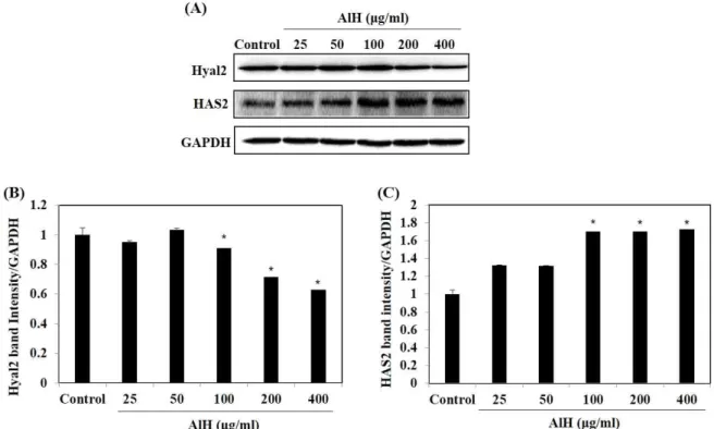
(7) J . Mar . Biosci. Biotechnol. 2018,. p.. 62 - 72. Vol.. 10,. No.. 2. [Research. Paper]. Figure 3. Effect of AlH on HAse-inhibition (A) and HA content (B). HA content of HaCaT cells treated with AlH was determined via an ELISA kit. Experiments were performed in triplicate and the data are expressed as mean ± SE. Significant differences from the control group were identified at *p < 0.05.. Effect of AlH on HAS2 and HYAL2 expression in HaCaT cells The effects of AlH on HAS2 and HYAL2 expression. selected concentrations and control. At 100, 200 and 400 µg/ml of AlH, the intensity band level of HYAL2. in HaCaT cells were investigated by western blot assay. Fig. 4 represents the levels of HAS2 and HYAL2 expressions compared between AlH-treated sample with. were significantly decreased whereas the significantly increased level of HAS2 expressions were observed, compared with the control.. Figure 4. Effect of AIH on the expression of HYAL2 and HAS2 in HaCaT cells (A) and the densitometry analysis (B). Experiments were performed in triplicate and the data are expressed as mean ± SE. Significant differences from the control group were identified at *p < 0.05.. - 68 -.
(8) J . Mar . Biosci. Biotechnol. 2018,. p.. 62 - 72. Vol.. Discussion. Recently decade, skin-aging problem is becoming more widely concerned throughout the world. According to that, active ingredients with HAse inhibitory effect from nature are required due to their preferable safeness and stability for humans [4]. Sea cucumbers are a well-known marine organism in countries like Japan, China and Korea. It has been used as a traditional remedy with various biological activities such as anti-cancer, anti-inflammation, anti-oxidation and anti-melanogenesis [11]. However, to date there has been a lack of information regarding the cosmeceutical activities of RSCs for their potential use as materials in cosmetics and cosmeceuticals. Hence, in this study, RSCs collected from Jeju Island were hydrolyzed by protease hydrolysis and challenged their HAse-inhibitory ability against premature-skin problems. During the last decade, enzymatic-assistant extraction has been successfully applied for the extraction of numerous biologically active natural products from a wide variety of organisms. This method does not use any toxic chemicals. In addition, this technique results in higher yields of bioactive compounds that also show enhanced biological activity compared with ingredients extracted by water or organic solvents [21 -23]. Therefore, in the present study, the RSCs was enzymatically hydrolyzed using the various commercial protease such as Al, Try, Neu, pep, Chy and Pro to make it acceptable as a cosmeceutical. According to Fig. 1A, all of the extraction yields, extracted by enzymatic hydrolysis (Al, Try, Neu, Pep, Chy and Pro), were shown at over than 75%, which was higher than the extraction yield achieved with the water extract. Since sea cucumber consists of 70% collagen protein of their total body wall, which is rich protein resources, the enzymatic hydrolysis can promote the extraction yields of RSCs [24]. Taken together, this data indicated that enzymatic extraction techniques for obtaining the bioactive components of RSCs may be more advantageous than. 10,. No.. 2. [Research. Paper]. water extraction. ORAC and TEAC are common methods used for measurement the total antioxidant capacity. ORAC assay is a method based on hydrogen atom transfer which measures the anti-oxidant capacity caused by peroxyl radicals (ROO-), but, TEAC assay or called ABTS assay is based on electron transfer [25]. In this study, we evaluated potential antioxidant effects of enzymatic hydrolysates of RSCs by measuring ORAC and TEAC. The results demonstrated that all enzymatic hydrolysates of RSCs showed good antioxidant activities. Among the tested enzymatic hydrolysates, AlH exhibited the highest antioxidant effectives for both assays with a 2-fold greater capacity (135.78±3.24 µM TE/mg) was obtained by TEAC, compared with the value detected by ORAC (66.59±0.78 µM TE/mg). Moreover, the highest HAse-inhibitory effect (45.35%) at 2 mg/ml of AlH were observed (Fig. 1). Since the greatest antioxidant activities and HAse inhibitory effect, AlH was selected to the further investigate protective effect against oxidative stress in HaCaT cell line, among six enzymatic hydrolysates. ROS is electron transport chain which is by product of aerobic organisms respiratory such as superoxide anion (O2−•), hydrogenperoxide (H2O2), hydroxylradical (OH•), and singlet oxygen(1O2). Elevated level of ROS induces oxidative stress which can leading to damaging lipids, protein, and DNA as well as cell death [26-29]. Skin-aging events such as melanocytic overproduction, breakdown of ECM, weakening of elastin and collagen, and mitochondria defection are subsequently taken placed due to the free radical generation [30]. Ching-You et al [31] investigated the relationship between number of peroxyl radical in the fibroblasts at different ages and the activities of free radical scavenging enzymes. The results showed the exceed ROS, occurred in cellular, play a major role in mtDNA deletion during the human aging process. Moreover, the shortening of telomeric DNA process per unit of division can be accelerated which refers to a shorter life span resulting from mtDNA deletions and mutations [26]. Hence, ROS is one of major harmful which. - 69 -.
(9) J . Mar . Biosci. Biotechnol. 2018,. p.. 62 - 72. Vol.. accelerate skin aging, skin disorders and skin diseases, since skin layers contain abundant level of lipids, protein, and DNA and all of which are susceptible to oxidative stress [30,32]. In this study, free radicals were generated by introducing the AAPH to HaCaT cell. AAPH, 2,2’-Azobis (2-amidinopropane) dihydrochloride, is commonly known as a peroxyl radical generators which the intracellular ROS are subsequently formed leading to DNA-damaged cell and cell death [33-34]. Therefore, in this study, ROS was generated by introducing the AAPH to HaCaT cell. For the In-vitro cell experiment, MTT assay were performed to measure cytotoxicity of AlH. The results indicated that the viability of HaCaT cells were not affected by AlH in range of 25-400 µg/ml (Fig. 2A). As expected, AlH not only prevented the cell deaths, but also suppressed number of intracellular ROS generations on HaCaT cells, suggesting that AlH protect HaCaTs from AAPH-induced oxidative stress (Fig. 2). Previously, Zhou et al. reported that enzymatic hydrolysates obtained by using five proteases from sea cucumber (Stichopus japonicus) showed strong anti-oxidative activity [11,35]. In the present study, we also found that to AlH acts as antioxidant to protect cell from ROS damage and cell death. The findings suggest that AlH might decelerate skin-aging and protect skin appearance as well. In mammals, there are three membrane-bound Hyaluronic acid synthases (HASs) classified as HAS-1, HAS-2, and HAS-3 [36]. HAS-2 is a newly identified vertebrate gene family identified that associates the transcriptional activation of HA synthesis. HA is a glycosaminoglycan polymer can be found in ECM under the skin which plays important roles in our body such as controlling moisture of the physiologic conditions, sustaining and supporting the ECM space, containing tissue hydration, tissue generation, and associates cell proliferation [11, 37-38]. On the other hands, the degradation of HA took place in the presence of hyaluronidases (HYAL-1 and HYAL-2) [39]. Zhang et al. demonstrated the proliferation effect of sulfated polysaccharide (HS) derived from body wall of sea. 10,. No.. 2. [Research. Paper]. cucumber (S. japonicus). The results showed the HS fibroblast growth factor-2 (FGF-2) synergistically promoted neural stem/progenitor cells proliferation without apoptosis [11, 40]. However, the HAse-inhibitory effects of RSC have not been yet examined, thus, the HAse inhibitory capacity of AlH were determined. In our study, AlH provided HAse inhibitory activity with the IC50 of 3.21 mg/ml and significantly boosted the HA contents at concentrations of 100, 200 and 400 µg/ml on HaCaT cell (Fig. 3). As a result, the increased level of HAS-2 expressions and the reduction of HYAL2 marker (Fig. 4) suggested that AlH promoted HA contents in HaCaT cell by supporting HA synthesis and inhibiting HA enzyme production in HaCaT cell. These data demonstrated that AlH can build up HA which has a major role that plays in skin moisturizing and anti-wrinkle formation providing a healthy and young skin. Conclusion. In summary, our study demonstrated that enzymatic hydrolysate derived from RSCs (Stiphopus japonicus) enhanced anti-oxidant activity and HAse inhibitory effect which its potential can be an alternatively anti-aging cosmeceuticals ingredient. Acknowledgement. This research was supported by a research grant funded by the Korea Institute of Ocean Science and Technology (PO01333), Republic of Korea.. References. 1. Mukherjee, P. K., Maity, N., Nema, N. K. and Sarkar, B. K. 2011. Bioactive compounds from natural resources against skin aging. Phytomedicine. 19, 64-73. 2. Ratnasooriya, W. D., Abeysekera, W. P. K. M. and Ratnasooriya, C. T. D. 2014. In vitro anti-hyaluronidase activity of Sri Lankan low grown orthodox orange pekoe grade black tea (Camellia sinensis L.). Asian Pac. J. Trop. Biomed. 4, 959-963. 3. Ndlovu, G., Fouche, G., Tselanyane, M., Cordier, W. and. - 70 -.
(10) J . Mar . Biosci. Biotechnol. 2018,. p.. 62 - 72. Vol.. Steenkamp, V. 2013. In vitro determination of the anti-aging potential of four southern African medicinal plants. BMC Complement. Altern. Med. 13, 304-311. 4. Maity, N., Nema, N. K., Abedy, Md. K., Sarkar, B. K. and Mukherjee, P. K. 2011. Exploring Tagetes erecta Linn flower for the elastase, hyaluronidase and MMP-1 inhibitory activity. J. Ethnopharmacol. 137, 1300-1305. 5. Tammi, R., Ripellino, J. A., Margolis, R. U. and Tammi Markku. 1988. Localization of epidermal hyaluronic acid using the hyaluronate binding region of cartilage proteoglycan as a specific probe. J. Invest. Dermatol. 90, 412-414. 6. Buhren, B. A., Schrump, H., Hoff, N. P., Bölke, E., Hilton, S. and Gerber, P. A. 2016. Hyaluronidase: from clinical applications to molecular and cellular mechanisms. Eur. J. Med. Res. 21, 5. 7. Uppala, L. 2015. A review on active ingredients from marine sources used in cosmetics. SOJ Pharm. Pharm. Sci. 2, 1-3. 8. Lindequist, U. 2016. Marine-derived pharmaceuticals challenges and opportunities. Biomol. Ther. 24, 561-571. 9. Sanjeewa, K. K. A., Kim, E. A., Son, K. T. and Jeon, Y. J. 2016. Bioactive properties and potentials cosmeceutical applications of phlorotannins isolated from brown seaweeds: A review. J. Photochem. Photobiol. B-Biol. 162, 100–105. 10. Wang, T., Sun, Y., Jin, L. J., Thacker, P., Li, S. and Xu, Y. 2013. Aj-rel and Aj-p105, two evolutionary conserved NF-kappa B homologues in sea cucumber (Apostichopus japonicus) and their involvement in LPS induced immunity. Fish Shellfish Immunol. 34, 17-22. 11. Oh, G. W., Ko, S. C., Lee, D. H., Heo, S. J. and Jung, W. K. 2017. Biological activities and biomedical potential of sea cucumber (Stichopus japonicus): A review. Fish. and Aquat. Sci. 20, 28. 12. Li, L. and Li, Q. 2010. Effects of stocking density, temperature, and salinity on larval survival and growth of the red race of the sea cucumber Apostichopus japonicus (Selenka). Aquac. Int. 18, 447-460. 13. Kanno, M., Suyama, Y., Li, Q. and Kijima, A. 2006. Microsatellite analysis of Japanese sea cucumber, Stichopus (Apostichopus) japonicus, supports reproductive isolation in color variants. Mar. Biotechnol. 8, 672-685. 14. Park, S. Y., Lim, H. K., Lee, S. J., Cho, S. K., Park, S. G. and Cho, M. J. 2011. Biological effects of various solvent fractions derived from Jeju Island red sea cucumber (Stichopus japonicas). J. Korean Soc. Appl. Biol. Chem. 54, 718-724. 15. Ko, J. Y., Lee J. H., Samarakoon, K., Kim, J. S. and Jeon, Y. J. 2013. Purification and determination of two novel antioxidant peptides from flounder fish (Paralichthys olivaceus) using digestive proteases. Food. 10,. No.. 2. [Research. Paper]. Chem. Toxicol. 52, 113-120. 16. Ou, B., Hampsch-Woodill, M. and Prior, R. L. 2001. Development and validation of an improved oxygen radical absorbance capacity assay using fluorescein as the fluorescent probe. J. Agric. Food Chem. 49, 4619-4626. 17. Lee, S. J., Jeong, D. S., Yang, H. C., Liu, K. Z. and Lee, N. H. 2001. Screening of the tyrosinase inhibition and hyaluronidase inhibition activities, and radical scavenging effects using plants in Cheju. Ko. J. Pharmacogn. 32, 175-180. 18. Mosmann, T. 1983. Rapid colorimetric assay for cellular growth and survival: Application to proliferation and cytotoxicity assays. J. Immunol. Methods. 65, 55-63. 19. Wang, H. and Joseph, J. A. 1999. Quantifying cellular oxidative stress by dichlorofluorescein assay using microplate reader. Free Radic. Biol. Med. 27, 612–616. 20. García, J. L., Asadinezhad, A., Pacherník, J., Lehocký, M., Junkar, I., Humpolíček P., Sáha, P. and Valášek, P. 2010. Cell Proliferation of HaCaT keratinocytes on collagen films modified by argon plasma treatment. Molecules. 15, 2845-2856. 21. Puri, M., Sharma, D. and Barrow, C. J. 2012. Enzyme-assisted extraction of bioactives from plants. Trends Biotechnol. 30, 37-44. 22. Lee, S. H., Yang, H. W., Ding, Y. L., Wang, Y., Jeon, Y. J., Moon, S. H., Jeon, B. T. and Sung, S. H. 2015. Anti-inflammatory effects of enzymatic hydrolysates of velvet antler in Raw 264.7 cells in vitro and zebrafish model. EXCLI J. 14, 1122-1132. 23. Lee, S. H., Park, M. H., Park, S. J., Kim, J., Kim, Y. T., Oh, M. C., Jeong, Y., Kim, M. S., Han, J. S. and Jeon, Y. J. 2012. Bioactive compounds extracted from Ecklonia cava by using enzymatic hydrolysis protects high glucose-induced damage in INS-1 pancreatic β-cells. Appl. Biochem. Biotechnol. 167, 1973-1985. 24. Saito, M., Kunisaki, N., Urano, N. and Kimura, S. 2006. Collagen as the major edible component of sea cucumber (Stichopus japonicus). J. Food Sci. 67, 1319-1322. 25. Zulueta, A., Esteve, M. J. and Frígola, A. 2009. ORAC and TEAC assays comparison to measure the antioxidant capacity of food products. Food Chem. 114, 310-316. 26. Lu, C. Y., Lee, H. C., Fahn, H. J. and Wei, Y. H. 1999. Oxidative damage elicited by imbalance of free radical scavenging enzymes is associated with large-scale mtDNA deletions in aging human skin. Mutat. Res. Fundam. Mol. Mech. Mutagen. 423, 11-21. 27. Hitoshi, M. 2010. Role of antioxidants in the skin: anti-aging effects. J. Dermatol. Sci. 58, 85-90. 28. Poljsak, B. and Dahmane, R. 2012. Free Radicals and extrinsic skin aging. Dermatol Res. Practi. 2012, 135206. 29. Schieber, M. and Navdeep, S. C. 2014. ROS function in redox signaling and oxidative stress. Curr. Biol. 24, R453-R462.. - 71 -.
(11) J . Mar . Biosci. Biotechnol. 2018,. p.. 62 - 72. Vol.. 30. Kammeyer, A. and Luiten, R. M. 2015. Oxidation events and skin aging. Ageing Res. Rev. 21, 16-29. 31. Raha, S. and Robinson B. H. 2000. Mitochondria, oxygen free radicals, disease and ageing. Trends Biochem. Sci. 25, 502-508. 32. Kohen, R. 1999. Skin antioxidants: Their role in aging and in oxidative stress new approaches for their evaluation. Biomed. Pharmacother. 53, 181-192. 33. Hochberg, M., Kohen, R. and Enk., C. D. 2006. Role of antioxidants in prevention of pyrimidine dimer formation in UVB irradiated human HaCaT keratinocytes. Biomed. Pharmacother. 60, 233-237. 34. Cui, Y., Kima, D., Park, S., Yoon, J., Kima, S., Kwon, S. and Park, K. 2004. Involvement of ERK and p38 MAP kinase in AAPH-induced COX-2 expression in HaCaT cells. Chem. Phys. Lipids. 129, 43-52. 35. Zhou, X. and Wang, C. 2012. Antioxidant peptides isolated from sea cucumber Stichopus Japonicus. Eur. Food Res. Technol. 234, 441-447.. 10,. No.. 2. [Research. Paper]. 36. Selbi, W., Day, A. J., Rugg, M. S., Fülöp, C., de la Motte, C. A., Bowen, T., Hascall, V. C. and Phillips, A. O. 2006. Overexpression of hyaluronan synthase 2 alters hyaluronan distribution and function in proximal tubular epithelial cells. J. Am. Soc. Nephrol. 17, 1553-1567. 37. Stern, R. and Maibach, H. I. 2008. Hyaluronan in skin: aspects of aging and its pharmacologic modulation. Clin. Dermatol. 26, 106-122. 38. Scotti, L., Singla, R. K., Ishiki, H. M., Mendonca, F. J., da Silva, M. S., Barbosa Filho, .J. M. and Scotti, M. T. 2016. Recent advancement in natural hyaluronidase Inhibitors. Curr. Top. Med. Chem. 16, 2525-2531. 39. Calve, S., Isaac, J., Gumucio, J. P. and Mendias, C. L. 2012. Hyaluronic acid, HAS1, and HAS2 are significantly upregulated during muscle hypertrophy. Am. J. Physiol. Cell Physiol. 303, C577-C588. 40. Zhang, Y., Song, S., Song, D., Liang, H., Wang, W. and Ji, A. 2010. Proliferative effects on neural stem/progenitor cells of a sulfated polysaccharide purified from the sea cucumber Stichopus japonicus. J. Biosci. Bioeng. 109, 67-72.. - 72 -.
(12)
수치
관련 문서
Catalogue of marine and freshwater red algae (Rhodophyta) of New South Wales, including Lord Howe Island, South-western.. discussion of apical organizations
Protein hydrolysates have properties such as inhibitory activity against angiotensin I converting enzyme (ACE), immunologi- cally regulating activity and
When the concentration of ozone exceeded the air quality standard over Jeju Island, the other gaseous pollutants were also increased together, and
In the research on the expression techniques, this study investigated the utilization of stone tools necessary for processing the Jeju stones, and synthesized
The northern sea area of Jeju Island exhibits a northeastward current regardless of season; the coastal sea area within seven miles from the northern part
The methodology for this study is historical investigation using old texts relating to Jeju such as Tamlaji (‘Records of an ancient Jeju Island’),
Through the contents analysis and survey for the plans to activate the Amenity development policy, the problems of developing Jeju Island are that
The purpose of this study is to understand the high saline water phenomenon of Handong-ri area in the eastern coast of Jeju Island, were investigate the
