Yonsei Medical Journal
Vol. 46, No. 6, pp. 862 - 865, 2005
Yonsei Med J Vol. 46, No. 6, 2005
Torsion of the gallbladder is a rare entity that is difficult to diagnose preoperatively. The condition occurs most often in the elderly. Although its etiology is unknown, a constant finding is the presence of the gallbladder on a mobile mesen- tery (floating gallbladder). Torsion, or volvulus, of the gall- bladder occurs when it twists axially, with the subsequent occlusion of bile and/or blood flow. Herein, a case of torsion of the gallbladder is presented where preoperative computed tomographic scan and laparoscopy were successfully used to diagnose and treat this condition without the usual requirement of open exploration. Given the possibility of laparoscopic cholecystectomy and the increasing incidence with which torsion of the gallbladder is being witnessed today, the impor- tance of a preoperative computed tomographic scan is emphasized when there is a high index of clinical suspicion.
Key Words: Torsion, gallbladder, computed tomography, x-ray, laparoscopy, cholecystectomy
INTRODUCTION
Torsion of the gallbladder (GB) is an uncom- mon occurrence, the exact etiology of which is poorly understood.1-11 What is certain is that prompt surgical intervention is imperative with this condition to avoid the potential risk and catastrophic consequence of gangrene and per- foration. The treatment of choice is an immediate cholecystectomy. Although most cases are diag- nosed at the time of surgery, earlier diagnosis in
light of the recent upward trend of reported cases is particularly important in the laparoscopic era, where the treatment strategies will be influenced.
Here, the case of an elderly patient with torsion of the GB, presenting with symptoms of acute acalculous cholecystitis, is reported. In our case, a preoperative computed tomographic (CT) scan provided an important diagnostic clue, and a laparoscopy was successfully used to treat this condition without the usual requirement of open exploration. Torsion of the GB remains a benign condition if diagnosed rapidly and treated appro- priately. Therefore, it should be included in the differential diagnosis of elderly patients pre- senting with symptoms of acute acalculous chole- cystitis.
CASE REPORT
A 94-year-old female presented at the emer- gency room with a 1-day history of abdominal pain. She had a medical history of hypertension, for which she was being treated with anti-hyper- tensive agents. The abdominal pain had started as epigastric discomfort, which increased in intensity and then became more localized to the right lower quadrant. The pain was sharp and constant in nature. There was no history of nausea, vomiting, fever, chills, diarrhea or constipation. Physical examination revealed a thin elderly woman (body weight, 40 kg) with a temperature of 36.3 , blood pressure of 200/90 mmHg and pulse of 94 beats/
min. The abdomen was soft and mildly distended, with a palpable mass and diffuse tenderness in
Torsion of the Gallbladder: Report of a Case
Yong-Pil Cho1, Hee-Jeong Kim2, Seung-Mun Jung3, Gil-Hyun Kang4, Myoung-Sik Han1, Hyuk-Jai Jang1, Yong-Ho Kim1, and Sung-Gyu Lee2
Departments of 1Surgery, 3Diagnostic Radiology and 4Diagnostic Pathology, University of Ulsan College of Medicine, Gangneung Asan Hospital, Gangneung-si, Korea;
2Department of Surgery, University of Ulsan College of Medicine, Seoul Asan Hospital, Seoul, Korea.
Received July 6, 2004 Accepted September 3, 2004
Reprint address: requests to Dr. Yong-Pil Cho, Department of Surgery, University of Ulsan College of Medicine, Gangneung Asan Hospital, 415 Bangdong-ri, Sacheon-myeon, Gangwon-do, Gangneung-si 210-711, Korea. Tel: 82-33-610-3229, Fax: 82-33- 641-8120, E-mail: [email protected]
Torsion of the Gallbladder
Yonsei Med J Vol. 46, No. 6, 2005
the right lower quadrant. No guarding and re- bound tenderness were appreciated. An initial complete blood count revealed hemoglobin of 10.1g/dL, hematocrit of 30.3% and white blood cell count of 6,600/ L. Other blood chemistryμ parameters were unremarkable. Routine chest and abdominal radiographs were also unremarkable.
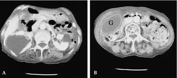
A contrast-enhanced axial CT scan demonstrated a massively distended “floating GB”, with a coni- cal structure connecting the GB to the liver (Fig.
1). No stones were identified. A presumptive diagnosis of torsion of the GB was considered,
with diagnostic laparoscopy performed to confirm the diagnosis. During the laparoscopic procedure, the GB was markedly distended, hemorrhagic and
“floating” away from the liver bed (Fig. 2). The GB was twisted around its axis at the level of the cystic duct and artery, forming a complete torsion of the GB. Three-port laparoscopic cholecystec- tomy was performed without complications. A pathologic specimen showed features of a trans- mural hemorrhage and congestion, compatible with torsion of the GB. The postoperative course was uneventful and the patient was discharged on postoperative day 6.
DISCUSSION
Torsion of the GB is an uncommon occurrence, with a reported incidence of one in 365,520 hospital admissions.1-10 The condition can occur at any age and in either sex, but has a predilection for the elderly, with a 3 : 1 female-to-male ratio.1-10 The etiology is unknown; however, several factors are postulated as playing causative roles.1-10
There are two requirements for torsion of the GB: an anatomic configuration, allowing rotatio- nal GB mobility, and a triggering event that re- sults in the GB twisting around the cystic duct as an axis point.1 The anatomic configurations neces- sary for torsion of the GB are well documented,
Fig. 1. A, B. Preoperative contrast-enhanced axial computed tomographic scan demonstrated a massively distended
“floating gallbladder” outside its fossa and inferior to the liver, with a conical structure (arrow), connecting the gallbladder to the liver (G; gallbladder). No stones were identified.
Fig. 2.During the laparoscopic procedure, the gallbladder was markedly distended, hemorrhagic and “floating”
away from the liver bed.
A B
Yong-Pil Cho, et al.
Yonsei Med J Vol. 46, No. 6, 2005
but the triggering event is poorly understood.
Two types of anomalies have been implicated in the majority of cases, as well as a third, less com- mon condition.1-3 The first may be related to the congenital deformity. Between the 4th and 7th weeks of embryological development, the pars cystica forms from the hepatic diverticulum.
Abnormal migration, with an absence of a GB mesentery, creates a “free-floating GB”. The second occurs by generalized visceroptosis. The mesen- tery of the GB and cystic duct relax and elongate, creating a mobile situation. Atrophy of the liver, loss of visceral fat and elasticity with aging, weight loss and spinal deformities may place the GB in a more dependent position, with a predis- position to torsion. Arteriosclerosis, tortuosity of the cystic duct, cholelithiasis with acute dilatation, violent movements and intense peristalsis of adjacent viscera may also contribute. The third and least common anatomic configuration in- volves a normal GB to liver attachment; however, the lobe of the liver itself lacks coronary and triangular ligaments, which allows the GB and liver lobe, as a unit, to undergo torsion around the cystic duct.
The two types of torsion that may occur are:
complete torsion, with a greater than 180-degree rotation, and incomplete torsion, with a rotation of less than 180 degrees.1-3 Complete torsion oc- cludes both the bile and blood flows, whereas incomplete torsion occludes only the bile flow.
Intense peristalsis by the stomach or duodenum has been implicated in clockwise rotation, where- as the transverse colon is implicated in coun- terclockwise rotation.1-3
Nonspecific symptoms make the preoperative diagnosis of torsion of the GB difficult on the basis of history and physical examination alone.1-10 Laboratory investigations are usually unhelpful.
In recent years, the preoperative diagnosis of this entity has been facilitated with the use of ultra- sound and CT scan.4Safadi et al. reported that the presence of the GB outside its fossa, and inferior to the liver, with an echogenic conical structure are specific sonographic signs of torsion.10Merine et al. also proposed that a massively distended GB, resembling a fluid-filled loop of the bowel with a circular high attenuation structure to the right of the GB on CT scan, were specific signs of
torsion of the GB.11 The diagnostic imaging cri- teria for torsion of the GB can be summarized as follows; fluid collection between the GB and the liver bed indicating a “floating GB”, a GB posi- tioned horizontally along its long axis indicating a free-lying GB, the presence of a well-enhanced cystic duct located on the right side of the GB visualized on CT scan and signs of inflammation, ischemia or necrotic change of the GB.4 In our case, a preoperative CT scan provided several important diagnostic clues; the presence of the GB outside its fossa and inferior to the liver, with a conical structure connecting the GB to the liver, fluid collection between the GB and the liver bed, indicating a “floating GB”, and a massively dis- tended GB with wall thickening.
When torsion of the GB is suspected, an emer- gency cholecystectomy should be performed.
Laparoscopic cholecystectomy is recommended for treating torsion of the GB, as the GB is mini- mally adhered to the liver bed, so can be per- formed easily, with minimal invasion.5 In our case, laparoscopic surgery was successfully per- formed to confirm the diagnosis and treat this condition without the usual requirement of open exploration.
In conclusion, a preoperative CT scan can pro- vide an important diagnostic clue, and laparos- copy successfully used to treat this condition without the usual requirement of open explora- tion. Torsion of the GB remains a benign condi- tion if diagnosed rapidly and treated appro- priately. Therefore, it should be included in the differential diagnosis when such findings are seen on a CT scan in elderly patients presenting with symptoms of acute acalculous cholecystitis.
REFERENCES
1. Schroder DM, Cusumano DA 3rd. Laparoscopic chole- cystectomy for gallbladder torsion. Surg Laparosc Endosc 1995;5:330-4.
2. Vosswinkel JA, Colantonio AL. Torsion of the gallblad- der: laparoscopic identification and treatment. Surg Endosc 1999;13:1154-6.
3. Losken A, Wilson BW, Sherman R. Torsion of the gall- bladder: a case report and review of the literature. Am Surg 1997;63:975-8.
4. Kalimi R, Zarcone J 3rd, McNelis J. Acute necrotizing
Torsion of the Gallbladder
Yonsei Med J Vol. 46, No. 6, 2005 torsion of the gallbladder. Am Surg 2001;67:748-51.
5. Nakao A, Matsuda T, Funabiki S, Mori T, Koguchi K, Iwado T, et al. Gallbladder torsion: case report and review of 245 cases reported in the Japanese literature.
J Hepatobiliary Pancreat Surg 1999;6:418-21.
6. Aibe H, Honda H, Kuroiwa T, Yoshimitsu K, Irie H, Shinozaki K, et al. Gallbladder torsion: case report.
Abdom Imaging 2002;27:51-3.
7. Lyons KP, Challa S, Abrahm D, Kennelly BM. Floating gallbladder: a questionable preclude to torsion: a case report. Clin Nucl Med 2000;25:182-3.
8. Nguyen T, Geraci A, Bauer JJ. Laparoscopic cholecys-
tectomy for gallbladder volvulus. Surg Endosc 1995;9:
519-21.
9. Gonzalez-Fisher RF, Vargas-Ramirez L, Rescala-Baca E, Dergal-Baude E. Gallbladder volvulus. HPB Surg 1993;7:147-8.
10. Safadi RR, Abu-Yousef MM, Farah AS, al-Jurf AS, Shirazi SS, Brown BP. Preoperative sonographic diag- nosis of gallbladder torsion: report of two cases. J Ultrasound Med 1993;12:296-8.
11. Merine D, Meziane M, Fishman EK. CT diagnosis of gallbladder torsion. J Comput Assist Tomogr 1987;11:
712-3.