C-reactive Protein and Lipid Profiles in Korean Patients With Normal Tension Glaucoma
Jaewan Choi, MD, PhD
1,2, Soo Geun Joe, MD
2, Mincheol Seong, MD
2,3, Jin Young Choi, MD
1, Kyung Rim Sung, MD
2, Michael S. Kook, MD
21
HanGil Eye Hospital, Incheon, Korea
2
Department of Ophthalmology, University of Ulsan, College of Medicine, Asan Medical Center, Seoul, Korea
3
Hanyang University Guri Hospital, Guri, Korea
Purpose: To compare high-sensitivity C-reactive protein (hsCRP) levels and lipid profiles between Korean normal tension glaucoma (NTG) patients and healthy controls.
Methods: This cross-sectional study included 38 Korean patients with NTG and 38 age- and sex-matched healthy control subjects. We excluded the patients with cardiovascular risk factors and other systemic diseases that might affect CRP levels and lipid profiles. Each patient underwent a Humphrey visual field examination and blood sam- pling for hsCRP and lipid profile analyses. Subsequently, the NTG patients were classified into two groups based on their untreated intraocular pressure (IOP) level: low NTG (LNTG) with IOP≤13 mmHg (13 subjects) and high NTG (HNTG) with relatively high IOP (>13 and ≤21 mmHg, 25 subjects). The hsCRP levels and lipid profiles were compared between NTG patients and healthy controls, and between LNTG, HNTG, and healthy controls.
Results: There were no significant differences in hsCRP and lipid profiles between either the NTG patients and healthy controls, or between the LNTG, HNTG, and controls (p>0.05) after exclusion of Korean patients with cardiovascular risk factors. There was no significant association between hsCRP and visual field indices (p>0.05).
Conclusions: High-sensitivity C-reactive protein-related vascular inflammatory conditions may not be directly asso- ciated with the development of NTG, regardless of the untreated IOP level.
Korean J Ophthalmol 2009;23:193-197 ⓒ 2009 by the Korean Ophthalmological Society.
Key Words: Atherosclerosis, C-reactive protein, Lipid profiles, Normal tension glaucoma
Received: October 6, 2008 Accepted: August 5, 2009
Reprint requests to Michael S. Kook, MD. Department of Ophthalmology, University of Ulsan, College of Medicine, Asan Medical Center, #388-1 Pungnap 2-dong, Songpa-gu, Seoul 138-736, Korea. Tel: 82-2-3010-3677, Fax: 82-2-470-6440, E-mail: [email protected]
* This article was presented as a poster at the Association for Research in Vision and Ophthalmology annual meeting, May 6 - May 10, 2007; Fort Lauderdale, Florida.
Normal tension glaucoma (NTG) is a clinical disease entity characterized by progressive thinning of the retinal nerve fiber layer and corresponding visual field loss in eyes with a normal range of intraocular pressure (IOP). Hypotheses associated with the development of NTG include vascular dysregulatory factors, such as systemic blood pressure changes, ocular blood flow abnormalities, or vasospasm.
1-3Many patients with glaucoma have an increased endothelin-1 plasma level.
4In patients with primary open angle glaucoma (POAG), both systemic arterio- sclerosis and sclerotic changes in the ocular vessels have been observed but also questioned.
5,6Theoretically, these sclerotic changes in ocular vasculature might result in the impairment of ocular perfusion, with thickening of the vascular endothelial wall.
C-reactive protein (CRP) is an early acute phase reactant of inflammation, increasing in response to active infections or acute inflammatory processes. Minor elevations of CRP are also present in chronic inflammatory situations, such as athe- rosclerosis.
7High-sensitivity CRP (hsCRP) is the most reliable and accessible marker of inflammation associated with athe- rosclerosis.
8It was also found to be correlated with impaired systemic endothelial vascular reactivity, and it was suggested that it may have a role in the pathogenesis of endothelial dys- function and atherogenesis.
9,10Risk factors for atherosclerosis are also important fields of interest. Any cardiovascular risk factor which can damage the vascular endothelial wall could increase the incidence of atherosclerotic change in the vessels.
Dyslipidemia is one of the most important risk factors for the development of atherosclerosis.
The vascular dysfunction observed in patients with NTG
may be a consequence of systemic vascular endotheliopathy.
11A population-based epidemiological survey in Japan revealed
that 92% of POAG patients had a normal IOP range.
12The
prevalence of NTG in the Japanese population is higher than
that of Western countries.
13,14Because of this, it could be
speculated that there might be a different underlying mecha-
nism of pathogenesis for NTG in Asians.
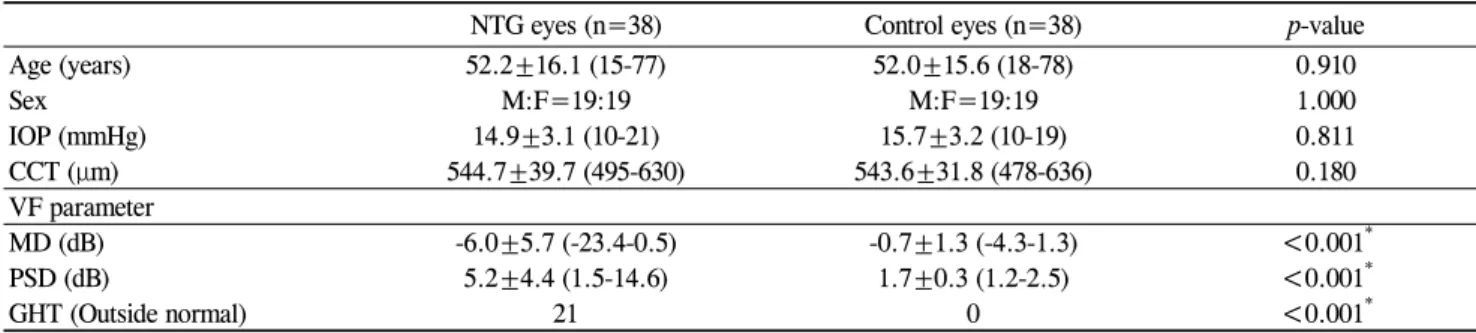
Table 1. Descriptive statistics for the demographics and clinical characteristics of the study patients
NTG eyes (n=38) Control eyes (n=38) p-value
Age (years) 52.2±16.1 (15-77) 52.0±15.6 (18-78) 0.910
Sex M:F=19:19 M:F=19:19 1.000
IOP (mmHg) 14.9±3.1 (10-21) 15.7±3.2 (10-19) 0.811
CCT (μm) 544.7±39.7 (495-630) 543.6±31.8 (478-636) 0.180
VF parameter
MD (dB) -6.0±5.7 (-23.4-0.5) -0.7±1.3 (-4.3-1.3) <0.001
*PSD (dB) 5.2±4.4 (1.5-14.6) 1.7±0.3 (1.2-2.5) <0.001
*GHT (Outside normal) 21 0 <0.001
*Data are expressed as number (percentage) of subjects except for age: mean±SD (range).
NTG=normal tension glaucoma; IOP=intraocular pressure; CCT=central corneal thickness; VF=visual field; MD=mean deviation; PSD=
pattern standard deviation; GHT=glaucoma hemifield test.
*
Statistical significance was found using an independent samples t-test (p<0.05). Chi-square tests were performed for the sex distribution and GHT.
The roles of atherosclerosis and dyslipidemia in the develop- ment of NTG have not yet been well defined. Leibovitch et al.
found that plasma hsCRP levels were significantly higher in the NTG cases than in the controls.
15However, Su et al. found no significant differences in hsCRP levels and lipid profiles in NTG patients when compared with age- and sex-matched controls.
16,17We compared hsCRP and lipid profiles between patients with NTG and healthy controls in a homogenous Korean po- pulation, in which NTG is presumably the most common type of open-angle glaucoma. We also investigated whether or not there was any difference in hsCRP and lipid profiles between NTG patients with high levels of IOP and those with low IOP levels at the initial diagnosis of glaucoma.
Materials and Methods
We performed an age- and sex-matched case-control study with 38 newly diagnosed NTG patients and 38 healthy control subjects. All subjects were Korean. Each subject underwent a complete slit-lamp examination, including gonioscopy, IOP evaluation by Goldmann applanation tonometry, Humphrey visual field 24-2 full threshold or SITA standard examination (Humphrey Visual Field Analyzer II Model 745i, Carl Zeiss Meditec, Dublin, CA), and fasting blood sampling for hsCRP and lipid profiles including total cholesterol, low density lipo- protein (LDL) cholesterol, high density lipoprotein (HDL) cho- lesterol, and triglycerides.
Normal tension glaucoma eyes (n=38) were defined as having:
(1) an untreated IOP not exceeding 22 mmHg at different times of the day, from 9 AM to 5 PM (2) open anterior chamber angles on gonioscopy (3) glaucomatous optic neuropathy with thinning or notching of the neuroretinal rim and (4) reliable glaucoma- tous visual field defects with Glaucoma Hemifield Test results (GHT) outside of 99% of the age-specific normal limits or a PSD outside of 95% of the normal limits.
Thirty-eight healthy volunteers were recruited from within the medical staff or clinic patients who had previously under- gone cataract surgery. All subjects had a best-corrected visual
acuity of 20/30 or better. The normal appearances of the optic nerve heads in both eyes were confirmed by a complete oph- thalmic examination. The control eyes had intact neuroretinal rims; there was no evidence of disc hemorrhage, notching, ex- cavation, or asymmetry of the vertical cup to disc ratio >0.2.
No other pathologic ocular condition other than cataract was noted. No subject in the control group had a history of IOP elevation above 21 mmHg or ocular trauma. The visual fields of the control subjects were all normal. The GHT result was within normal limits, and the field did not meet any of the following criteria described above.
Subjects with systemic diseases that might be associated with an increased level of CRP (hypertension, hypercholeste- rolemia, diabetes mellitus, cerebral vascular accident, infection, chronic pulmonary obstructive disease or autoimmune diseases) were excluded from the study, thus none of our subjects were taking systemic medication that might affect CRP level, such as anti-hypertensives, cholesterol-lowering agents, or aspirin.
Normal subjects were selected to match NTG patients by age and sex. For both NTG and control subjects, if both eyes met the inclusion criteria for the study, one eye was selected at random. The affected eye was selected in unilaterally di- seased NTG patients. NTG patients were initially classified into two groups based on their untreated IOP levels: one with low IOP (≤13 mmHg low normal tension glaucoma, LNTG n=13), and the other with relatively high IOP (>13 and ≤ 21 mmHg high normal tension glaucoma, HNTG n=25). Data are expressed as the mean±standard deviation. Continuous variables were compared between the NTG and control groups using the unpaired Student’s t-test and were compared among the LNTG, HNTG, and controls with the Kruskal-Wallis test.
Categorical variables between groups were compared with the
Chi-square test. Differences with a value of p<0.05 were con-
sidered statistically significant. Subsequently, NTG patients
were classified based on the IOP cutoff point of 16 mmHg for
the comparison of hsCRP and lipid profiles (IOP ≤16 mmHg,
n=23 >16 and ≤21 mmHg, n=15). All applicable institutional
and governmental regulations concerning the ethical use of
human volunteers were followed during this study.
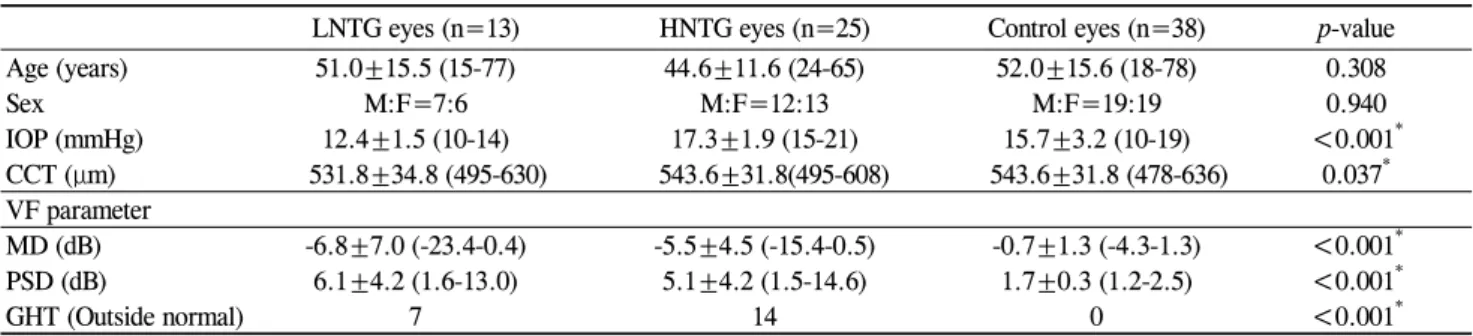
Table 2. Descriptive statistics for the demographics and clinical characteristics of LNTG, HNTG, and control patients
LNTG eyes (n=13) HNTG eyes (n=25) Control eyes (n=38) p-value
Age (years) 51.0±15.5 (15-77) 44.6±11.6 (24-65) 52.0±15.6 (18-78) 0.308
Sex M:F=7:6 M:F=12:13 M:F=19:19 0.940
IOP (mmHg) 12.4±1.5 (10-14) 17.3±1.9 (15-21) 15.7±3.2 (10-19) <0.001
*CCT (μm) 531.8±34.8 (495-630) 543.6±31.8(495-608) 543.6±31.8 (478-636) 0.037
*VF parameter
MD (dB) -6.8±7.0 (-23.4-0.4) -5.5±4.5 (-15.4-0.5) -0.7±1.3 (-4.3-1.3) <0.001
*PSD (dB) 6.1±4.2 (1.6-13.0) 5.1±4.2 (1.5-14.6) 1.7±0.3 (1.2-2.5) <0.001
*GHT (Outside normal) 7 14 0 <0.001
*Data are expressed as number (percentage) of subjects except for age: mean ±SD (range).
NTG=normal tension glaucoma; IOP=intraocular pressure; CCT=central corneal thickness. VF=visual field; MD=mean deviation; PSD=
pattern standard deviation; GHT=glaucoma hemifield test.
*