Original Articles Korean Circulation J 1999;;;;29((((7))))::::712-721
임상 증상이 심하지 않은 심낭 삼출 환자의 장기 추적 결과
안동성소병원 순환기내과,
1계명대학교 의과대학 내과학교실 순환기내과,
2생화학교실
3배장호
1
·김기식2
·강미숙2
·남명희2
·김미정2
·김민수2
김기영2
·남창욱2
·김윤년2
·김권배2
·정시전1
·김여희3
Long-term Follow-Up Results of the Patients with Clinically Inapparent Pericardial Effusion
Jang-Ho Bae, MD
1, Kee-Sik Kim, MD
2, Mi-Sook Kang, MT
2, Myung-Hee Nam, MT
2, Mi-Jung Kim, MD
2, Min-Soo Kim, MD
2, Ki-Young Kim, MD
2, Chang-Wook Nam, MD
2, Yoon-Nyun Kim, MD
2, Kwon-Bae Kim, MD
2, Shee-Juhn Chung, MD
1and You Hee Kim, MD
31
Division of Cardiology, Department of Internal Medicine, Andong Presbyterian Hospital, Andong,
2
Division of Cardiology, Department of Internal Medicine,
3Biochemistny, School of Medicine, Keimyung University, Taegu, Korea
ABSTRACT
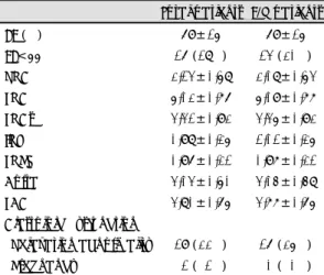
Background:Pericardiocentesis is not routinely recommended in most patients with pericardial effusion (PE), except for patients with cardiac tamponade. However, the long-term follow-up results in patients with clinically not significant PE are few. Methods:Sixty-five consecutive patients (mean age;57 yrs, 26 males) out of 87 patients with PE, who were clinically not serious, were studied prospectively once in every two month for mean 6 months (2-12 months) withoutt any specific treatment. Theamount of PE was measured at the enddiastole period of parasternal long axis view and apical four chamber view. Results:
The incidence of insignificant PE in our echocardiographic laboratory is 3.4% (n=87 from 2461). The maximal accumulation site of PE was posterior (n=51, 79%). The next is anterior (n=11, 17%) and right ventricular side (3, 5%). The amount of PE is less (0.37±0.17cm vs 0.64±0.54cm, p=0.018) in localized PE (n=24, 37%) than that of diffuse form (n=41, 63%), which spreads to more than 2 chambers. The presumptive etiologies of PE were unknown (n=41), heart failure (n=5), myocardial infarction (n=6), viral (n=3), and others (n=10). The amount of PE was decreased from 0.54±0.46 cm to 0.30±0.26 cm, 0.23±0.24 cm, and 0.21±0.23 cm 2, 4, and 6 months after intial evaluation, respectively, without any complication. Conclusion:The patients with PE, not combining with cardiac tamponade, can be followed by regular eechocardiographicexamination without any specific treatment. ((((Korean Circulation J 1999;
29((((7)))):712-721))))
KEY WORDS:Pericardial effusion·Treatment·Echocardiography.
논문접수일:1999년 4월 2일 심사완료일:1999년 6월 9일
교신저자:김기식, 700-712 대구광역시 중구 동산동 194 계명대학교 의과대학 내과학교실 순환기내과 전화:(053) 250-7379・전송:(053) 250-7434
E-mail:[email protected]