Corresponding Author: Man-Bin Yim, M.D., Department of Neurosurgery, Keimyung University School of Medicine 56 Dalseong-ro, Jung-gu, Daegu 700-712, Korea
Tel: +82-53-250-7332 E-mail: [email protected]
Abstract
To obtain the successful management of complex aneurysm require multimodality methods, including clipping and/or bypass procedure and/or endovascular method. In this paper, author’s management experiences of complex aneurysms and key points of surgical and management strategies for those lesions are described.
During the past 26 years, 2,821 cases of cerebral aneurysm were managed with surgical and/or endovascular methods. Among these patients, one hundred eleven cases are regarded as complex aneurysm (large size, complex wide neck, intra-aneurysmal thrombus, fusiform or bizarre shape, aneurysm associated with vessel anomaly). The treatment strategies, surgical complications and outcome of those cases are analyzed and, based on those experiences, the author would like to suggest key points of the management methods for those aneurysms. The aneurysms were managed by direct clipping of aneurysmal neck in 79 cases, coiling with Guglielmi detachable coil (GDC) in 11, aneurysm trapping or clipping followed by extracranial- intracranial (EC-IC) bypass in 7, excision of aneurysm with or without end-to-end anastomosis of parent artery in 6, proximal clipping or coiling of parent artery in 3, and aneurysmorrhaphy, bypass, or wrapping only in 5.
Management complications occurred in 28 cases. Distal thromboembolism, parent vessel stenosis and important vessel injury after clipping or endovascular management of aneurysm are the main complications.
The author obtained good outcome in 91 (82.0%) and poor outcome in 11 (9.9%) cases. The author concludes that selection of suitable management method for each case, high quality of surgical and endovascular technique, and combination of endovascular, clipping and bypass methods are important key points for the successful treatment of complex aneurysm.
Key Words : Bypass, Clipping, Coiling, Complex aneurysm, Management, Outcome Department of Neurosurgery,
Keimyung University School of Medicine, Daegu, Korea Man-Bin Yim, M.D.
Management of Complex Cerebral Aneurysm
Introduction
Treatment of patients with complex aneurysm is o n e o f t h e m o s t c h a l l e n g i n g w o r k s f o r neurosurgeons and it requires the use of more than one treatment modality [1,2].
Large/giant aneurysm of cavernous sinus internal carotid artery (ICA), clinoid segment of ICA aneurysm, large/giant aneurysm of anterior or posterior circulation, bizarre shaped aneurysm, aneurysm associated with vessel anomaly and reoperation after surgical clipping or endovascular coiling of aneurysms are regarded as complex aneurysm. The treatment of these aneurysms are required open surgery, endovascular treatment, or a combination of these modalities in a one-step or staged fashion [2].
The successful treatment of large/giant symptomatic cavernous sinus ICA aneurysm is usually required a high flow bypass from cervical ICA or external carotid artery (ECA) to middle cerebral artery (MCA) using radial artery or saphenous vein followed by an obliteration of ICA [3,4].
The treatment of clinoid segment of large ICA aneurysm is usually required orbitozygomatic osteotomy, complete removal of anterior clinoid process (ACP) and optic strut, opening of optic canal, circumferential section of dural ring and clipping of aneurysm with using multiple fenestrated clips [4].
The paraclinoid large/giant aneurysm usually required retrograde suction decompression by blood withdraw from cervical ICA [5]. The aneurysm was clipped using multiple fenestrated clips in tandem fashion [6]. After clipping, the patency and blood flow of parent artery should be confirmed by applying of microDoppler probes on the wall of ICA or intraoperative angiography [6].
The tension of the large and giant aneurysmal
wall is very high. Reducing the intra-aneurysm pressure with temporary arterial clipping may facilitate adequate placement of clips. Spinal needle aspiration of aneurysm sac or thrombectomy with ultrasonic aspirator in thrombosed aneurysm after temporary trapping of aneurysm is very useful method for surgery of large/ giant aneurysm [5,7,8].
The clips should be placed parallel with the long axis of the parent vessel to prevent kinking of the parent artery.
In case of bizarre shaped peripheral aneurysm, resection of aneurysm followed by superficial temporal artery (STA) to peripheral intracranial vessel anastomosis can be performed. The integration of endovascular and open surgical strategies may permit treatment of certain large/giant aneurysms that are not safely or effectively treated by either modality alone.
This article describes author’s experiences of multimodality treatment methods for complex aneurysms.
Material & Methods
A retrospective review of intracranial aneurysms which are regarded as complex aneurysm as well as managed at our institute is performed. During from the September 1982 to December 2009, 2,821 aneurysms were managed by surgical and/or endovascular methods. Among these aneurysms, one hundred eleven cases are regards as complex aneurysm. Of these, one hundred four cases are saccular, three cases are fusiform or bizarre shaped aneurysm, and four cases are aneurysm associated with vessel anomaly (Table 1). The treatment strategies, surgical complications and outcome of those cases are analyzed. The outcome after treatment is classified as follows [9] 1) good outcome: resumption of normal life despite minor
deficits, 2) fair outcome: moderate to severe disability, 3) poor outcome: persistent vegetative state, 4) death.
Results
The most frequent treatment method of complex aneurysm is directing clipping of aneurysm neck (79
cases), followed by endovascular coiling of aneurysm sac with GDC coil (11 cases), aneurysm trapping or clipping followed by EC-IC bypass (7 cases), excision of aneurysm with or without end- to-end anastomosis of parent artery (6 cases), proximal clipping or coiling of parent artery (3 cases), and aneurysmorrhaphy, bypass, or wrapping only (5 cases) (Table 2).
Management complications occurred in 28 cases.
Distal thromboembolism (6 cases) and parent vessel stenosis or important vessel injury (6 cases) after clipping or endovascular management of aneurysm are the main complications. The other complications are accidental cutting of posterior communicating artery, occlusion of anterior choroidal artery, injury of perforators, injury of optic nerve, occurrence of postoperative intracerebral hematoma, temporary paralysis of lower cranial nerves, etc. (Table 3).
The author obtained good outcome in 91 (82.0%), fair outcome in 9 (8.1%), poor and death outcome in 11 (9.9%) cases (Table 4).
Table 1. The surgical and/or endovascular management of cerebral aneurysms
*Total treated number of aneurysm from September, 1982 to December, 2009.
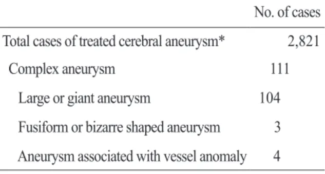
No. of cases Total cases of treated cerebral aneurysm* 2,821
Complex aneurysm 111
Large or giant aneurysm 104 Fusiform or bizarre shaped aneurysm 3 Aneurysm associated with vessel anomaly 4
Table 2. The management methods of complex aneurysms
SVG=saphenous vein graft; STA=superficial temporal artery; VA=vertebral artery; PICA=posterior inferior cerebellar artery; An=aneurysm; GDC=Guglielmi detachable coil; ICA=internal carotid artery; Bif=bifurcation.
No. of cases
Direct clipping of aneurysm neck 79
GDC coil embolization 11
Aneurysm trapping or clipping combined with bypass (SVG or STA) 7
Excision of aneurysm with/without anastomosis or bypass 6
Proximal parent vessel clipping (VA-PICA An) or coil embolization 3
Aneurysmorrhaphy (ICA Bif An) 1
Bypass or wrapping of aneurysm only 4
Discussion
1. The large aneurysm of cavernous sinus portion of the ICA
Cavernous sinus region aneurysms account for approximately 3% to 5% of all intracranial aneurysms, and aneurysms with intractable facial
pain or progressive neurological symptoms are indicated for surgical intervention [4]. In case of aneurysm which is not able to tolerable to balloon occlusion test (BOT) can be treated by a saphenous vein graft (SVG) bypass from the cervical ICA or ECA to the MCA followed by ICA occlusion [3,4,10]
(Illustrative case 1).
The normal blood flow of the MCA is about 250 mL/minute to the cerebral hemisphere. The average blood flow through STA graft, radial artery (RA) graft and a SVG are 10 to 70 mL/minutes in STA graft, 50-150 mL/minutes in RA graft and 100 to 200 mL/minute in SVG [11-13]. Therefore, the blood flow through the STA graft is not enough, but SVG or RA graft is enough to support the circulation in an entire major arterial territory at a level well above the ischemic threshold [12,14].
The advantages of SVG compare to RA are sufficient length, less occurrence of spasm and immediate acquirement of high blood flow after bypass. The disadvantages of that graft are Table 4. The outcomes* after management of complex
cerebral aneurysms
*: Good = resumption of normal life despite minor deficit; Fair = moderate to severe disability; Poor = persistent vegetative state.
Case (%)
Good 91 (82.0)
Fair 9 (8.1)
Poor 6 (5.4)
Death 5 (4.5)
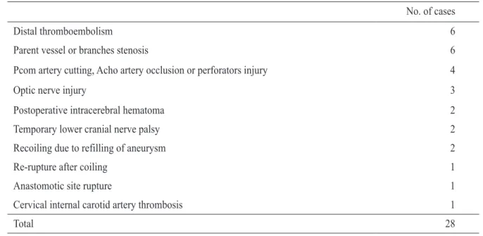
Table 3. The complications of surgical and/or endovascular management of complex aneurysms
Pcom=posterior communicating; Acho=anterior choroidal.
No. of cases
Distal thromboembolism 6
Parent vessel or branches stenosis 6
Pcom artery cutting, Acho artery occlusion or perforators injury 4
Optic nerve injury 3
Postoperative intracerebral hematoma 2
Temporary lower cranial nerve palsy 2
Recoiling due to refilling of aneurysm 2
Re-rupture after coiling 1
Anastomotic site rupture 1
Cervical internal carotid artery thrombosis 1
Total 28
technically more difficult to perform anastomosis due to the thicker wall of the saphenous vein than the intracranial vessels, more prone to kinking at the distal anastomotic site [11,15]. Because of the high flow through the SVG, there may be a flow mismatch when it is anastomosed into vessels smaller than 2 mm in diameter, which could lead to turbulence and graft flow problems [13]. Key operative tips of SVG include (1) occlusion and removal of the vein immediately before the anastomosis to minimize endothelial damage, (2) minimal pressure distension of the vein when checking for holes, (3) preventing the vein from twisting during tunneling, (4) anastomosis end-to- side to the ECA to preserve collaterals [16] and finally, (5) early occlusion of ICA (operative or endovascular) to ensure a flow demand for the bypass, which promote patency [17]. In terms of the ICA occlusion, the occlusion by clip placement is preferred to endovascular coiling to prevent of perforating impairment caused by embolic or thrombotic event [18].
Illustrative case 1
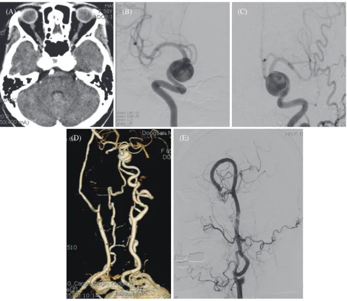
This 58-year-old woman suffered from sudden development of diplopia and severe facial pain on right side. Neurological examination revealed total opthalmoplegia (full dilatation of pupil, loss of light reflex, fixed eyeball movement) on right side.
Enhanced brain computed tomography (CT) showed bilateral large round aneurysms at the cavernous portion of the ICAs (Fig. 1A). Bilateral ICA angiograms also revealed large aneurysms at the cavernous portion of the ICAs (Fig. 1B & 1C).
During performing of BOT of the right ICA, she showed some deterioration of consciousness. About 3 weeks after BOT, interpositional SVG between ECA to MCA followed by surgical ICA occlusion was performed. Postoperative cervical CT
angiogram and common carotid artery (CCA) angiograms revealed a good patience of vein graft from ECA to MCA and disappearance of aneurysm of the right cavernous portion of the ICA (Fig. 1D &
1E). About 6 months later after operation, facial pain and total ophthalmoplegia improved near completely.
2. Clinoid segment ICA aneurysm [4,19,20]
Two types of clinoid segment aneurysm can be divided according to their site of origin and direction of projection. The anterolateral type arises from the anterolateral surface of the clinoid segment as it obliquely ascends toward the dural ring underneath the ACP. The medial type extends from the medial surface of the clinoid segment, and enlarges toward the sphenoid sinus and sella.
Kobayashi et al. [21] described variant of medial type as carotid cave aneurysm which locate intradurally at the dural penetration of the ICA on the ventromedial side and carry a risk for intracranial hemorrhage. Small clinoid segment aneurysms are good candidate for treatment with endovascular coiling, because most have a narrow neck. In case of large aneurysms with broad neck require surgical clipping of aneurysmal neck [4].
In the surgical management, cervical ICA prior to craniotomy is exposed to gain proximal control, and take some fat tissue from abdomen for packing at the dural defect site after closing of dura.
Orbitozygomatic bone flap is usually made in one piece. This approach provides more easily removal of ACP extradually. The ACP is internally hollowed out with a high-speed diamond drill, and the remaining thin remnants are carefully removed with small rongeurs. Bleeding is easily controlled with bone wax and by packing a small amount Surgicel.
After opening of dura, circumferential section of dural ring performs to allow complete mobilization
of the ICA (Fig. 2D) and viewing of the entire segment. The anterolateral type can be clipped with using a gently curved or side-angled clip along the long axis of the ICA, paralleling the curve of the clinoidal ICA. Medial type aneurysms usually can be clipped with using a fenestrated clip placed parallel to the ICA with the tips abutting or extending pass
the proximal dural ring (carotid-oculomotor membrane) (Fig. 2E). After clipping of aneurysm, the dural leaves are closed with watertight suture, followed by packing of fat tissue in order to sealing of dural defect.
(D) (E)
Fig. 1. (A) Enhanced brain CT, axial view, showing large aneurysms at the bilateral cavernous portion of the ICAs. (B & C) Bilateral cervical ICA angiograms, A-P views, revealing large aneurysms at the bilateral cavernous portion of the ICAs. (D & E) Postoperative cervical CT angiogram, A-P view, and CCA angiogram, lateral view, revealing a good patience of vein graft from ECA to MCA and disappearance of aneurysm of the right cavernous portion of the ICA.
(A) (B) (C)
Illustrative case 2
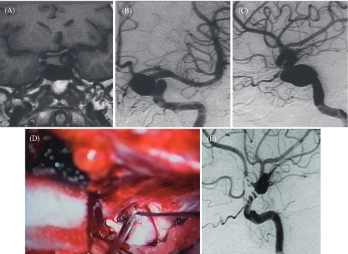
This 54-year-old woman had been suffered from headache and palpitation for one week . She visited local radiologic clinic where checked sella brain magnetic resonance image (MRI) which revealed large aneurysm of clinoid segment of left ICA (Fig.
2A). This aneurysm elevated the left side of optic chiasm. Left ICA angiogram, anterior-posterior (A-P) view (Fig. 2B) and lateral view(Fig. 2C) also
demonstrated large medial directed aneurysm originated from clinoid segment of ICA. After making orbitozygomatic osteotomy, the bone flap removed in a one piece followed by removal of ACP completely. Circumferential section of dural ring (Fig. 2D) performs and clipped the aneurysm using several fenestrated clips placed parallel to the ICA. Postoperative ICA angiogram, lateral view, showed complete obliteration of the aneurysm (Fig.
2E).
(D) (E)
Fig. 2. (A) Brain sella MRI, coronal view, showing a large aneurysm of clinoid segment of left ICA which elevates the left side of optic chiasm. (B & C) Left carotid angiogram, A-P (B) and lateral views (C), also demonstrating large medial directed aneurysm originated from clinoid segment of ICA. (D) Operation view showing circumferential section of dural ring. (E) Postoperative ICA angiogram, lateral view, showing complete obliteration of the aneurysm using multiple fenestrated clips placed parallel to the ICA.
(A) (B) (C)
3. Large, giant and bizarre shaped aneurysm
The successful management of large and giant aneurysm is very difficult due to several structural characteristics such as high aneurysm wall tension, thick and/or calcified aneurysm wall, frequent occurrence of intra-aneurysm thrombus, wide neck, frequent involvement of parent vessel or branches into aneurysm neck [4] and frequent recanalization of aneurysm sac after endovascular treatment [7].
Because of the aneurysm’s enormous bulk, it is often difficult to dissect the aneurysm neck from the neural or vascular structures as well as proximal and distal control of vessel during surgery [22]. The size of neck becomes wide-based and bulbous, often incorporating the parent artery or perforating arteries. The parent artery may enter and exit the aneurysm separately. These morphological features may make it extremely difficult to dissect the neck completely and clip it safely. The clip will Fig. 3. (A) Parallel clipping of large aneurysm to the parent vessel with tandem clip placement. Tandem clips can
be used to reinforce on another to cover a wide aneurysm neck. [6]. (B) Right ICA angiogram, A-P view, showing a medial directed large paraclinoid aneurysm. (C) Operation view showing a large aneurysm which elevated optic nerve. The aneurysm was clipped using multiple fenestrated clips in tandem fashion with suction decompression through blood withdraw from cervical ICA [5]. (D) Postoperative right ICA angiogram, A-P view, demonstrating complete disappearance of aneurysm with maintenance patency of ICA lumen by using tandem clips placement.
(A)
(C)
(B)
(D)
frequently slide down, compromising the parent vessel or critical perforating arteries.
Laminated thrombus is common in giant aneurysm, and can obstruct complete clipping of aneurysm neck. The lumen demonstrated angiographically is frequently not an accurate reflection of the true aneurysm size and shape. CT or MRI is preferable for delineating the aneurysm
architecture and its relationship to surrounding structures. It is necessary in these cases to perform a thrombectomy with using ultrasonic aspirator after applying of temporary clips on proximal and distal vessels to the aneurysm.
The presence of intraluminal thrombus, platelet aggregates, and fibrin debries increase the danger of distal embolization into the parent vessel during Fig. 4. Aneurysm clip closing force. Graphs illustrating the lever law (top) and spring law (bottom). The
aneurysm clip closing force is inversely proportional to the distance along the blades from the fulcrum, according to the lever law (A-C), and directly proportional to the gap between the blades, according to the spring law (X-Z) [19].
surgery. The judicious use of temporary trapping with heparinization may prevent such peripheral embolization.
The tension of the large and giant aneurysmal wall is very high due to the law of Laplace as T = PR, where T = tension of aneurysmal wall, P = transmural pressure and R = vessel radius [23].
Reducing the intra-aneurysm pressure with temporary arterial clipping may facilitate adequate placement of clips. Spinal needle aspiration of aneurysm sac or retrograde suction decompression
(Illustrative case 3) after temporary trapping of aneurysm is very useful method for surgery of large and giant aneurysm [5,7,8,24].
The optimal skull base approach to a giant aneurysm offers a wide degree of exposure with full visualization of the aneurysm’s origin, outflow, associated perforators, adjacent vessels, and nearby neural structures. Orbitozygomatic pterional approache is one of the most commonly used skull base approaches to large or giant aneurysm of anterior circulation. It provides a lower trajectory Fig. 5. (A) Axial view of MRI showing large round low signal mass at left temporal lobe. (B) Left ICA
angiogram, A-P view, demonstrating a large aneurysm originated from trifurcation of MCA. The aneurysm has a broad neck and temporal branch of MCA involves in neck of aneurysm. (C) Postoperative left ICA angiogram, A-P view, showing disappearance of aneurysm with occlusion of the temporal branch of MCA. (D) Postoperative left ECA angiogram, lateral view, showing well blood supply to distal portion of temporal branch through superficial temporal artery.
(A)
(C)
(B)
(D)
along the skull base, reducing the need for cerebral retraction [6]. It is especially ideal approach for ophthalmic, paraclinoid, superior hypophysial, posterior communicating, anterior choroidal, and ICA bifurcation large and giant aneurysms. It is also useful approach for large and giant aneurysm of MCA, because the approach trajectory to the proximal M1 segment and the aneurysm neck is optimized [6].
Because of the high aneurysm wall tension or thick, laminated clot in the wall of the aneurysm
neck, even long, strong aneurysm clip often will not close the aneurysm neck completely. It is necessary in these cases to clip the aneurysm neck with using combined fenestrated and short blade clips or tandem clip placement (Fig. 3A) because the aneurysm clip closing force is inversely proportional to the distance along the blades from the fulcrum and directly proportional to the gap between the blades [19]. The lowest closing force along the clip is located at its tip [6] (Fig. 4). Clip application is usually performed beyond the true neck of the Fig. 6. (A) An enhanced brain CT, axial view, showing partial thrombosed large aneurysm at left temporal lobe. (B & C) 3-dimensional left ICA angiogram, A-P view (B) and oblique view (C), revealing a fusiform shaped aneurysm at the left M2 portion of the MCA. (D & E) The postoperative left ICA and ECA angiograms, lateral views, showing complete disappearance of aneurysm as well as well blood supplying to distal M2 portion of the MCA through the anastomsed STA.
(A)
(D)
(B) (C)
(E)
lesion (surgical vs. anatomical neck) so as to prevent kinking and other compromise of the parent vessel as well as thrombus and wall atheroma extend into the neck of parent vessel and compromise the ostia of vital arterial branches [7].
The clips should be placed parallel with the long axis of the parent vessel to prevent kinking of the parent artery (Fig. 3A)[22]. Various combinations of straight and angled aperture clips in tandem fashion can be positioned to obliterate the wide neck of large and giant aneurysm (Illustrative case 3).
Although externally the clip may seem to be in good
position, the aneurysm may continue to fill due to intermittent, slight opening of the clip. Complete obliteration of the aneurysm in such case may be accomplished by a series of clips stacked one on top of another above the original clip (Illustrative case 4) [22].
Microvascular bypass followed by open surgical or endovascular parent vessel occlusion has been employed in a wide variety of circumstances. This combined approach has been used for serpentine or fusiform intracranial aneurysm. In case of peripheral serpentine or fusiform intracranial Fig. 7. (A) A T1-weighted MRI revealing a thrombosed giant aneurysm which compressing the brainstem.
(B) Right VA angiogram, A-P view, reveals partial thrombosed giant aneurysm at the right VA. (C) Left VA angiogram after packing of right VA aneurysm sac and parent artery with coils, A-P view, reveals complete disappearance of aneurysm. (D & E) The follow-up CT and VA angiogram after 6 months following coiling of aneurysm and parent artery show partial filling of aneurysm through distal right VA from the contralateral left VA. (F) The follow-up CT, taken 5 months after trapping of aneurysm, reveals near same size of aneurysm. (G) The follow-up CT after aneurysmectomy and removal of the thrombus shows the decreased size of aneurysm.
(D) (E) (F) (G)
(A) (B) (C)
aneurysm, resection of aneurysm followed by STA to peripheral intracranial vessel anastomosis can be performed [25] (Illusrtative case 5).
The integration of endovascular and open surgical strategies may permit treatment of certain giant aneurysms that are not safely or effectively treated by either modality alone [26] (Illustrative case 6). Partial surgical clipping of a large or giant aneurysm followed by endovascular coil embolization of the patent remnant is another important combined approach [7].
Illustrative case 3
This 64-year-old woman suffered from visual disturbance for 6 months. Neurological examination revealed bitemporal hemianopsia. She was checked MRI at another hospital which revealed a round mass at sella region. Right ICA angiogram, A-P view, demonstrated a medial directed large paraclinoid aneurysm (Fig. 3B). The aneurysm was clipped using multiple fenestrated clips in tandem fashion (Fig. 3A) with retrograde suction decompression through blood withdraw from cervical ICA (Fig. 3C)). The ICA patency and blood flow after clipping of aneurysm was confirmed by applying of microDoppler probes on the wall of ICA. Postoperative angiogram demonstrated complete disappearance of aneurysm with maintenance patency of ICA lumen (Fig. 3D).
Illustrative case 4
This 55-year-old woman suffered from transient weakness on right side with brief period of dysarthria. She was checked brain CT and MRI which revealed round homogenous enhanced mass in CT and large round low signal mass in axial view of MRI at left temporal lobe (Fig. 5A). The left ICA angiogram demonstrated a large aneurysm
originated from trifurcation of MCA. The aneurysm had a broad neck and involved the temporal branch of MCA in neck of aneurysm (Fig. 5B). After making an orbitozygomatic osteotomy, STA was anastomosed to distal part of temporal branch of the MCA initially to take precaution against occlusion or stenosis of temporal branch after clipping of broad neck of aneurysm. The aneurysm was clipped using multiple types of clips after internal decompression with opening of aneurysm sac after transient trapping of aneurysm. During trapping of aneurysm, heparin 5,000 unit, phenytoin 200 mg, pentothal 200 mg were infused through intravenous route. Postoperative left ICA angiogram showed disappearance of aneurysm with occlusion of the temporal branch of MCA (Fig. 5C). The distal part of occluded temporal branch received blood through anastomosed STA (Fig. 5D).
Illustrative case 5
This 68-year-old man suffered from sudden onset of severe vertigo. The enhanced brain CT showed partial thrombosed large aneurysm at left temporal lobe (Fig. 6A). 3-dimensional left ICA angiogram, A-P view (Fig. 6B) and oblique view(Fig. 6C), revealed a fusiform shaped aneurysm at the left M2 portion of MCA. The aneurysm took out followed by a STA anastomosing to M2 portion of MCA distal to aneurysm and cutting the M2 portion of the MCA proximal to aneurysm. The postoperative left ICA and ECA angiograms, lateral views, showed complete disappearance of aneurysm (Fig. 6D) and well blood supplying to distal M2 portion of the MCA through STA (Fig. 6E).
Illustrative case 6
A 63-year-old woman harboring a partially thrombosed giant aneurysm of the vertebral artery
(VA) (Fig. 7A & B) presented with lower cranial nerve palsies and cerebella ataxia. The author initially attempted to reduce the mass effect by obliterating the lumen of the aneurysm as well as by trapping of the parent artery with coils (Fig. 7C).
Although there was no angiographically demonstrated evidence of filling of aneurysm after initial coiling, the follow-up CT and vertebral angiogram after 6 months showed partial filling of aneurysm through right distal VA from the contralateral left VA (Fig. 7D & E). The trapping of aneurysm was performed by obliteration of distal VA with surgical clipping. Follow-up MRI, taken 5 months after trapping of aneurysm, revealed near same size of aneurysm (Fig. 7F). Aneurysmectomy and removal of the coils were performed and resulted in decreased size of aneurysm (Fig. 7G).
Summary
The author concludes that selection of suitable management method for each case, high quality of s u r g i c a l t e c h n i q u e, a n d c o m b i n a t i o n o f endovascular, clipping and bypass methods are important key points for the treatment of complex aneurysm. The aneurysm decompression during dissection and clipping of aneurysm, and avoiding the narrowing of parent vessel after clipping of aneurysm are the main technical maneuvers used to avoid complications of complex large and giant aneurysms surgery.
The author also stress that cerebrovascular neurosurgeon should be familiar with a cerebral revascularization and endovascular techniques in order to successful management in some cases of complex aneurysm.
References
1. Jin SC, Kwon DH, Song Y, Kim HJ, Ahn JS, Kwun BD.
Multimodal treatment for complex intracranial aneurysms: clinical research. J Korean Neurosurg Soc 2008;44:314-9.
2. Eckardstein KL, Pollock BE, Lanzino G, Meyer FB.
Multimodality management of complex cerebrovascular lesions. Winn HR, editor. Youmans neurological surgery.
6th ed. Philadelphia: Elsevier; 2011. p. 3991-9.
3. Barrow D. Cavernous segment giant aneurysms. In:
Awad IA, Barrow DL, editors. Giant intracranial aneurysm. Illinois: American Association of Neurological Surgeons; 1995. p. 117-30.
4. Zipfel GJ, Day AL. Surgical treatment of intracavernous and paraclinoid internal carotid artery aneurysm. In:
Winn HR, editor. Youmans neurological surgery. 5th ed.
Philadelphia: Saunders; 2004. p. 1895-913.
5. Batjer HH, Samson DS. Retrograde suction decompression of giant paraclinoidal aneurysms.
Technical note. J Neurosurg 1990;73:305-6.
6. Lemole GM, Henn JS, Spetzle RF, Riina HA. Giant aneurysms. In: Winn HR, editor. Youmans neurological surgery. 5th ed. Philadelphia: Saunders; 2004. p. 2079- 99.
7. Chaloupka JC, Awad IA. Therapeutic strategies and armamentarium of treatment options. In: Awad IA, Barrow DL, editors. Giant intracranial aneurysm.
Illinois: American Association of Neurological Surgeons; 1995. p. 91-116.
8. Scott JA, Horner TG, Leipzig TJ. Retrograde suction decompression of an ophthalmic artery aneurysm using balloon occlusion. Technical note. J Neurosurg 1991;75:146-7.
9. Jennett B, Bond M. Assessment of outcome after severe brain damage. A practical scale. Lancet 1975;1:480-4.
10. Park EK, Ahn JS, Kwon DH, Kwun BD. Results of extracranial-intracranial bypass surgery in the treatment of complex intracranial aneurysms: outcomes in 15 cases. J Korean Neurosurg Soc 2008;44:228-33.
11. Baaj AA, Agazzi S, van Loveren H. Graft selection in cerebral revascularization. Neurosurg Focus 2009;26:E18.
12. Martin NA, Kureshi I, Coiteiro D. Revascularization techniques for complex aneurysms and skull base tumors. In: Winn HR, editor. Youmans Neurological Surgery. 5th ed. Philadelphia: Saunders; 2004. p. 2107- 19.
13. Mohit AA, Sekhar LN, Natarajan SK, Britz GW, Ghodke B. High-flow bypass grafts in the management of complex intracranial aneurysms. Neurosurgery 2007;60:ONS105-22; discussion ONS22-3.
14. lshikawa T, Kamiyama H, Kobayashi N, Tanikawa R, Takizawa K, Kazumata K. Experience from "double- insurance bypass." Surgical results and additional techniques to achieve complex aneurysm surgery in a safer manner. Surg Neurol 2005;63:485-90; discussion 90.
15. Kocaeli H, Andaluz N, Choutka O, Zuccarello M. Use of radial artery grafts in extracranial-intracranial revascularization procedures. Neurosurg Focus 2008;24:1-9.
16. Bisson EF, Visioni AJ, Tranmer B, Horgan MA.
External carotid artery to middle cerebral artery bypass with the saphenous vein graft. Neurosurgery 2008;62:ONS 134-9.
17. Russell SM, Post N, Jafar JJ. Revascularizing the upper basilar circulation with saphenous vein grafts: operative technique and lessons learned. Surg Neurol 2006;66:285-97.
18. Murakami K, Shimizu H, Matsumoto Y, Tominaga T.
Acute ischemic complications after therapeutic parent artery occlusion with revascularization for complex internal carotid artery aneurysms. Surg Neurol 2009;71:434-41; discussion 41.
19. Roberts GA, Dacey Jr GD. General techniques of
aneurysm surgery. In: Le Roux PD, Winn HR, Newell DW, editors. Management of cerebral aneurysm.
Philadelphia: Saunders; 2004. p. 563-82.
20. Zipfel GJ, Day AL. Surgical treatment of clinoidal and ophthalmic segment internal carotid aneurysms. In: Le Roux PD, Winn HR, Newell DW, editors. Management of cerebral aneurysms. Philadelphia: Saunders; 2004. p.
731-45.
21. Kobayashi S, Kyoshima K, Gibo H, Hegde SA, Takemae T, Sugita K. Carotid cave aneurysms of the internal carotid artery. J Neurosurg 1989;70:216-21.
22. Steinberg GK, Chung M. Morphology and structural pathology In: Awad IA, Barrow DL, editors. Giant intracranial aneurysm. Illinois: American Association of Neurological Surgeons; 1995. p. 1-11.
23. Bederson JB. Hemodynamics and pathophysiology of giant intracranial aneurysms. In: Awad IA, Barrow DL, editors. Giant intracranial aneurysm. Illinois:
American Association of Neurological Surgeons; 1995.
p. 13-22.
24. Levy ML, Day JD, Giannotta SL. Giant aneurysms of the paraclinoid ophthalmic segment of the internal carotid artery: intradural approaches. In: Awad IA, Barrow DL, editors. Giant intracranial aneurysm.
Illinois: American Association of Neurological Surgeons; 1995. p. 131-42.
25. Park SH, Yim MB, Lee CY, Kim EM, Son EI.
Intracranial fusiform aneurysms: it's pathogenesis, clinical characteristics and managements. J Korean Neurosurg Soc 2008;44:116-23.
26. Iihara K, Murao K, Sakaki N, Soeda A, Ishibashi-Ueda H, Yutani C, et al. Continued growth of and increased symptoms from a thrombosed giant aneurysm of the vertebral artery after complete endovascular occlusion and trapping: the role of vasa vasorum. Case report. J Neurosurg 2003;98:407-13.