201
Stemphylium lycopersici에 의한 고려엉겅퀴 점무늬병의 발생
최효원
1* · 김석구
2· 홍성기
3· 이영기
1· 이재금
1· 김효원
1· 이은형
11국립농업과학원 작물보호과, 2영월군 농업기술센터, 3국립농업과학원 유해생물팀
Occurrence of Leaf Spot Caused by Stemphylium lycopersici on Cirsium setidens in Korea
Hyo-Won Choi
1*, Seok Gu Kim
2, Sung Kee Hong
3, Young Kee Lee
1, Jae Guem Lee
1, Hyo Won Kim
1and Eun Hyeong Lee
11Crop Protection Division, National Institute of Agricultural Sciences, Wanju 55365, Korea
2Yeongwol Agricultural Technology Center, Yeongwol 26231, Korea
3Microbial Safety Team, National Institute of Agricultural Sciences, Wanju 55365, Korea
ABSTRACT : In August 2015, leaf spot symptoms were observed on Korean gondre thistle (Cirsium setidens) in Youngwol, Korea.
During the early stage, the symptoms appeared as one or more small gray-brown to brown spots on plant leaves. The spots showed extensive enlargement over time and eventually became large dark brown to black lesions on the whole leaf.
Stemphylium species were consistently isolated from affected leaves. All isolates were identified as S. lycopersici, S. solani, or S.
xanthosomatis based on morphological and cultural characteristics. The isolates were confirmed as S. lycopersici based on a multi- locus sequence analysis using the ribosomal internal transcribed spacer (ITS) region, elongation factor 1, GAPDH (glyceraldehyde- 3-phosphate dehydrogenase), and the noncoding region between the vacuolar membrane ATPase catalytic subunit A gene and a gene involved in vacuolar biogenesis. Pathogenicity was tested by spore suspension inoculation on wounded or unwounded gondre leaves. The lesions were observed on inoculated leaves within 3 days after inoculation, regardless of wound. To our knowledge, this is the first report of the leaf spot on gondre thistle caused by S. lycopersici in Korea or elsewhere.
KEYWORDS : Gondre, Leaf spot, Stemphylium lycopersici
‘곤드레’라는 이름으로 잘 알려진 고려엉겅퀴(Cirsium se- tidens L.)는 ‘도깨비엉겅퀴’ 또는 ‘구멍이’라고 불리며 국화 과에 속하는 다년생 초본식물로 전국 각지에 분포하는 우 리나라 자생식물이다[1]. 주로 강원도 지역에서 재배되며,
특히 영양적으로 우수하고, 다양한 약리성분이 있는 것으 로 알려져 있어 식용으로 이용되거나 지혈, 소염, 해열 및 고혈압의 치료에 이용되어왔다. 2015년 8월경, 강원도 영월 지역의 고려엉겅퀴 재배 농가에서 잎에 점무늬 증상이 발 생하였다. 작은 점무늬 증상이 있는 잎을 수확한 뒤 3~4일 이 지나면 잎 전체가 검게 변하여 상품성을 떨어뜨리는 피 해를 나타냈다. 병든 잎에서 병원균을 순수 분리하여 균학 적 특성 및 DNA 염기서열 분석을 수행한 결과, Stemphy- lium lycopersici (Enjoji) W. Yamam.으로 동정되었으며, 고 려엉겅퀴 잎에 접종하여 병원성을 확인하였다.
국내에서 고려엉겅퀴에 발생하는 병으로는 Septoria cirsii Niessi에 의한 점무늬병과 Sphaerotheca fusca (Fr.) S. Blu- mer에 의한 흰가루병 등 2개의 진균병이 보고되어 있다[2].
Stemphylium lycopersici는 전 세계적으로 토마토, 가지, 고 추, 상추, 마늘, 감자 등 다양한 채소 작물을 가해하는 병원 균으로 알려져 있고[3], 국내에서는 고추[4], 토마토[5], 칼
*Corresponding author E-mail: [email protected] Received August 24, 2016 Revised September 17, 2016 Accepted September 24, 2016
This is an Open Access article distributed under the terms of the Creative Commons Attribution Non-Commercial License (http://
creativecommons.org/licenses/by-nc/3.0/) which permits unrestricted non-commercial use, distribution, and reproduction in any medium, provided the original work is properly cited.
Kor. J. Mycol. 2016 September, 44(3): 201-205 http://dx.doi.org/10.4489/KJM.2016.44.3.201 pISSN 0253-651X • eISSN 2383-5249
© The Korean Society of Mycology
랑코에[6], 가지[7], 큰조롱(백하수오)[8] 등의 작물에 주로 점무늬 증상 혹은 잎마름 증상을 나타내는 것으로 보고되 었다.
본 연구에서는 고려엉겅퀴 잎에 발생한 점무늬병의 병징 과 병원균의 균학적 특성, 염기서열 분석 결과 및 병원성 검정에 대한 결과를 보고하고자 한다.
병징
초기에는 고려엉겅퀴 잎에 회갈색 내지 갈색의 작은 점 이 찍히며, 병이 진전되면 부정형의 진한 갈색의 병반으로 커지면서 병반 중앙부가 흰색 내지 회색으로 변하면서 움 푹 파이거나 구멍이 생긴다(Fig. 1A). 반점 주변에는 황색 의 달무리(halo) 증상이 나타나기도 하고, 계속해서 병이 진행되면 병반이 크게 확대되면서 합쳐지고, 결국 잎 전체 가 진한 갈색 내지 흑색으로 변하며 수침상으로 물러지는 등 상품성이 전혀 없게 된다(Fig. 1B).
균학적 특성 및 염기서열 분석
병원균을 분리하기 위하여 시료의 병든 조직과 건전 조 직의 경계부위를 5 × 5 mm 크기로 절단하여 1% 차아염소 산나트륨(NaOCl) 용액으로 표면살균하고, 멸균수로 3회 세척한 후 물기를 제거하고 물한천 배지(water agar)에 치 상하였다. 치상 3~5일 후, 자라난 균총에서 포자를 관찰하 여 Stemphylium균을 확인하였고, 이 중 5개 균주를 단포자 분리하여 감자한천 배지(potato dextrose agar, PDA)에 옮
겨 배양하고, 이 균주를 10°C에 보관하면서 실험에 사용하 였다. 분리한 Stemphylium 균의 균학적 특성은 V8 juice agar (V8A)배지에서 25°C, near ultra-violet(NUV)/암조건 (12시간/12시간)으로 배양하여 조사하였고, 배양적 특성은 PDA배지와 V8A배지에서 25°C, 암조건으로 7일간 배양하 여 조사하였다.
분생포자경은 직선형으로, 단생하거나 다발로 형성되고, 여러 개의 격막이 있고, 매끄러우며, 담갈색 내지 갈색으로 원통형이며, 분생포자 형성세포(conidiogenous cells)의 정 단부위는 약간 부풀어 있다. V8A 배지에서 형성된 분생포 자경의 크기는 130.7~242.2 × 4.6 μm으로 조사되었다(Fig.
1E). 분생포자는 단생으로 주로 장타원형이며, 선단부분은 대체로 둥근 편이지만 간혹 뾰족한 경우도 있고, 담갈색 내 지 갈색이고, 매끄러운 편이며, 횡격막은 5~7개, 종격막은 1~3개 있고, 크기는 34.5~56.1 × 15.7~23.5 μm이었다(Fig.
1D). 각각의 크기는 20개의 분생포자경과 30개의 분생포자 를 대상으로 조사하였다. 균총은 25°C에서 7일간 암상태로 배양했을 때, PDA배지에서는 43~49 mm, V8A 배지에서 는 41~47 mm로 생장하였다. 균총 색깔은 연황색 내지 연 회색을 나타내었고, 색소는 두 개의 배지 모두에서 연황색 을 나타내었다(Fig. 1F, 1G). 이전에 보고된 Stemphylium종 과 비교해볼 때, 포자 및 분생포자경의 크기 등 형태적 특 성에 의해 S. lycopersici, S. solani, S. xanthosomatis종과 비 슷한 형태적 특성을 나타내었으며, 연황색 색소를 형성하는 배양적 특징으로는 S. lycopersici종과 유사하였다(Table 1).
Fig. 1. Symptoms and mycological characteristics of leaf spot of Cirsium setidens leaves caused by Stemphylium lycopersici. A, Small spots on leaves at the early stage; B, Enlarged lesions at the late stage; C, Symptoms on wounded (left side of the leaf) or unwounded (right side of the leaf) gondre leaves artificially inoculated with two S. lycopersici isolates (middle: NC15-393 and right: NC15-378) and control (left); D, Conidiophores arising in fascicles; E, Conidia with trasversal and longitudinal septa produced on V8 juice agar (V8A); F, G, Seven-day-old colonies grown on potato dextrose agar at 25°C (scale bar = 20 μm).
이와 같은 균학적 특성에 의한 종 동정의 한계로 최근에 는 DNA 염기서열 분석에 의한 계통분류를 사용하고 있기 때문에 본 연구에서는 분리 균주의 정확한 동정을 위하여 다자위 염기서열 분석(multi-locus sequence analysis)을 수 행하였다[8, 9]. Potato dextrose broth (PDB) 배지에서 배 양한 균사를 동결 건조하여 마쇄한 후 CTAB-phenol법으 로 genomic DNA를 분리하였다[10]. 염기서열 분석을 위한 유전자 부위는 ribosomal internal transcribed spacer (ITS) 영역, elongation factor 1 alpha (EF-1α), glyceraldehydes- 3-phosphate dehydrogenase (gpd), vacuolar membrane ATPase catalytic subunit A gene (vmaA)와 vacuolar bio- genesis gene (vpsA) 사이의 noncoding 영역을 대상으로 하 였으며, 각각 ITS1/ITS4 [11], EF1-688F/EF1-1251R [12], gpd f/gpd r [9], ATPF2/GTPr [13] 프라이머를 이용하여 polymerase chain reaction (PCR) 증폭하였다. 증폭된 산물 은 Wizard SV Gel & PCR Clean-up System kit (Promega,
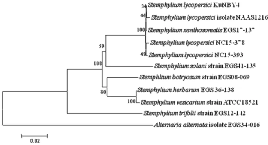
San Luis Obispo, CA, USA)로 정제한 후, direct sequenc- ing을 통해 염기서열 분석을 실시하였다. 분석된 염기서열 은 Clustal W 소프트웨어를 이용하여 정렬하였고, nucleo- tide의 유사도를 계산하였으며, 4개 유전자 부위의 염기서 열을 종합하여 MEGA 6.0 프로그램을 이용하여 neighbor- joining법을 통해 계통수를 작성하였다(Fig. 2). 계통수 작성 을 위해 사용된 관련 균주와 유전자 정보는 Table 2에 기 재하였으며, 계통분석을 위한 outgroup으로는 Alternaria alternata를 사용하였다. 그 결과, 고려엉겅퀴 분리균은 S.
lycopersici, S. xanthosomatis와 하나의 그룹을 이루었으며, 형태적, 배양적 특성이 유사한 S. solani와는 구분되는 것으 로 나타났다. 국내 큰조롱에서 분리한 S. lycopersici 균주는 S. xanthosomatis와 염기서열에 차이가 많지 않아, 이들 두 종은 종내(intra-species) 변이가 하나의 같은 종으로 판단 된다고 하였으며[8], 이는 본 연구 결과에서도 일치하는 것 으로 나타나 두 종에 대해 면밀한 분류학적 연구가 필요할 Table 1. Comparison of morphological characteristics between the present isolate obtained from infected leaves of Cirsium setidens and Stemphylium species described previously
Structurea
Characteristics b Present isolate
NC15-378
Stemphylium lycopersici
Stemphylium solani
Stemphylium xanthosomatis Conidia size (μm) 34.5~56.1 × 15.7~23.5 50~74 × 16~23 35~55 × 18~28 30~57.5 × 11~17
L/W Ratioc 2.3:1 3:1 or more 2:1 2.9:1
No. of transverse septa 5~7 1~8 3~6 -
Conidiophore length (μm) 130.7~242.2 67~285 104~234.5 73~312
aAll structures were investigated on V8 juice agar (V8A) plates incubated in alternating cycles of 12 hr near ultra-violet (NUV) light and 12 hr darkness at 25oC for 7 days.
bStemphylium species described by CMI description [5] and Câmara et al. [9].
cRatio of length (L) to width (W).
Table 2. Isolates and GenBank accession numbers used in the phylogenetic analysis of Stemphylium species and Alternaria alternate
Species Isolate Genbank accession numbera
gpd vma ITS EF-1α
S. lycopersici KuNBY4 AB704322* AB828251 AB704311 AB828256
S. lycopersici NAAS12164 KC160509 KC160512 JX845138 KC160506
S. solani EGS 41-135 AY317018 AY329290 AY329214 AY324759
S. xanthosomatis EGS 17-137 AT317010 AT329284 AT329206 AT324758
S. trifolii EGS 12-142 AY317022 AY329292 AY329218 AY324744
S. botryosum EGS 08-069 AY316968 AY329271 AY329168 AY324751
S. herbarum EGS 36-138 AF081398 AY329272 AF442785 AY324675
S. vesicarium ATCC 18521 AY278821 AB828248** AF229484 JQ672392
A. alternata EGS 34-016 AF081400 AY329324 FJ717729 AH013339
gpd, glyceraldehydes-3-phosphate dehydrogenase; vma, vacuolar membrane ATPase catalytic subunit; ITS, internal transcribed spacer;
EF-1α, elongation factor 1 alpha.
aAB704322* and AB828248** indicated Genbank accession numbers of the relevant genes of S. lycopersici MAFF306895 and S. vesicar- ium EGS 37-067, respectively.
것으로 생각된다. 여기서 분석된 NC15-378과 NC 15-393 균주의 각 유전자 부위는 NCBI GenBank에 KU59 9917~
KU599924의 accession number로 등록하였다.
병원성 검정
고려엉겅퀴 잎을 대상으로 병원성을 확인하기 위하여 건 전한 고려엉겅퀴 잎에 70% 에탄올을 뿌려 무균대에서 건조 시켰다. 고려엉겅퀴에서 분리한 NC15-378과 NC15-393 균 주를 각각 V8A 배지에 10일간 배양하고, 멸균수로 포자를 회수하여 1 × 10⁶ spores/mL의 농도로 포자현탁액을 만들 었다. 멸균한 바늘로 고려엉겅퀴 잎을 찔러 상처를 낸 잎과 상처가 없는 잎에 포자현탁액 20 μL를 떨어뜨려 접종하였 고, 대조구는 상처 및 무상처 잎에 멸균수를 처리하였다.
접종한 잎을 습실처리한 플라스틱 박스에 넣고 25oC의 배 양기에 두면서 습도와 온도를 유지시켰다. 접종 3일 후부터, 상처의 유무에 관계없이 병원균을 접종한 부위에서 병반이 관찰되었고, 7일 후에는 갈색의 점무늬 병반이 1 cm 이상 진전되었으며, 대조구에서는 병징이 나타나지 않았고, 병반 부위로부터 Stemphylium균이 재분리됨을 확인하였다(Table 3, Fig. 1C). 큰조롱에서 분리한 S. lycopersici 균주를 대상으 로 고추, 토마토, 담배에 대한 병원성을 조사한 결과, 고추 와 토마토 잎에 병원성이 있었고 담배에는 병원성이 없는 것으로 나타났으며, 따라서 본 연구에서 분리한 균주에 대 해서도 기주범위를 조사하기 위하여 다른 작물에 대한 병 원성 검정을 수행할 필요가 있을 것으로 생각된다[8].
고려엉겅퀴는 다른 엉겅퀴류와는 달리 식용으로만 이용 되는데, 특히 잎과 줄기가 그 대상이 된다. 잎에 발생하는 점무늬병과 같은 병해는 고려엉겅퀴 재배 농가에 직접적인 피해를 주게 되므로 이 병해에 대한 발생생태와 친환경 방 제법에 관한 연구가 필요할 것으로 생각된다.
이상과 같이 고려엉겅퀴 잎에 발생한 점무늬 증상에서 분리한 병원균은 형태적, 배양적 특성 및 DNA 염기서열 분석에 의해 S. lycopersici로 동정되었고, 인공 접종을 통해 고려엉겅퀴에 병원성이 있음이 확인되었다. 따라서 본 결 과를 토대로 국내 고려엉겅퀴 잎에 발생하는 점무늬병균으 로 Stemphylium lycopersici (Enjoji) W. Yamam.를 보고하 고자 한다.
적 요
2015년 8월경 강원도 영월군의 고려엉겅퀴 재배 농가에 서 잎에 소형 점무늬 증상이 발생하였다. 초기에는 고려엉 겅퀴 잎에 회갈색 내지 갈색의 작은 점이 나타나며, 병이 진 전되면 부정형의 진한 갈색의 병반으로 커지면서 크게 확 대되고 결국 잎 전체가 진한 갈색 내지 흑색으로 변하였다.
병든 잎에서 Stemphylium균이 분리되었으며, 분생포자 크 Fig. 2. Phylogenetic tree based on a combined alignment of internal transcribed spacer, glyceraldehydes-3-phosphate dehydro- genase, elongation factor 1 alpha, and vacuolar membrane ATPase catalytic subunit A gene-gene involved in vacuolar biogenesis sequence data for Stemphylium lycopersici and related species. The tree was generated using neighbor-joining analysis, with Kimura 2-parameter model. Bar represents the number of nucleotide substitutions per site.
Table 3. Pathogenicity of Stemphylium lycopersici isolates on leaves of Cirsium setidens by artificial inoculation
Isolate Pathogenicitya of tested isolates on gondre leaves
Wounded Unwounded
NC15-378 + ++
NC15-393 ++ ++
Control − −
++, above 10 mm lesion diameter; +, 5~8 mm lesion diameter; −, no symptom.
aPathogenicity was carried out by inoculation on detached leaves of gondre plants with 20 μL of spore suspensions of two Stemphy- lium isolates. The rated based on the lesion diameter seven days after inoculation.
기와 분생포자경의 길이 등을 고려한 균학적 특성에 의해 Stemphylium lycopersici 혹은 Stemphylium solani, Stemphy- lium xanthosomatis와 유사한 것으로 나타났다. Ribosomal internal transcribed spacer (ITS) 영역, elongation factor 1 alpha (EF-1α), glyceraldehydes-3-phosphate dehydrogenase (gpd), vacuolar membrane ATPase catalytic subunit A gene (vmaA)와 vacuolar biogenesis gene (vpsA) 사이의 noncoding 영역을 대상으로 한 다자위 염기서열 분석에 의 해 분리균은 모두 S. lycopersici로 확인되었다. 고려엉겅퀴 잎을 대상으로 상처구와 무상처구로 나누어 분리균의 포자 현탁액을 접종하여 병원성을 확인한 결과, 접종 3일 후부터 상처의 유무에 관계없이 분리균을 접종한 부위에서 병반이 관찰되었다. 따라서 본 결과를 토대로 본 병을 S. lycoper- sici에 의한 고려엉겅퀴 점무늬병으로 명명하며, 고려엉겅퀴 점무늬병의 발생을 국내 최초로 보고한다.
Acknowledgements
This study was supported by a grant (Project No. PJ01 0004) from Rural Development Administration, Republic of Korea.
REFERENCES
1. Chang SY, Song JH, Kwak YS, Han MJ. Quality characteristics of Gondre tofu by the level of Cirsium setidens powder and storage. J Korean Soc Food Cult 2012;27:737-42.
2. The Korean Society of Plant Pathology. List of plant diseases in Korea. 5th ed. Seoul: Korean Society of Plant Pathology;
2009.
3. Nasehi A, Kadir JB, Esfahani MN, Mahmodi F, Golkhandan
E, Akter S, Ghadirian H. Cultural and physiological charac- teristics of Stemphylium lycopersici causing leaf blight disease on vegetable crops. Arch Phytopathol Plant Prot 2014;47:1658- 65.
4. Kim BS, Yu SH, Cho HJ, Hwang HS. Gray leaf spot in peppers caused by Stemphylium solani and S. lycopersici. Plant Pathol J 2004;20:85-91.
5. Min JY, Kim BS, Cho KY, Yu SH. Grey leaf spot caused by Stemphylium lycopersici on tomato plants. Korean J Plant Pa- thol 1995;11:282-4.
6. Kwon JH, Jeong BR, Yun JG, Lee SW. Leaf Spot of Kalanchoe (Kalanchoe blossfeldiana) caused by Stemphylium lycopersici.
Res Plant Dis 2007;13:122-5.
7. Yu SH, Cho HS. Korean species of Alternaria and Stemphylium.
Suwon: National Institute of Agricultural Science and Tech- nology; 2001.
8. Hong SK, Choi HW, Lee YK, Shim HS, Lee SY. Leaf spot and stem rot on Wilford Swallowwort caused by Stemphylium lyco- persici in Korea. Mycobiology 2012;40:268-71.
9. Câmara MP, O'Neill NR, Van Berkum P. Phylogeny of Stem- phylium spp. based on ITS and glyceraldehyde-3-phosphate dehydrogenase gene sequences. Mycologia 2002;94:660-72.
10. Choi HW, Kim JM, Hong SK, Kim WG, Chun SC, Yu SH.
Mating types and optimum culture conditions for sexual state formation of Fusarium fujikuroi isolates. Mycobiology 2009;
37:247-50.
11. White TJ, Bruns TD, Lee SB, Taylor JW. Amplification and direct sequencing of fungal ribosomal RNA genes for phylo- genetics. In: Innis MA, Gelfand DH, Sninsky JJ, editors. PCR protocols: a guide to methods and applications. San Diego:
Academic Press; 1990. p. 315-22.
12. Alves A, Crous PW, Correia A, Phillips AJ. Morphological and molecular data reveal cryptic speciation in Lasiodiplodia theobromae. Fungal Divers 2008;28:1-13.
13. Inderbitzin P, Harkness J, Turgeon BG, Berbee ML. Lateral transfer of mating system in Stemphylium. Proc Natl Acad Sci USA 2005;102:11390-5.