• 검색 결과가 없습니다.

Genetic positioning of Korean viral hemorrhagic septicemia virus (VHSV) from cultured and wild marine fishes

N/A
N/A
Protected

Academic year: 2021

Share "Genetic positioning of Korean viral hemorrhagic septicemia virus (VHSV) from cultured and wild marine fishes"

Copied!
9
0
0

로드 중.... (전체 텍스트 보기)

전체 글

(1)

Genetic positioning of Korean viral hemorrhagic septicemia virus (VHSV) from cultured and wild marine fishes

Wi Sik Kim, Sung Ju Jung * , Jong Oh Kim * , Du Woon Kim ** , Jeong Ho Kim *** and Myung Joo Oh *†

The Fisheries Science Institute, Chonnam National University, Yeosu 556-901, Korea

* Department of Aqualife Medicine, College of Fisheries and Ocean Science, Chonnam National University, Yeosu 550-749, Korea

** Department of Food Science and Technology, and Functional Food Research Center,Chonnam National University, Gwangju 500-757, Korea

*** Faculty of Marine Bioscience and Technology, Gangneung-Wonju National University, Gangneung 210-702, Korea

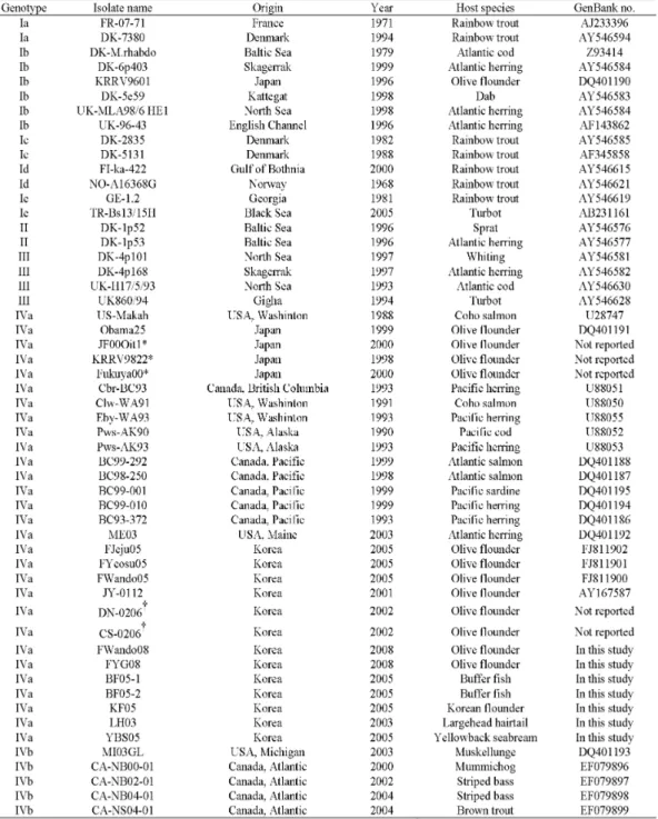
Viral haemorrhagic septicaemia virus (VHSV) is an epidemic virus in olive flounder Paralichthys olivaceus farms in Korea, since the virus have first isolated in 2001. In the present study, partial glycoprotein (G) gene nucleotide sequences of seven Korean VHSV from cultured olive flounder and wild marine fishes in coastal areas of Korea were analyzed to evaluate their genetic relatedness to worldwide isolates. Phylogenetically, all Korean VHSV formed only one minor cluster including Japanese isolates, in genotype IVa, while the North America isolates formed a different minor cluster in genotype IVa. These results suggest that Korean VHSV could be an indigenous virus in Korean and Japanese coastal areas, but have not been introduced from North America.

1)

Key words :

Viral hemorrhagic septicemia virus, Glycoprotein gene, Phylogeny, Korea

Viral haemorrhagic septicaemia (VHS) is an infectious disease causing extensive losses in rainbow trout Oncorhynchus mykiss farms in European countries (Wolf, 1988; Smail, 1999), and it is listed as notifiable disease by the Aquatic Animal Health Code of the World Organization for Animal Health (OIE). VHS virus (VHSV), the causative agent of VHS, was first isolated from freshwater-cultured rainbow trout in Denmark in 1963 (Jensen, 1963). Over the succeeding four decades, VHSV has been isolated as the predominant pathogen of not only rainbow trout but other fish species including

†Corresponding Author : Myung-Joo Oh Phone: +82-61-659-3173 E-mail: ohmj@chonnam.ac.kr

marine fishes in European countries (Wolf, 1988;

Mortensen et al ., 1999; Skall et al ., 2005). At the end of the 1980s, VHSV was also isolated from anadromous salmon and marine fish on the west coast of North America, marking the first detection of VHSV on that continent (Winton et al ., 1989; Meyers and Winton, 1995;

Meyers et al ., 1999). Moreover, it has also been found in olive flounder Paralichthys olivaceus and other marine fish in Far Eastern Asia (Takano et al ., 2000; Isshiki et al ., 2001; Watanabe et al ., 2002; Kim et al ., 2009).

Thus, VHSV is ubiquitous in a variety of freshwater and marine fishes throughout the northern hemisphere, including North America, Asia and Europe.

As a member of the genus Novirhabdovirus in the

(2)

family Rhabdoviridae , VHSV has a negative- and single- stranded RNA genome with approximately 11.1 kb containing six genes, nucleocapsid (N), phosphoprotein (P), matrix protein (M), glycoprotein (G), non-virion protein (NV) and polymerase (L) genes, in the order 3’-N-P-M-G-NV-L-5’ (Trdo et al ., 2005). Based on phylogenetic analyses with nucleotide sequences of N and G genes, worldwide isolates of VHSV were classified into four genotypes. Genotype I includes a wide range of isolates originating in freshwater and marine fish species in continental Europe, genotype II is composed of marine isolates recovered from the Baltic Sea, genotype III comprises isolates distributed in the North Sea coastal area of the United Kingdom and Ireland, and genotype IV is comprised of isolates from Japan, and the Pacific coast and Great Lakes regions of North America (Benmansour et al ., 1997; Stone et al ., 1997; Snow et al ., 1999; Nishizawa et al ., 2002;

Einer-Jensen et al ., 2004; Snow et al ., 2004; Skall et al ., 2005; Elsayed et al ., 2006; Nishizawa et al ., 2006;

Gagné et al ., 2007). It is considered that genetic differences of VHSV are more related to geographic areas than to host fish species.

VHSV was not detected in Korea before 2000. In 2001, VHSV infection occurred in juveniles of farmed olive flounder at Pohang, in eastern Korea (Kim et al ., 2003). Thereafter, more widespread VHS outbreaks occurred in Korean olive flounder farms (Kim et al ., 2009), with VHS becoming an endemic problem in the Korean aquaculture industry. Furthermore, VHSV has been detected from wild marine fishes in Korean coastal areas (Kim and Park, 2004; Lee et al ., 2007). Our hypothesis asserts that the original source of VHSV in

Korea may be introduced from outside of Korea by the movement of VHSV-contaminated fish in nature or imported fish from many countries as a feed source in aquaculture industry, because the disease was not reported prior to 2001. However, there is no evidence to support this hypothesis. Thus, in the present study, we analyzed partial nucleotide G gene sequences of seven Korean VHSV from cultured olive flounder and wild marine fishes in coastal areas of Korea, and compared the sequences with those of existing Korean, Japanese and worldwide isolates for evaluation of the origin of Korean VHSV.

Materials and Methods

VHSV for nucleotide sequence analysis

Two isolates of VHSV from cultured olive flounder and five VHSV from apparently healthy wild marine fishes were subjected to nucleotide sequence analysis of partial G gene (Fig. 1). The FWando08 and FYG08 were isolated from moribund juveniles of olive flounder involved in VHS epidemic outbreaks at Wando and Yeonggwang in 2008. BF05-1 and BF05-2 were detected from wild buffer fish Pampus argenteus, while KF05 was from wild Korean flounder Glyptocephalus stelleri;

the fish were captured at a coastal area of the western

part of Korea in 2005. LH03 and YBS05 were detected

from wild largehead hairtail Trichiums lepturus and

yellowback seabream Dentex tumifrons captured at a

coastal area of the southern part of Korea in 2003 and

2005, respectively. FWando08 and FYG08 isolates were

propagated in fathead minnow (FHM) cells, which were

maintained at 20°C with Eagle’s MEM supplemented with

(3)

10% fetal bovine serum, 100 IU/ml penicillin G and 100 μg/ml streptomycin sulfate. The isolates used for nucleotide sequence analysis had undergone two passages in cell culture. In addition, each 0.2 g tissues (spleen and kidney) from wild marine fishes were homogenized in a 4-fold volume of TE buffer (10 mM Tris-HCl, 1 mM EDTA, pH 8.0) and centrifuged at 2500 g for 10 min at 4°C. The supernatants were used for reverse transcription (RT)-polymerase chain reaction (PCR) of VHSV.

Fig. 1. Locations where VHSV have been detected in cultured olive flounder or wild marine fish from or near coastal areas of Korea. Stars indicate locations of Korean VHSV detected in this study, and circles indicate locations of Korean VHSV reported previously (Kim et al., 2003; Kim et al., 2009).

RT-PCR

Viral RNA was extracted from the tissue homogenates and viral-culture fluid using TRIZOL reagent (Gibco, Gaithersburg, MD, USA) according to the manufacturer’s instructions for RT-PCR amplification with two PCR primer sets. PCR primers VG1 (5’-ATGGAATGGAAC ACTTTTTTC-3’)-VD3 (5’-TGTGATCATGGGTCCT GGTG-3’) (one-step PCR) and VD5 (5’-TCCCGCTAT CAGTCACCAG-3’)-VD3 (nested PCR) were used for amplification of a 697 and 444 base-region of the VHSV G gene, respectively (Miller et al., 1998). Briefly, total RNA was isolated using TRIZOL reagent, precipitated

with isopropanol, and resuspended with 20 μl of diethyl pyrocarbonate (DEPC)-DW (Bioneer, Daejon, Korea).

Viral genomic RNA was denatured at 95°C for 5 min, and then incubated at 42°C for 30 min in 10 μl of RT buffer (10 mM Tris-HCl, pH 8.3, 50 mM KCl) containing 100 U M-MLV reverse transcriptase (Bioneer, Daejon, Korea), 2.5 μM forward primer, 1 mM dNTP and 5 mM MgCl 2 for reverse transcription. The synthesized cDNA was amplified in 50 μl of PCR buffer containing 0.5 μM each PCR primer, 1.25 U Ex-Taq DNA polymerase (Takara, Shiga, Japan), 0.2 mM dNTP and 2 mM MgCl 2

using a thermal cycler (MyGenie 96 thermal block, Bioneer, Daejon, Korea) with 30 cycles (95°C for 1 min, 54°C for 1min, and 72°C for 1min). When PCR using primers VG1 and VD3 was negative at first amplification, a nested-PCR was performed using the same conditions as above.

Nucleotide sequence analysis

PCR products were analyzed by 1.5% agarose gel

electrophoresis. PCR products were purified with QIAquick

gel extraction kit (Qiagen, Valencia, CA, USA) for nucleotide

sequence analysis using an ABI PRISM 3730 XL DNA

Analyzer (Applied Biosystems, Foster City, CA, USA),

according to the manufacturer’s instructions. The resulting

sequences were assembled with Genetyx Win Ver. 5.1

software and construction of multiple alignments was

done to infer geneticrelationships with neighbor-joining

criteria using Clustal X software (Thompson et al.,

1997). The final phylogenetic tree was drawn with the

MEGA4 program (Tamura et al., 2007). The determined

nucleotide sequences were registered in GenBank as

accession numbers GU265809 - GU265815. The seven

Korean VHSV in the present study were compared by

(4)

Table 1. Data for the 54 VHSV used in this study for phylogenetic analysis

other 41 of worldwide isolates and other 6 Korean isolates reported in olive flounder (Table 1).

* Same nucleotide sequences as Obama25

† Same nucleotide sequences as JY-0112

(5)

Results and Discussion

PCR products of approximately 690 bp were obtained by RT-PCR with the primers VG1 and VD3 (one-step PCR) for the G gene from two Korean VHSV isolates (FWando08 and FYG08) from cultured olive flounder, while approximately 440 bp of PCR products were amplified with the primers sets (VG1-VD3 and VD5-VD3;

nested PCR) from five Korean VHSV (LH03, BF05-1, BF05-2, KF05 and YBS05) from wild marine fish (data not shown). The PCR products were sequenced and the determined nucleotide sequences of the partial G gene (nt 273-677) without the PCR primer sequences were compared with those of 47 worldwide VHSV isolates.

The nucleotide sequences showed more than 99.5%

identity among Korean VHSV, and more than 78.3%

identity among 54 worldwide VHSV (data not shown).

A radial tree of phylogeny based on the determined G gene nucleotide sequences revealed the four major

clusters for genotypes I-IV, as described in previous studies (Fig. 2) (Benmansour et al., 1997; Stone et al., 1997; Nishizawa et al., 2002; Einer-Jensen et al., 2004;

Elsayed et al., 2006; Gagné et al., 2007). Moreover, two minor clusters for classes IVa and IVb were observed for genotype IV. Genotype IVa represented isolates in Japan and in the Pacific coast areas of North America, and IVb included isolates originating from the eastern Atlantic coast of Canada and the Great Lakes of the North America. All eight Korean isolates (JY-0112, DN-0206, CS-0206, FJeju05, FYeosu05, FWando05, FWando08 and FYG08) from cultured olive flounder were classified into genotype IVa in the present tree (Fig. 2) and were closely related to isolates from the Pacific coast areas of North America and Japan. Also, all five Korean VHSV (BF05-1, BF05-2, KF05, LH03 and YBS05) from wild marine fish grouped in genotype IVa. Thus, VHSV belonging to genotype IVa may be responsible for the epizootic episodes of VHS in olive flounder farms in Korea.

Fig. 2a

(6)

Fig. 2b

Fig. 2. Molecular phylogenetic tree showing the genetic relationships among 54 VHSV based on the nucleotide sequence of the partial G gene (nt 273-677) (a) and a phylogram using isolates belong to genotype IV (b). Bootstrap values at 1000 times of construction are shown at major nodes in the tree. Distance marker refers to the expected number of substitutions per site.

A recent study of VHSV in Japan suggests at least two different origins for VHSV in Japan (Nishizawa

et al., 2002). In one route, Japanese isolates could be

an indigenous virus in Japanese coastal areas, but not

(7)

have been introduced from North America because the Japanese isolates fall into one minor cluster in genotype IVa, which does not contain any American isolates.

Alternatively, Japanese isolate may have originated outside of Japan because the isolate (KRRV9601) falls into the traditional European genotype Ib. However, the situation for Korean VHSV seemed different from that of Japanese isolates, because Korean VHSV was not included in the genotypes I, II and III. In addition, all Korean VHSV including the oldest Korean isolate (JY-0112) formed only one minor cluster including Japanese isolates, in genotype IVa, while the North America isolates formed a different minor cluster in genotype IVa (Fig. 2b). These results suggest that the Korean VHSV could be an indigenous virus in Korean and Japanese coastal areas, indicating that the origin of Korean VHSV may be from coastal water sharing Korea and Japan.

VHSV has been isolated from several wild marine fish species (Stone et al., 1997; Skall et al., 2005).

Feeding of VHSV-infected marine fish to domestic stock may be a route of infection (Stone et al., 1997; Skall et al., 2005). In Korea, fresh minced marine fish containing of herring, chub mackerel Scomber japonicus, horse mackerel Trachurus japonicas, etc are commonly used in the flounder-farming industry as feed. Additionally, VHSV has been isolated or detected from several species of wild marine fish including chub mackerel in coastal area of Korea (Kim and Park 2004; present study). The previous and present data highlight the potential threat to the flounder farming industry from marine fish reservoirs of VHSV, and in particular the use of untreated trash fish as feed.

Acknowledgment

This research was supported by Basic Research Program through the National Research Foundation of Korea (NRF) funded by the Ministry of Education, Science and Technology (2009-0087136).

References

Benmansour, A., Basurco, B., Monnier, A.F., Vende, P., Winton, J.R. and de Kinkelin, P.: Sequence variation of the glycoprotein gene identifies three distinct lineages within field isolates of viral haemorrhagic septicaemia virus, a fish rhabdovirus. J. Gen. Virol., 78:2837-2846, 1997.

Einer-Jensen, K., Ahrens, P., Forsberg, R. and Lorenzen, N.:

Evolution of the fish rhabdovirus viral haemorrhagic septicaemia virus. J. Gen. Virol., 85:1167-1179, 2004.

Elsayed, E., Faisal, M., Thomas, M., Whelan, G., Batts, W. and Winton, J.: Isolation of viral haemorrhagic septicaemia virus from muskellunge, Esox masqu- inongy(Mitchill), in Lake St Clair, Michigan, USA reveals a new sublineage of the North American genotype. J. Fish Dis., 29:611-619, 2006.

Gagné, N., MacKinnon, A.M., Boston, L., Souter, B., Cook-Versloot, M., Griffiths, S. and Olivier, G.:

Isolation of viral haemorrhagic septicaemia virus from mummichog, stickleback, striped bass and brown trout in eastern Canada. J. Fish Dis., 30:213-223, 2007.

Isshiki, T., Nishizawa, T., Kobayashi, T., Nagano, T. and

Miyazaki, T.: An outbreak of VHSV (viral hemorrhagic

(8)

septicemia virus) infection in farmed Japanese flounder Paralichthys olivaceus in Japan. Dis.

Aquat. Org., 47:87-99, 2001.

Jensen, M.H.: Preparation of fish tissue cultures for virus research. Bull. Off. Int. Epizoot., 59:131-134, 1963.

Kim, S.M. and Park, S.I.: Detection of viral hemorrhagic septicemia virus (VHSV) in wild marine fishes in the coastal region of Korea. J. Fish Pathol., 17:1-10, 2004.

Kim S.M., Lee, J.I., Hong, M.J., Park, H.S. and Park, S.I.:

Genetic relationship of the VHSV (viral hemorrhagic septicemia virus) isolated from cultured olive flounder, Paralichthys olivaceus in Korea. J. Fish Pathol., 16:1-12, 2003.

Kim, W.S., Kim, S.R., Kim, D.W., Kim, J.O., Park, M.A., Kitamura, S.I., Kim, H.Y., Kim, D.H., Han, H.J., Jung, S.J. and Oh, M.J.: An outbreak of VHSV (viral haemorrhagic septicaemia virus) infection in farmed olive flounder Paralichthys olivaceus in Korea. Aquaculture, 296:165-168, 2009.

Lee, W.L., Yun, H.M., Kim, S.R., Jung, S.J. and Oh, M.J.:

Detection of viral haemorrhagic septicaemia virus from marine fish in the South Western coastal area and East China sea. J. Fish Pathol., 20:201-209, 2007.

Meyers, T.R. and Winton, J.R. Viral hemorrhagic septicemia virus in North America. Annu. Rev. Fish Dis., 5:3-24, 1995.

Meyers, T.R., Short, S. and Lipson, K.: Isolation of the North American strain of viral hemorrhagic septicemia virus (VHSV) associated with epizootic mortality in two new host species of Alaskan marine fish.

Dis. Aquat. Org., 38:81-86, 1999.

Miller, T.A., Rapp, J., Wastlhuber, U., Hoffmann, R.W.

and Enzmann, P.J.: Rapid and sensitive reverse transcriptase-polymerase chain reaction based detection and differential diagnosis of fish pathogenic rhabdoviruses in organ samples and cultured cells.

Dis. Aquat. Org., 34:13-20, 1998.

Mortensen, H.F., Heuer, O.E., Lorenzen, N., Otte, L. and Olesen, N.J.: Isolation of viral haemorrhagic septicaemia virus (VHSV) from wild marine fishspecies in the Baltic Sea, Kattegat, Skagerrak and the North Sea. Virus Res., 63:95-106, 1999.

Nishizawa, T., Iida, H., Takano, R., Isshiki, T., Nakajima, K. and Muroga, K.: Genetic relatedness among Japanese, American and European isolates of viral hemorrhagic septicemia virus (VHSV) based on partial G and P genes. Dis. Aquat. Org., 48:143-148, 2002.

Nishizawa, T., Savaş, H., Işidan, H., Üstündağ, C., Iwamoto, H. and Yoshimizu, M.: Genotyping and pathogenicity of viral hemorrhagic septicemia virus from free- living turbot (Psetta maxima) in a Turkish coastal area of the Black Sea. Appl. Environ. Microbiol., 72:2373-2378, 2006.

Skall, H.F., Olesen, N.J. and Mellergaard, S.: Viral haemorrhagic septicaemia virus in marine fish and its implications for fish farming - a review. J.

Fish Dis., 28:509-529, 2005.

Smail, S.A.: Viral haemorrhagic septicaemia. In Woo, P.T.K.

and et Bruno, D.W., editors. Fish diseases and disorders, Vol. 3: Viral, bacterial and fungal infections. CAB International, New York, USA.

pp.123-146, 1999.

Snow, M., Bain, N., Black, J., Taupin, V., Cunningham,

(9)

C.O., King, J.A., Skall, H.F. and Raynard, R.S.:

Genetic population structure of marine viral haemorrhagic septicaemia virus (VHSV). Dis.

Aquat. Org., 61:11-21, 2004.

Snow, M., Cunningham, C.O., Melvin, W.T. and Kurath, G.: Analysis of the nucleoprotein gene identifies distinct lineages of viral haemorrhagic septicaemia virus within the European marine environment.

Virus Res., 63:35-44, 1999.

Stone, D.M., Way, K. and Dixon, P.F.: Nucleotide sequence of the glycoprotein gene of viral haemorrhagic septicaemia (VHS) viruses from different geographic areas: a link between VHS in farmed fish species and viruses isolated from North Sea cod (Gadus morhua L.). J. Gen. Virol., 78:1319-1326, 1997.

Takano, R., Nishizawa, T., Arimoto, M. and Muroga, K.:

Isolation of viral haemorrhagic septicaemia virus (VHSV) from wild Japanese flounder, Paralichthys olivaceus. Bull. Eur. Assoc. Fish Pathol., 20:186-192, 2000.

Tamura, K., Dudley, J., Nei, M. and Kumar, S.: MEGA4:

Molecular Evolutionary Genetics Analysis (MEGA) software version 4.0. Mol. Biol. Evol., 24:1596-1599, 2007.

Thompson, J.D., Gibson, T.J., Plewniak, F., Jeanmougin, F. and Higgins, D.G.: The Clustal X windows interface: flexible strategies for multiple sequence

alignment aided by quality analysis tools. Nucleic Acids Res., 25:4876-4882, 1997.

Trdo, N., Benmansour, A., Calisher, C., Dietzgen, R.G., Fang, R.X., Jackson, A.O., Kurath, G., Nadin-Davis, S., Tesh, R.B. and Walker, P.J.: Family Rhabdo- viridae. In Fauquet, C.M., Mayo, M.A., Maniloff, J., Desselberger, U. and Ball, L.A., editors. Virus taxonomy: Eighth report of the international committee on taxonomy of viruses. Elsevier/Academic Press, London, United Kingdom. pp.623-644, 2005.

Watanabe, L., Pakingking, R.J., Iida, H., Nishizawa, T., Iida, Y., Arimoto, M. and Muroga, K.: Isolation of aquabirnavirus and viral hemorrhagic septicemia virus (VHSV) from wild marine fishes. Fish Pathol., 37:189-191, 2002.

Winton, J.R., Batts, W.N., Nishizawa, T. and Stehr, C.M.:

Characterization of the first North American isolates of viral hemorrhagic septicemia virus. Newsl. Am.

Fish Soc. Fish Health Sec., 17:2-3, 1989.

Wolf, K.: Viral hemorrhagic septicemia. In Wolf, K., editor.

Fish viruses and fish viral diseases. Cornell University Press, Ithaca, New York, USA. pp.

217-249, 1988.

Manuscript Received : February 8, 2011

Revised : March 17, 2011

Accepted : March 18, 2011

수치

Fig. 1. Locations where VHSV have been detected in cultured olive flounder or wild marine fish from or near coastal areas of Korea
Table 1. Data for the 54 VHSV used in this study for phylogenetic analysis

참조

관련 문서

A summer marine benthic algal flora and community of uninhabited islands in Haenamgun, southern coast of Korea. Seasonal Variations in the Macroalgal Flora and

Sequencing results of groEL gene of Anaplasma phagocytophilum detected in blood, kidney and spleen of wild rodents captured in Jeollanam-do area using a

Regarding the development plan of marine architectures and the waterfront, this study has focused on the planning of housing complexes located in

In this way, it need to investigate and research the risk element for the safety of marine traffic to prevent marine pollution, life and property resulted from marine

H, 2011, Development of Cascade Refrigeration System Using R744 and R404A : Analysis on Performance Characteristics, Journal of the Korean Society of Marine Engineering, Vol.

A Systematics and Ecological Notes on Marine Polychaetes (Annelida, Polychaeta) from Korea, with the Secondary Production of Hediste japonica Population in Suncheon

Study of diabetes and the nutrition education and awareness of nutrition labels in Korean adults: Using data from the fifth Korea National Health and Nutrition Examination

Full genome cloning and nucleotide sequence analysis of hepatitis C virus from sera of chronic hepatitis patients in Korea. Detection of hepatitis C virus RNA