바이러스성출혈성패혈증(viral hemorrhagic sep- ticemia, VHS)은 유럽에서 양식되고 있는 무지개송 어(Oncorhynchus mykiss)에 발생하는 질병으로 알 려져 있다(Wolf, 1988; Skall et al., 2005; Egusa et al., 2006). 본 질병은 1950년 무렵부터 덴마크에서 양식되고 있는 무지개송어에서 발생된 후(Jensen, 1965), 원인 병원체인 VHS virus (VHSV)가 유럽, 미국, 일본 등 다양한 나라의 자연산과 양식산 담
수어 및 해산어 약 80여종으로부터 검출되었다 (Isshiki et al., 2001; Meyers and Winton, 1995;
Mortensen et al., 1999; OIE, 2017). 우리나라에서는 2001년 양식산 넙치(Paralichthys olivaceus)에서 VHSV가 처음으로 검출되었고(Kim et al., 2003), 그 후 다양한 지역에 위치한 넙치 양식장에서 VHS 가 발생하였다(Kim et al., 2009; Kim et al., 2012).
VHS의 전형적인 임상증상으로는 체색흑화, 복수 로 인한 복부팽만, 간 울혈, 신장과 비장의 비대가 나타나며 간혹 근육의 점상출혈이 관찰된다(Isshiki et al., 2001).
VHSV는 랩도바이러스과(Rhabdoviridae) 노비랩
넙치 유래 viral hemorrhagic septicemia virus (VHSV, genotype IVa)에 대한 단클론 항체 생산
정하나·장민석·오명주·김위식
†
전남대학교 수산생명의학과
Production of monoclonal antibodies against viral hemorrhagic septicemia virus (VHSV, genotype IVa) from olive flounder
Ha-Na Jeong, Min-Seok Jang, Myung-Joo Oh and Wi-Sik Kim
†Department of Aqualife Medicine, College of Fisheries and Ocean Science, Chonnam National University, Yeosu 59626, Korea
Mouse monoclonal antibodies (MAbs) were produced by using viral hemorrhagic septicemia virus (VHSV, genotype IVa) as an immunogen, isolated from diseased olive flounder (Paralichthys oliva- ceus). Four hybridoma clones secreting MAbs against VHSV were established. The MAbs were recog- nized the nucleoprotein (MAb 4), phosphoprotein (MAb 1) and matrix protein (MAbs 2 and 3) of VHSV by western blot analysis. Among them, the MAbs 1 and 4 strongly reacted with the VHSV-in- fected FHM cells, but not normal FHM cells. In enzyme linked immunosorbent assay, the four MAbs reacted with the VHSV, but not different six fish viruses (infectious hematopoietic necrosis virus, hirame rhabdovirus, spring viraemia of carp virus, infectious pancreatic necrosis virus, marine birnavi- rus and nervous necrosis virus). These results indicate that the MAbs are useful for diagnosis of VHSV infection.
Key words: Viral hemorrhagic septicemia virus, VHSV, Monoclonal antibody, Olive flounder
†
Corresponding author: Wi-Sik Kim
Tel: +82-61-659-7177, Fax: +82-61-659-7177
E-mail: wisky@jnu.ac.kr
도바이러스속(Novirhabdovirus)에 속하며, 6개의 gene (3’-N-P-M-G-NV-L-5’)으로 구성된 약 11.1 kbp의 negative-sense RNA를 가지고 있다(Tordo et al., 2005; Egusa et al., 2006). VHSV는 G 또는 N gene을 사용한 계통발생학적 분석을 통해 4개의 유전자형(genotype I-IV)으로 구분된다(Stone et al., 1997; Snow et al., 1999; Nishizawa et al., 2002; Skall et al., 2005; Kim et al., 2011). 국내에서 분리된 VHSV 분리주들은 일본 분리주들이 속한 genotype IVa에 속하며, 미국 및 유럽 분리주들과는 유전적 으로 차이를 보인다(Kim et al., 2011).
VHSV를 검사하는 방법으로는 어류주화세포를 이용한 분리배양법, 유전자를 이용한 분자생물학 적 방법(reverse transcriptase polymerase chain re- action (RT-PCR), real time PCR), 항체를 이용한 면 역학적인 방법(enzyme-linked immunosorbent assay (ELISA), fluorescent antibody test (FAT)) 등이 사용 되고 있다(OIE, 2017). 우리나라에서는 VHSV가 감 염된 넙치를 검사하는 방법으로 RT-PCR과 분리배 양법이 주로 사용되고 있으나, 면역학적 검사법은 VHSV에 대한 특이 항체가 보급되어 있지 않아 사 용되지 않고 있다. 본 연구에서는 면역학적 검사에 사용가능 한 VHSV에 대한 단클론 항체 (monoclonal antibody, MAb)를 생산하고자 한다.
재료 및 방법
VHSV는 2005년 전남 여수시에 위치한 양식장 에서 사육중인 넙치로부터 분리한 분리주 FYeosu 05를 사용하였다(Kim et al., 2009). 대량으로 배양 하기 위하여 75 cm2 tissue culture flask (Nunc, Denmark)에 fathead minnow cell line (FHM)을 단층 으로 배양 후 VHSV를 접종하여 15℃에서 10일간 배양하면서 cytopathic effect를 관찰하였다. 세포의 90% 이상이 용해되면 세포 배양액을 취해 4℃에 서 12,000 rpm으로 30분간 원심 분리하여 세포 잔 여물을 제거한 후 상층액을 분리하였다. 바이러스 를 농축하기 위해 바이러스 상층액에 polyethylene glycol (PEG)-6000 (Sigma, USA)과 NaCl을 각각 7.5% (w/v), 2.3% (w/v)로 첨가한 후 4℃에서 over- night 하였다(Nishizawa et al., 1991). PEG가 처리된
바이러스 배양액을 4℃에서 12,000 rpm으로 30분 간 원심 분리한 후, pellet을 phosphate buffer saline (PBS: 0.13 M NaCl, 2.7 mM KCl, 4.3 mM Na2HPO4, 1.4 mM KH2PO4) 완충용액으로 현탁하였다. 현탁 액은 30,000 rpm에서 2시간 동안 초원심을 실시한 후 pellet을 PBS로 재부유시켜 바이러스를 농축하 였다. 바이러스를 정제하기 위해, 농축된 바이러스 를 step sucrose gradient (20%, 35%, 50% sucrose 용 액(w/w)) 위에 넣은 후 21,000 rpm으로 2시간 동안 초원심을 실시하였다. 20%와 35% 경계 지점에 형 성된 바이러스로 추정되는 band를 주사기를 이용 하여 취한 후, PBS로 현탁하여 2시간 동안 초원심 분리하였다. 원심 분리 후 얻어진 침전물은 PBS로 다시 재현탁한 후, 초음파 기기(Branson, USA)를 사용하여 40 kHz로 1시간 처리한 후 사용하기 전 까지 -80℃에 보존하였다. VHSV에 대한 단클론 항 체를 생산하기 위해, 정제 후 초음파 처리한 VHSV (약 100 ug)와 incomplete freund’s adjuvant를 동량 으로 섞어 BALB/c 마우스의 발바닥에 1차 접종하 였고, 2주 후에 동일한 VHSV로 2차 접종하였다.
2차 접종 1주 후에 동일한 VHSV로 3차 면역을 하 였다. 3일 후 마우스의 림프절을 분리한 후 PEG (Roche, Germany)를 사용하여 SP2/0 myeloma cell 세포와 융합시킨 후 fetal bovine serum이 10% 첨가 된 hypoxanthine-aminopterin- thymidine (HAT) 배지 (0.1 mM hypoxanthine, 4×10⁻⁴ mM aminopterin, 1.6×
10-2 mM thymidine in Dulbecco’s modified eagle me- dium)로 현탁 시킨 후 96 well plate에 분주하여 3 7℃로 설정된 CO2 배양기에서 배양하였다(Liddell and Cryer, 1991). 양성 hybridoma는 정제된 VHSV 를 항원으로 사용하여 ELISA법으로 선별하였고 3회 이상 제한 희석법으로 클로닝 하였다(Liddell and Cryer, 1991). 선별된 단클론 항체의 isotyping은 Pierce rapid ELISA mouse mAb isotyping kit (Thermo, USA)를 사용하여 결정하였다. VHSV에 대한 항체의 인식부위을 확인하기 위해 sodium do- decyl sulfate polyacrylamide gel electrophoresis (SDS- PAGE)와 western blot을 실시하였다(Laemmli, 1970;
Towbin et al., 1979). 정제된 VHSV를 12% poly- acrylamide gel에 loading한 후 30 mA에서 전기영동 하였다. 전기영동 후, gel에 있는 단백질을 transblot
장치(ATTO, Japan)를 이용하여 144 mA에서 1시간 동안 nitrocellulose membrane (Bio-Rad, USA)에 blotting하였다. 2% skim milk로 1시간 동안 block- ing한 후, 본 연구에서 제작한 MAb(1차 항체)와 al- kaline phosphatase (AP)가 붙어있는 goat anti- mouse IgG serum (novus, USA, 2차 항체, 1,000배 희석액 사용)으로 반응시켰다. 발색제(100 mM Tris- HCl, 100 mM NaCl, 50 mM MgCl2 (pH 9.5) 20 ml, NBT (75 mg/ml 4-nitrotetrazolium blue chloride) 90
㎕, BCIP (50 mg/ml 5-Bromo-4-chloro-3-indolyl phos- phate p-toluidine salt/dimethyl-formamide) 70 ㎕)로 발색하여 육안으로 확인한 후, 발색 정지액(1 mM EDTA, 10 mM Tris-HCl)을 첨가하였다. 양성 대조 구로는 본 연구실에서 토끼로부터 제작한 VHSV 에 대한 polyclonal antibody를 사용하였다. VHSV 감염 세포에 대한 반응을 확인하기 위해, VHSV에 감염된 FHM 세포와 정상 FHM 세포를 사용하여 western blot을 실시하였다. VHSV에 감염된 세포 는 VHSV를 FHM에 접종한 후 3일째 SDS-sample buffer를 첨가하여 세포를 lysis 시킨 후 100℃에서 3분간 열처리하여 준비하였다. 제작된 MAb의 특 이도를 평가하기 위해 7종의 바이러스를 사용하여 ELISA를 실시하였다. VHSV (109.3 tissue culture in- fective dose (TCID)50/ml), 전염성조혈기괴사증바 이러스(infectious hematopoietic necrosis virus, IHNV:
107.3 TCID50/ml), 넙치랩도바이러스(hirame rhabdo- virus, HIRRV: 108.3 TCID50/ml), 잉어봄바이러스 (spring viraemia of carp virus, SVCV: 107.55 TCID50/ ml), 전염성췌장괴사증바이러스(infectious pancre- atic necrosis virus, IPNV: 109.55 TCID50/ml), 해양버 나바이러스(marine birnavirus, MABV: 109.3 TCID50/ ml) 및 신경괴사증바이러스(nervous necrosis virus, NNV: 108.05 TCID50/ml) 상층액을 증류수로 320배 희석하여 96 well ELISA microplates (Greiner bio- one, Germany)에 각각 50 ㎕씩 분주한 후 37℃에서 overnight하여 항원을 코팅하였다. T-PBS (0.05%
Tween-20/PBS (v/v))로 3회 세정한 후, 5% skim milk를 380 ㎕씩 분주하여 25℃에서 1시간 동안 blocking하였다. 1차 항체로는 본 연구에서 제작한 MAb를 50 ㎕씩 분주하였으며, 2차 항체는 horse- radish peroxidase (HRP)가 표식되어 있는 goat anti-
mouse IgG serum (Youngin, Korea)를 5% skim milk 로 1,000배 희석하여 well 당 50 ㎕씩 분주였다.
ELISA 발색액(100 mM Na2HPO4, 50 mM citric acid, 1 mg/ml o-phenylen diamine, 0.1% H2O2)을 well 당 50 ㎕씩 분주하여 발색하였다. 각 well에 1N H2SO4
를 50 ㎕씩 넣어 발색을 중지시킨 후, 492 nm에서 흡광도(opical density, OD)를 측정하였다.
결과 및 고찰
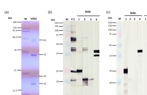
본 연구에서는 VHSV를 특이적으로 인식하는 MAb를 생산하고자 하였다. 정제한 VHSV를 사용 하여 SDS-PAGE를 실시한 결과, 약 62 kDa, 42 kDa, 29 kDa, 25 kDa의 분자량이 확인되었다(Fig.
1a). 넙치 유래 VHSV의 바이러스 구조 단백질은
>140 kDa (polymerase, L), 61.6 kDa (glycoprotein, G), 43.1 kDa (nucleocapsid protein, N), 28.4 kDa (phosphoprotein, P), 24 kDa (matrix protein, M)으로 보고되어 있다(Takano et al., 2000). 본 연구에서 정제한 VHSV의 SDS-PAGE 패턴은 기존에 보고된 결과와 거의 유사하였다.
초음파 처리된 VHSV를 마우스에 면역시킨 후 마우스의 림프절과 SP2/0 myeloma 세포를 융합시 켜 hybridoma를 제작하였다. Hybridoma로부터 생 성되는 항체를 ELISA법으로 선별한 후, 제한 희석 법으로 클로닝하여 최종적으로 4개의 MAb를 선 별하였다. 4개의 MAb와 정제된 VHSV를 사용하여 western blot을 실시한 결과, 양성 대조구(항 VHSV 토끼 혈청)에서는 약 61-66 kDa, 44와 46 kDa, 25 kDa 및 21 kDa에서 밴드가 관찰되었다(Fig. 1b).
본 결과는 정제된 VHSV의 SDS-PAGE 결과와 유 사한 패턴을 보여 위의 반응은 각각 G, N, P, M protein을 인식하는 것으로 사료된다. 25 kDa과 21 kDa의 경우, SDS-PAGE 결과와 약간의 차이를 보 였는데 이는 분자량 측정 시 발생하는 오류로 생각 된다. MAb 1은 약 26 kDa을 강하게 인식하였고, MAb 2는 약 21 kDa을 강하게 인식할 뿐만 아니라 약 46와 44 kDa을 약하게 인식하였다. MAb 3은 약 21 kDa을 약하게 인식하였고, MAb 4는 약 41 kDa과 36 kDa을 강하게 인식하였다. VHSV에 감염 된 FHM 세포와 정상 FHM 세포에 대한 MAb의
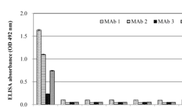
반응을 조사한 결과, MAb 1과 4번은 VHSV에 감 염된 FHM 세포에서 각각 약 26 kDa과 43 kDa의 밴드를 강하게 인식하였고, MAb 2에서는 약 21 kDa을 약하게 인식하였다. MAb 3은 VHSV에 감염 된 세포에 반응하지 않았으며, MAb 1~4는 정상세 포에 모두 반응하지 않았다(Fig. 1c). 제작된 MAb 의 특이도를 확인하기 위해 7종의 바이러스를 이 용하여 ELISA를 실시한 결과, 4개의 MAb 모두 VHSV에만 반응(OD: 0.23-1.6)하였고, 6종의 어류 바이러스(IHNV, HIRRV, SVCV, IPNV, MABV, NNV)에서는 반응하지 않았다(Fig. 2). MAb 1, 2, 4는 각각 1.6, 1.1, 0.7의 OD값을 보였고, MAb 3은 0.23의 OD값을 보였다. 이상의 결과, 제작된 MAb 1, 2와 4는 ELISA 상에서 VHSV에 강하게 반응하 는 것이 확인되었다. 4개의 선별된 MAb의 isotyp- ing 결과, H chain은 IgG1, L chain은 모두 kappa를 인식하는 것으로 확인되었다(data not shown).
본 연구에서는 VHSV에 대한 특이적으로 반응
하는 4개의 MAb를 생산하였다. 4개 MAb는 west- ern blot에서 MAb 1은 P, MAb 2와 3은 M, MAb 4는 N을 인식하였다. 또한 MAb 1과 4는 VHSV에 감염된 FHM 세포에서 강하게 반응하였다. ELISA 에서는 MAb 1, 2 및 4는 VHSV에 반응하였고(MAb 1과 2가 높게 반응함) 6종의 어류바이러스에는 반 응하지 않았다. 이상의 결과, MAb 1과 4는 western blot에서, MAb 1과 2는 ELISA에서 VHSV를 검출 하는데 유용하게 사용될 수 있을 것으로 사료된다.
본 연구실에서는 기존에 본 연구와 동일한 방법으 로 VHSV를 정제한 후(초음파 미처리 VHSV 사용) MAb를 제작한 결과, 5개의 MAb를 생산할 수 있었 는데 이들 MAb는 G (4개)와 N (1개)를 인식하였다 (data not shown). 본 연구에서는 초음파 처리된 VHSV를 사용하여 MAb를 제작한 결과, G를 인식 하는 항체는 얻을 수 없었으나 N뿐만 아니라 P, M을 인식하는 항체를 얻을 수 있었다. 본 방법은 1회 실험한 결과로서 비교하기에는 한계가 있으
(a) (b) (c)
Fig. 1. SDS-PAGE and western blot analysis. (a) SDS-PAGE analysis of the structural protein of VHSV. M: molecular marker (Bio-rad, USA), VHSV: purified VHSV particles. (b) Western blot analysis using purified VHSV and four monoclonal antibodies (MAbs). M: molecular marker (Bio-rad, USA), P.C: Anti-VHSV polyclonal antibody, 1-4:
MAbs. (c) Western blot analysis using VHSV-infected FHM cells and normal FHM cells. M: molecular marker,
1-4: Mabs.
나, VHSV의 초음파 처리 방법을 통해 VHSV의 다 양한 구조 단백질을 인식하는 항체를 생산할 수 있었다.
감사의 글
본 연구는 2016년 해양수산부 재원으로 해양수 산과학기술진흥원의 지원을 받아 수행되었습니다 (수산동물 바이러스 전염병 진단용 항체 생산).
References