Tumor-suppressor Protein p53 Sensitizes Human Colorectal Carcinoma HCT116 Cells to 17α-estradiol-induced Apoptosis via Augmentation of Bak/Bax Activation
Cho Rong Han
1, Ji Young Lee
1, Dongki Kim
2, Hyo Young Kim
2, Se Jin Kim
2, Seokjoon Jang
2, Yoon Hee Kim
1,2, Do Youn Jun
1and Young Ho Kim
1*
1
Laboratory of Immunobiology, School of Life Science and Biotechnology, College of Natural Sciences, Kyungpook National University, Daegu 702-701, Korea
2
Daegu Science High School, Daegu 706-852, Korea
Received September 23, 2013 /Revised Ocotber 23, 2013 /Accepted Ocotber 24, 2013
The regulatory effect of the tumor-suppressor protein p53 on the apoptogenic activity of 17α-estradiol (17α-E
2) was compared between HCT116 (p53
+/+) and HCT116 (p53
-/-) cells. When the HCT116 (p53
+/+) and HCT116 (p53
-/-) cells were treated with 2.5~10 μM 17α-E
2for 48 h or with 10 μM for various time periods, cytotoxicity and an apoptotic sub-G
1peak were induced in the HCT116 (p53
+/+) cells in a dose- and time-dependent manner. However, the HCT116 (p53
-/-) cells were much less sensitive to the apoptotic effect of 17α-E
2. Although 17α-E
2induced aberrant mitotic spindle organization and in- complete chromosome congregation at the equatorial plate, G
2/M arrest was induced to a similar ex- tent in both cell types. In addition, 17α-E
2-induced activation of Bak and Bax, Δψm loss, and PARP degradation were more dominant in the HCT116 (p53
+/+) than in the HCT116 (p53
-/-) cells. In accord- ance with enhancement of p53 phosphorylation (Ser-15) and p53 levels, p21 and Bax levels were ele- vated in the HCT116 (p53
+/+) cells treated with 17α-E
2. The HCT116 (p53
-/-) cells exhibited barely or undetectable levels of p21 and Bax, regardless of 17α-E
2treatment. On the other hand, although the level of Bcl-2 was slightly lower in the HCT116 (p53
+/+) than in the HCT116 (p53
-/-) cells, it remained relatively constant after the 17α-E
2treatment. Together, these results show that among the components of the 17α-E
2-induced apoptotic-signaling pathway, which proceeds through mitotic spindle defects causing mitotic arrest, subsequent activation of Bak and Bax and the mitochondria-dependent caspase cascade, leading to PARP degradation, 17α-E
2-induced activation of Bak and Bax is the upstream tar- get of proapoptotic action of p53.
Key words : 17α-estradiol, mitotic arrest, activation of Bak and Bax, p53 phosphorylation (Ser-15), mitochondrial apoptosis
*Corresponding author
*Tel : +82-53-950-5378, Fax : +82-53-955-5522
*E-mail : [email protected]
This is an Open-Access article distributed under the terms of the Creative Commons Attribution Non-Commercial License (http://creativecommons.org/licenses/by-nc/3.0) which permits unrestricted non-commercial use, distribution, and reproduction in any medium, provided the original work is properly cited.
Journal of Life Science 2013 Vol. 23. No. 10. 1230~1238 DOI : http://dx.doi.org/10.5352/JLS.2013.23.10.1230
Introduction
Estrogens can exert biological effect on target cells by the intracellular estrogen receptor (ER)-mediated genomic mechanism, the plasma membrane ER-mediated non- genomic mechanism associated with cell signaling path- ways, or an ER-independent mechanism [5, 30]. The ER- mediated genomic action of estrogens is elicited by binding with the nuclear receptors estrogen receptor α (ERα) and β (ERβ), and the subsequent transcriptional regulation of gene expression [13]. The plasma membrane ER-mediated action
of estrogens rapidly triggers second messenger signaling events, in which activated ERs do not directly alter the ex- pression of target genes. The ER-independent action of estro- gens is induced at pharmacological concentrations (in the micromolar range) and is not blocked by ER antagonists such as ICI 182,780 or tamoxifen [31].
As the predominant and most biologically active estrogen,
17β-estradiol (17β-E
2) is known to reduce neuronal apopto-
sis at physiological concentrations (in the nanomolar range)
in various in vivo and in vitro neurodegenerative conditions
[4, 31]. However, 17β-E
2induces apoptotic cell death in os-
teoclasts and thymocytes, suggesting that the apoptotic regu-
latory activity of 17β-E
2may differ depending upon the tar-
get cell types [21, 25]. On the other hand, although 17α-estra-
diol (17α-E
2), which is a stereoisomer of 17β-E
2and fails to
interact effectively with the ER, has been considered hormo-
nally inactive and little attention has been given to its roles,
17α-E
2turns out to be as potent as 17β-E
2in protecting neu-
rons from toxic stress conditions [12]. This neuroprotective action of 17α-E
2appears to be mediated by ER-independent nongenomic mechanisms. In this regard, the clinical applica- tion of 17α-E
2, as a neuroprotective therapeutic agent, is ex- pected to be more beneficial than 17β-E
2, in that 17α-E
2pos- sesses low genomic effects and equipotent nongenomic ef- fects compared with 17β-E
2, thus avoiding the adverse ef- fects of 17β-E
2.
In relation to the cytoprotective or apoptogenic effect of 17β-E
2toward malignant tumor cells, a number of studies have indicated that 17β-E
2stimulates cancer cell survival and proliferation at a physiological dose through an ER-de- pendent mechanism, whereas 17β-E
2inhibits cancer cell pro- liferation at a pharmacological dose by inducing apoptosis or microtubule disruption independent of the ER. Recently, several studies have reported that an endogenous metabolite of 17β-E
2, 2-methoxyestradiol (2-MeO-E
2) can be a promising anticancer drug candidate [24]. The anticancer actions of 2-MeO-E
2at pharmacological concentrations are exerted by inducing apoptosis, arresting cell growth at the G
1/S boun- dary and/or G
2/M boundary, or inhibiting angiogenesis [3, 15, 28]. However, little information is known regarding the effect of 17α-E
2on tumor cell proliferation and apoptosis.
Recently, we have shown that a pharmacological dose (5~10 μM) of 17α-E
2, but not 17β-E
2, can induce apoptosis in human acute leukemia Jurkat T cells, which are known to not express ERs [9], via a mitochondria-dependent cas- pase cascade activation [20]. The 17α-E
2-induced apoptosis occurs mainly in G
2/M-arrested cells and is accompanied by an increase in Bcl-2 phosphorylation at Thr-56 [20], a known site that can be phosphorylated by the Cdk1/cyclin B1 kinase during G
2/M phase [14]. More recently, we have reported that the apoptogenic activity of 17α-E
2was due to the impaired mitotic spindle assembly causing prometaphase arrest and prolonged Cdk1 activation, phosphorylation of the Bcl-2 family members (Bcl-2, Mcl-1 and Bim), Bak activation, and mitochondria-dependent caspase cascade activation [18].
In the present study, to elucidate further how 17α-E
2-in- duced apoptotic events are regulated by the tumor sup- pressor protein p53, we investigated the effect of 17α-E
2(2.
5~10 μM) on mitotic arrest, microtubule network organ- ization, and mitochondria-dependent apoptotic signaling pathway using human colorectal carcinoma cell clones HCT116 (p53
+/+) and HCT116 (p53
-/-).
Materials and Methods Reagents, antibodies, and cells
An ECL Western blotting kit was purchased from Amersham (Arlington Heights, IL, USA), and Immobilon-P membrane was obtained from Millipore Corporation (Bed- ford, MA, USA). The anti-caspase-3 antibody was purchased from Pharmingen (San Diego, CA, USA), and anti-p53, an- ti-poly (ADP-ribose) polymerase (PARP), anti-Bak, anti-Bax, anti-Bcl-2, and anti-β-actin were purchased from Santa Cruz Biotechnology (Santa Cruz, CA, USA). The anti-caspase-9 was purchased from Cell Signaling Technology (Beverly, MA, USA). The anti-Bak (Ab-1) and anti-Bax (6A7) were ob- tained from Calbiochem (San Diego, CA, USA). The an- ti-phopho-p53 (Ser-15) was obtained from Cell Signaling (Beverly, MA, USA). The monoclonal anti-p21 antibody was purchased from Neomarkers (Freemont, CA, USA). Human colorectal carcinoma cell lines HCT116 (p53
+/+) and HCT116 (p53
-/-) were provided by Dr. B. Vogelstein (Johns Hopkins University, Baltimore, MD, USA). Both HCT116 cells were maintained in DMEM (Hyclone, (Gaithersburg, MD, USA) containing 10% FBS (Hyclone), and 100 μg/ml gentamycin.
Cytotoxicity assay
The cytotoxic effect of 17α-E
2on HCT116 cells was ana- lyzed by 3-(4,5-dimethylthiazol-2-yl)-2,5-diphenyltetrazolium bromide (MTT) assay as described previously [20]. Briefly, cells (1×10
4/well) were incubated with a serial dilution of 17α-E
2in 96-well plates. At 44 h after incubation, 50 μl of MTT solution (1.1 mg/ml) was added to each well and in- cubated for an additional 4 h. The colored formazan crystal produced from MTT was dissolved in DMSO and OD values of the solutions were measured at 540 nm by a plate reader.
Flow cytometric analysis
Flow cytometric analysis for the cell cycle of HCT116 cells exposed to 17α-E
2was done as described elsewhere [19].
Changes in the mitochondrial membrane potential (Δψm) following treatment with 17α-E
2were measured after stain- ing with 3,3'dihexyloxacarbocyanine iodide (DiOC
6) [26].
Activation of Bak and Bax in HCT116 cells following treat- ment with 17α-E
2was measured by flow cytometry as pre- viously described [22].
Immunofluorescence Microscopy
HCT116 cells adhered onto glass cover slips were fixed
Fig. 1. Cytotoxic effect of 17α-E2on human colorectal carcinoma cell clones HCT116 (p53+/+) and HCT116 (p53-/-) cells.
For cell viability analysis, continuously growing cells (1×10⁴/well) were incubated at the indicated concen- trations of 17α-E2in a 96-well plate for 24 h and the final 4 h was incubated with MTT. The cells were sequentially processed to assess the colored formazan crystal pro- duced from MTT as an index of cell viability. Each value is expressed as the mean±SD (n=3 with three replicates per independent experiment). *
p
<0.05 compared with control. A representative study is shown and two addi- tional experiments yielded similar results.with cold methanol for 3 min [22]. The cells were rinsed four times with cold PBS containing 0.5% Triton X-100, and blocked with 10% goat serum for 30 min. The cells were then incubated with monoclonal anti-α-tubulin (1:2,500) overnight at 4
oC. For detection, the cells were treated with Alexa Fluor 488-labeled goat anti-mouse IgG (1:1,000) for 1 h at room temperature. Thereafter, the cells were stained with 4',6-diamidino-2-phenylindole (DAPI) to label the nuclei.
Images were visualized and photographed using a Carl Zeiss MicroImaging Confocal Laser Scanning Microscope.
Preparation of cell lysate and western blot analysis
Cell lysates were prepared by suspending 3×10
6HCT116 cells in 200 μl of the lysis buffer (137 mM NaCl, 15 mM EGTA, 1 mM sodium orthovanadate, 15 mM MgCl
2, 0.1%
Triton X-100, 25 mM MOPS, 1 mM PMSF, and 2.5 μg/ml proteinase inhibitor E-64, pH 7.2). Cells were disrupted by sonication and extracted for 30 min at 4
oC. An equivalent amount of protein lysate (15~20 μg) was electrophoresed on a 4~12% NuPAGE gradient gel and then electro- transferred to an Immobilon-P membrane. The detection of each protein was carried out with an ECL Western blotting kit, according to the manufacturer's instructions.
Statistical analysis
Unless otherwise indicated, each result in this paper is representative of at least three separate experiments.
Results and Discussion
Comparison of cytotoxic effect of 17α-E
2between HCT116 (p53
+/+) and HCT116 (p53
-/-) cells
To examine whether 17α-E
2-induced apoptotic cell death is affected by tumor suppressor protein p53, the cytotoxicity of 17α-E
2(2.5~10 μM), the cytotoxic effects of 17α-E
2on HCT116 (p53
+/+) and HCT116 (p53
-/-) cells were compared.
As determined by MTT assay, the viabilities of HCT116 (p53
+/+) cells treated for 24 h with 17α-E
2at concentrations of 2.5 µM, 5 µM, and 10 µM were 96%, 80%, and 61.6%, respectively (Fig. 1). Under the same conditions, the cell via- bilities of HCT116 (p53
-/-) cells were not affected within the range of 2.5~5 μM, and declined to the level of 78.9% in the presence of 10 μM. When HCT116 (p53
+/+) and HCT116 (p53
-/-) cells were treated with 10 μM 17α-E
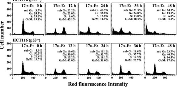
2for various time periods, the apoptotic sub-G
1peak was detected in HCT116 (p53
+/+) cells as early as 12 h after the treatment and then
the ratio of apoptotic sub-G
1cells was elevated up to the level of 74.1% at 48 h, whereas the apoptotic sub-G
1peak in HCT116 (p53
-/-) cells was first detected at 12 h and the time-dependent elevation of apoptotic sub-G
1peak was not so significant as compared with HCT116 (p53
+/+) cells (Fig.
2). In addition, an elevation in the ratio of G
2/M cells were observed in both HCT116 (p53
+/+) and HCT116 (p53
-/-) cells at 12 h following 10 μM 17α-E
2, and then the ratio of G
2/M cells appeared to decrease, possible due to elimination of G
2/M-arrested cells by inducing apoptotic cell death.
These results show that HCT116 (p53
+/+) cells were more sensitive to the cytotoxicity of 17α-E
2as compared with HCT116 (p53
-/-) cells, and suggest that the apoptotic effect of 17α-E
2, which is responsible for the cytotoxicity, could be augmented in the presence of the tumor suppressor protein p53.
Effect of 17α-E
2on the cellular microtubule network in HCT116 (p53
+/+) and HCT116 (p53
-/-) cells
The 17α-E
2-induced apoptotic signaling pathway in Jurkat
T cells has previously been shown to proceed through direct
inhibition of microtubule formation causing prometaphase
arrest, prolonged activation of Cdk1, and subsequent media-
tion of mitochondria-dependent apoptotic events including
Fig. 2. Flow cytometric analysis of the cell cycle distribution in HCT116 (p53+/+) and HCT116 (p53-/-) cells following exposure to 17α-E2for various time periods. The cells were incubated at a density of 2.5×10⁵/ml with 10 μM 17α-E2for 12, 24, 36, or 48 h. The analysis of cell cycle distribution was performed on an equal number of cells (2×10⁴) by flow cytometry after staining of DNA by PI as described in Materials and Methods. A representative study is shown and two additional experiments yielded similar results.
Fig. 3. Effect of 17α-E2on the organization of microtubule net- work in HCT116 (p53+/+) and HCT116 (p53-/-) cells. After treatment of cells with 10 μM 17α-E2for 12 h, the cells were fixed with
cold methanol
for 3 min, per- meabilized, incubated with mouse monoclonal anti-α-tu- bulin, and then treated with Alexa Fluor 488-conjugated goat anti-mouse immunoglobulin. The cells were then stained with 4',6-diamidino-2-phenylindole (DAPI) to la- bel the nuclei. Images were obtained using a confocal laser scanning microscope. Symbols: white arrowhead, incomplete/complete chromosome congression at the equatorial plate. A representative study is shown; two additional experiments yielded similar results.Bak activation, Δψm loss, and resultant caspase cascade acti- vation [18]. Because 17α-E
2-induced apoptotic sub-G
1peak was more apparent in HCT116 (p53
+/+) than in HCT116 (p53
-/-) cells, we decided to examine whether the impairment of mitotic spindle network, which can be caused by 17α-E
2, is modulated by the presence of p53. The effect of 17α-E
2on the organization of the microtubule network in HCT116 (p53
+/+) and HCT116 (p53
-/-) cells was investigated by im- munofluorescence microscopy using anti-α-tubulin antibody.
Although the microtubule network in continuously growing HCT116 (p53
+/+) and HCT116 (p53
-/-) cells showed a normal arrangement with the majority of cells in interphase, both cell types exhibited, in common, an aberrant bipolar array of microtubules after treatment with 10 μM 17α-E
2for 12 h (Fig. 3). In addition, DAPI staining revealed that while some chromosomes in the cells treated with 10 µM 17α-E
2were aligned at the equator of the mitotic spindle, other chromosomes failed to localize at the equator, regardless of the presence of p53.
These results indicate that the 17α-E
2-induced defect in the organization of mitotic spindle network, which is known to be the primary target for the apoptotic action of 17α-E
2[24], and subsequent mediation of incomplete alignment of the chromosomes at the equatorial plate, leading to prom- etaphase arrest of the cell cycle, were not influenced by the presence or absence of p53.
Effect of p53 on 17α-E
2-induced activation of Bak and Bax, and mitochondrial membrane potential (Δψm) loss in HCT116 (p53
+/+) and HCT116 (p53
-/-) cells
To examine whether p53 contributes to modulation of the
A B
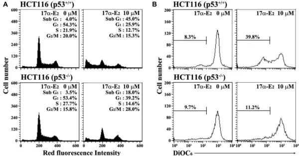
Fig. 4. Apoptotic changes in the cell cycle distribution (A), and mitochondrial membrane potential (∆ψm) loss (B) in HCT116 (p53+/+) and HCT116 (p53-/-) cells after exposure to 17α-E2. After the cells were incubated at a density of 2.5×10⁵/ml with 10 μM 17α-E2for 24 h, cell cycle distribution and ∆ψm loss of the cells were determined through flow cytometric analysis of PI staining and DiOC6staining, respectively. A representative study is shown and two additional experiments yielded similar results.
downstream events of the 17α-E
2-inducecd mitotic arrest, re- sulting from the mitotic spindle defects, the 17α-E
2-induced activation of Bak and Bax, and mitochondrial membrane po- tential (∆ψm) loss were compared between HCT116 (p53
+/+) and HCT116 (p53
-/-) cells. As shown in Fig. 4A, although continuously growing HCT116 (p53
+/+) and HCT116 (p53
-/-) cells untreated with17α-E
2showed barely detectable levels of apoptotic-sub-G
1cells, the apoptotic sub-G
1cells follow- ing treatment with 10 μM 17α-E
2for 24 h appeared to in- crease to a level of 45.0% in HCT116 (p53
+/+) cells and 18.0%
in HCT116 (p53
-/-) cells. Under these conditions, when the Δψ m loss of cells treated with 17α-E
2was measured by DiOC
6staining, the ratio of negative fluorescence in HCT116 (p53
+/+) cells treated with 10 µM 17α-E
2were 39.8% (Fig. 4B).
However, the ratio of negative fluorescence in HCT116 (p53
-/-) cells treated with 10 µM 17α-E
2was only 11.2%. Since current results demonstrated that 17α-E
2-induced Δψm loss was more dominant in HCT116 (p53
+/+) cells than in HCT116 (p53
-/-) cells, it was likely that the 17α-E
2-induced mitochon- drial damage and subsequent mitochondria-dependent acti- vation of caspase cascade might be positively modulated by p53.
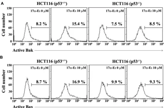
To examine this prediction further, 17α-E
2-induced activa- tion of the pro-apoptotic multidomain Bcl-2 family members, Bak and Bax, were compared between HCT116 (p53
+/+) and HCT116 (p53
-/-) cells. As shown in Fig. 5, both Bak and Bax
activations, as evidenced by their N-terminal conformational changes detected using an active conformation-specific an- ti-Bak antibody (Ab-1) and an active conformation-specific anti-Bax antibody (6A7), were more significantly observed in HCT116 (p53
+/+) cells than in HCT116 (p53
-/-) cells.
Previously, it has been reported that either Bak activation or Bax activation can mediate permeabilization of the mi- tochondrial outer membrane (MOM) to trigger mitochon- drial cytochrome c release into cytoplasm, leading to the cas- pase cascade activation [6, 8]. Consequently, current and pre- vious results indicated that p53-mediated enhancement in the 17α-E
2-induced mitochondrial membrane potential (Δψ m) loss was associated with the elevation in the level of Bak and Bax activations in the presecen of p53.
Numerous studies have reported that chemotherapeutic agents-induced apoptotic signaling pathways are frequently associated with mitochondria-dependent apoptotic events [2, 10, 11]. In addition, it has been shown that an alteration in the expression ratio of Bak to Bcl-2 and/or Bax to Bcl-2, re- sulting in an enhancement in the ratio of Bak to Bcl-2 and/or Bax to Bcl-2, is often prerequisite for provoking the activa- tion of Bak and/or Bax during the mitochondria-dependent apoptosis induced by chemotherapeutic agents [1, 6, 7, 16].
To examine the upstream pro-apoptotic events that mediate
17α-E
2-induced activation of Bak and Bax, the levels of Bcl-2
family proteins, such as the pro-apoptotic Bcl-2 family mem-
A
B
Fig. 5. Flow cytometric analysis of Bak activation (A) and Bax activation (B) in HCT116 (p53+/+) and HCT116 (p53-/-) cells after 17α-E2 treatment. The cells were incubated at the concentration of 2.5×10⁵/ml with 10 μM 17α-E2for 24 h, and subjected to flow cytometric analysis of the activation of Bak and Bax as described in Materials and Methods. A representative study is shown and two additional experiments yielded similar results.
bers (Bak and Bax) and anti-apoptotic Bcl-2, and the activa- tion of caspase-9, and -3, and PARP degradation were com- pared by western blot analysis between HCT115 (p53
+/+) and HCT116 (p53
-/-) cells treated with 17α-E
2.
The p53 expression was easily detected in HCT116 (p53
+/+), but was not detected in HCT116 (p53
-/-) cells (Fig.
6A). In HCT116 (p53
+/+) cells treated with 5~10 μM 17α-E
2, the level of phosphorylated p53 at Ser-15 and total p53 were enhanced dose-dependently. Although the up-regulation of p21, which act as a negative cell cycle regulator for G
1arrest [23, 29], was remarkable in HCT116 (p53
+/+) after 17α-E
2treatment, it was barely detected in HCT116 (p53
-/-) cells.
Under these conditions, caspase-9 and -3 activations, and PARP degradation were more significantly observed in HCT116 (p53
+/+). As shown in Fig. 6B, a time-kinetic analysis revealed that the levels of anti-apoptotic Bcl-2 and pro-apop- totic Bak appeared to remain relatively constant in HCT116 (p53
+/+) and HCT116 (p53
-/-) cells following exposure to 10 μ M 17α-E
2, whereas that of Bax appeared to increase in both cell types at 24~48 h, with more dominant increase in the presence of p53. In addition, the expression of p53 and phos- phorylated p53 at Ser-15, both of which were detected only in HCT116 (p53
+/+) cells, were enhanced time-dependently after 17α-E
2treatment. Un the these conditions, the level of
p21 was enhanced in HCT116 (p53
+/+) cells, whereas the en- hancement was barely or not detected in HCT116 (p53
-/-) cells. These results suggested that 17α-E
2-induced Bax activa- tion might be due to up-regulation in its expression level, whereas 17α-E
2-induced Bak activation might not require the up-regulation of its expression level. Recently, it has been shown that p53 can exert a pro-apoptotic role at the mi- tochondria through direct interaction of p53 with anti-apop- totic Bcl-2, which results in induction of mitochon- dria-dependent apoptosis [17, 27]. It is noteworthy that the differential expression pattern of Bcl-2, which exhibited a rel- atively lower level in HCT116 (p53
+/+) than in HCT116 (p53
-/-) cells, might also contribute to render HCT116 cells more susceptible to the onset of Bak and Bax activations.
Consequently, these previous and current results indicated that the 17α-E
2-induced activation of Bak and Bax, which occurred more dominantly in the presence of p53, was ex- erted by not only a transcription-dependent mechanism of p53 via up-regulation of Bax level, but also a transcription- independent mechanism of p53 via direct binding to Bcl-2.
In conclusion, these results show that HCT116 (p53
+/+)
cells were more sensitive to the cytotoxicity of 17α-E
2, which
is attributable to more potent induction of apoptosis, as com-
pared with HCT116 (p53
-/-) cells. Although the 17α-E
2-induced
A B
Fig. 6. Western blot analysis of dosage effect of 17α-E2 on mitochondria-dependent caspase cascade (A), and kinetic analysis of 17α-E2-induced apoptotic events (B) in HCT116 (p53+/+) and HCT116 (p53-/-) cells. HCT116 (p53+/+) and HCT116 (p53-/-) cells were incubated at a density of 2.5×10⁵/ml with 17α-E2for 48 h, or with 10 μM 17α-E2for various time periods before preparation of the cell lysates. Western blot analysis was performed, as described in Materials and Methods, in order to assess p53 phosphorylation at Ser-15, p53, p21, Bak, Bax, Bcl-2, activation of caspase-9 and -3, PARP cleavage, and β-actin in HCT116 (p53+/+) and HCT116 (p53-/-) cells treated with 17α-E2. A representative study is shown and two additional experi- ments yielded similar results.
defect in the organization of mitotic spindle network and subsequent mediation of incomplete alignment of the chro- mosomes at the equatorial plate, causing G
2/M arrest, were not influenced by p53, the downstream events including both Bak and Bax activations and Δψm loss were observed more potently in the presence of p53. In HCT116 (p53
+/+) cells following 17α-E
2treatment, the levels of phosphory- lated p53 at Ser-15, total p53, and Bax were markedly en- hanced in a dose- and time-dependent manner, suggesting that 17α-E
2-induced activation of Bak and Bax might be pos- itively modulated by the pro-apoptotic action of p53. These results provide insight into the molecular and cellular mech- anism underlying the pro-apoptotic role of p53 in the Bak and Bax activations, provoked by a microtubule-targeting drug, 17α-E
2.
Acknowledgement
This work was supported by the Kyungpook National University Research Fund, 2010, Republic of Korea.
References
1. Adams, J. M. and Cory, S. 2007. Bcl-2-regulated apoptosis:
mechanism and therapeutic potential.
Curr Opin Immunol
19, 488-496.
2. Ashkenazi, A. and Dixit, V. M. 1999. Apoptosis control by death and decoy receptors.
Curr Opin Cell Biol
11, 255-260.3. Batsi, C., Markopoulou, S., Kontargiris, E., Charalambous, C., Thomas, C., Christoforidis, S., Kanavaros, P., Constanti- nou, A. I., Marcu, K. B. and Kolettas, E. 2009. Bcl-2 blocks 2-methoxyestradiol induced leukemia cell apoptosis by a p27Kip1-dependent G1/S cell cycle arrest in conjunction with NF-kB activation.
Biochem Pharmacol
78, 33-44.4. Behl, C. 1998. Effects of glucocorticoids on oxidative stress- induced hippocampal cell death: implications for the patho- genesis of Alzheimer's disease.
Exp Gerontol
33, 689-696.5. Behl, C. and Holsboer, F. 1999. The female sex hormone estrogen as a neuroprotectant.
Trends Pharmacol Sci
20, 441- 6. Chipuk, J. E. and Green, D. R. 2008. How do BCL-2 proteins444.induce mitochondrial outer membrane permeabilization?
Trends Cell Biol
18, 157-164.7. Chipuk, J. E., Moldoveanu, T., Llambi, F., Parsons, M. J. and Green, D. R. 2010. The BCL-2 family reunion.
Mol Cell
37, 299-310.8. Czabotar, P. E., Colman, P. M. and Huang, D. C. 2009. Bax activation by Bim?
Cell Death Differ
16, 1187-1191.9. Danel, L., Menouni, M., Cohen, J. H., Magaud, J. P., Lenoir, G., Revillard, J. P. and Saez, S. 1985. Distribution of an- drogen and estrogen receptors among lymphoid and hae- mopoietic cell lines.
Leuk Res
9, 1373-1378.10. Desagher, S. and Martinou, J. C. 2000. Mitochondria as the central control point of apoptosis.
Trends Cell Biol
10, 369-377.
11. Desagher, S., Osen-Sand, A., Nichols, A., Eskes, R., Montes- suit, S., Lauper, S., Maundrell, K., Antonsson, B. and Martinou, J. C. 1999. Bid-induced conformational change of Bax is responsible for mitochondrial cytochrome c release during apoptosis.
J Cell Bio1
144, 891-901.12. Dykens, J. A., Moos, W. H., Howell, N., Dykens, J. A., Moos, W. H. and Howell, N. 2005. Development of 17α-estradiol as a neuroprotective therapeutic agent: rationale and results from a phase I clinical study.
Ann NY Acad Sci
1052, 116-135.13. Evans, R. M. 1988. The steroid and thyroid hormone re- ceptor superfamily.
Science
240, 889-895.14. Furukawa, Y., Iwase, S., Kikuchi, J., Terui, Y., Nakamura, M., Yamada, H., Kano, Y. and Matsuda, M. 2000. Phosphor- ylation of Bcl-2 protein by CDC2 kinase during G2/M phas- es and its role in cell cycle regulation.
J Biol Chem
275, 21661-21667.15. Gao, N., Rahmani, M., Dent, P. and Grant, S. 2005. 2- Methoxyestradiol-induced apoptosis in human leukemia cells proceeds through a reactive oxygen species and Akt- dependent process.
Oncogene
24, 3797-809.16. Gross, A., McDonnell, J. M. and Korsmeyer, S. J. 1999. BCL-2 family members and the mitochondria in apoptosis.
Genes Dev
13, 1899-1911.17. Ha, J. H., Shin, J. S., Yoon, M. K., Lee, M. S., He, F., Bae, K. H., Yoon, H. S., Lee, C. K., Park, S. G., Muto, Y. and Chi, S. W. 2013. Dual-site interactions of p53 protein trans- activation domain with anti-apoptotic Bcl-2 family proteins reveal a highly convergent mechanism of divergent p53 pathways.
J Biol Chem
288, 7387-7398.18. Han, C. R., Jun, D. Y., Kim, Y. H., Lee, J. Y. and Kim, Y.
H. 2013. Prometaphase arrest-dependent phosphorylation of Bcl-2 family proteins and activation of mitochondrial apop- totic pathway are associated with 17α-estradiol-induced apoptosis in human Jurkat T cells.
Biochim Biophys Acta
183, 2220-2232.19. Jun, D. Y., Kim, J. S., Park, H. S., Han, C. R., Fang, Z., Woo, M. H., Rhee, I. K. and Kim, Y. H. 2007. Apoptogenic activity of auraptene of
Zanthoxylum schinifolium
toward human acute leukemia Jurkat T cells is associated with ER stress-mediated caspase-8 activation that stimulates mi- tochondria-dependent or -independent caspase cascade.Carcinogenesis
28, 1303-1313.20. Jun, D. Y., Park, H. S., Kim, J. S., Kim, J. S., Park, W., Song, B. H., Kim, H. S., Taub, D. and Kim, Y. H. 2008. 17α-estra-
diol arrests cell cycle progression at G2/M and induces apoptotic cell death in human acute leukemia Jurkat T cells.
Toxicol Appl Pharmacol
231, 401-412.21. Kameda, T., Mano, H., Yuasa, T., Mori, Y., Miyazawa, K., Shiokawa, M., Nakamaru, Y., Hiroi, E., Hiura, K., Kameda, A., Yang, N. N., Hekeda, Y. and Kumegawa, M. 1997.
Estrogen inhibits bone resorption by directly inducing apop- tosis of the bone-resorbing osteoclasts.
J Exp Med
186, 489-495.22. Lee, J. W. and Kim, Y. H. 2011. Activation of pro-apoptotic multidomain Bcl-2 family member Bak and mitochondria- dependent caspase cascade are involved in
p
-coumaric acid- induced apoptosis of Jurkat T cells.J Life Sci
21, 1678-1688.23. Lepley, D. M. and Pelling, J. C. 1997. Induction of p21/
WAF1 and G1cell-cycle arrest by the chemopreventive agent apigenin.
Mol Carcinog
19, 74-82.24. Mueck, A. O. and Seeger, H. 2010. 2-Methoxyestradiol- Biology and mechanism of action.
Steroids
75, 625-631.25. Okasha, S. A., Ryu, S., Do, Y., McKallip, R. J., Nagarkatti, M. and Nagarkatti, P. S. 2001. Evidence for estradiol-in- duced apoptosis and dysregulated T cell maturation in the thymus.
Toxicology
163, 49-62.26. Park, H. S., Jun, D. Y., Han, C. R., Woo, H. J. and Kim, Y. H. 2011. Proteasome inhibitor MG132-induced apoptosis via ER stress-mediated apoptotic pathway and its potentia- tion by protein tyrosine kinase p56lck in human Jurkat T cells.
Biochem Pharmacol
82, 1110-1125.27. Tomita, Y., Marchenko, N., Erster, S., Nemajerova, A., Dehner, A., Klein, C., Pan, H., Kessler, H., Pancoska, P. and Moll, U. M. 2006. WT p53, but not tumor-derived mutants, bind to Bcl2 via the DNA binding domain and induce mi- tochondrial permeabilization.
J Biol Chem
281, 8600-8606.28. Verenich, S. and Gerk, P. M. 2010. Therapeutic promises of 2-methoxyestradiol and its drug disposition challenges.
Mol Pharm
7, 2030-2039.29. Vermeulen, K., Van Bockstaele, D. R. and Berneman, Z. N.
2003. The cell cycle: a review of regulation, deregulation and therapeutic targets in cancer.
Cell Prolif
36, 131-149.30. Wise, P. M. 2003. Estrogens: protective or risk factors in brain function?
Prog Neurobiol
69, 181-191.31. Wise, P. M., Dubal, D. B., Wilson, M. E., Rau, S. W. and Bottner, M. 2001. Minireview: neuroprotective effects of es- trogen-new insights into mechanisms of action.
Endocrinology
142, 969-973.초록:17α-Estradiol에 의한 인체 대장암 세포주 HCT116의 에폽토시스에 수반되는 Bak/Bax의 활 성화에 미치는 종양억제단백질 p53의 강화효과
한초롱
1․이지영
1․김동기
2․김효영
2․김세진
2․장석준
2․김윤희
1,2․전도연
1․김영호
1*(1경북대학교 자연과학대학 생명과학부, 2대구과학고등학교)