pISSN 2466-1384 eISSN 2466-1392 大韓獸醫學會誌 (2016) 第 56 卷 第 3 號 Korean J Vet Res(2016) 56(3) : 193~195 http://dx.doi.org/10.14405/kjvr.2016.56.3.193
193
<Case Report>
Persistent left cranial vena cava with congenital heart defect in two dogs
Yawon Hwang
1,†, Hyejong Oh
1,†, Dongwoo Chang
2, Gonhyung Kim
1,*
1
Laboratory of Veterinary Surgery, and
2Laboratory of Veterinary Radiology, Veterinary Medical Center, College of Veterinary Medicine, Chungbuk National University, Cheongju 28644, Korea
(Received: May 5, 2016; Accepted: June 9, 2016)
Abstract: The purpose of this report is to introduce persistent left cranial vena cava (PLCVC) with persistent right aortic arch (PRAA) and patent ductus arteriosus (PDA). Case 1 was a Cocker Spaniel with PRAA and case 2 was a Maltese with PDA. PLCVC was enclosed at the sites of PRAA and PDA surgery; therefore, it was lifted dorsally during PDA and PRAA surgery. Surgery to repair congenital heart defects including PRAA and PDA is recommended for dogs that do not die of PLCVC at a young age.
Keywords: congenital heart defect, patent ductus arteriosus, persistent left cranial vena cava, persistent right aortic arch, thoracotomy
During fetal life, dogs normally have bilateral symmetrical cranial and caudal cardinal veins. As fetus developed, the right cranial cardinal veins direct fused and transformed into right cranial vena cava [3, 5]. Left cranial cardinal veins usu- ally become atrophied and left caudal cardinal veins develop into coronary sinus. But in persistent left cranial vena cava (PLCVC) there is abnormal development of the sinus veno- sus and left cranial cardinal veins are not atrophied and remain the left common cardinal vein at the coronary sinus [3]. There are two types of PLCVC reported in the dog, a complete one in which the non-atrophied left cranial cardi- nal vein retains its embryological connection with the coro- nary sinus [11] and an incomplete one where the distal portion of the left cardinal vein atrophies as normal, but the proximal portion of the left cranial vena cava receives a vein which drains the left costocervical vertebral trunk [5]. PLCVC is an uncommon vascular anomaly but coexists with other congenital heart defects [6, 12]. PLCVC itself did not show significant clinical signs in most cases but mostly detected when cardiovascular imaging performed for other reasons [1, 6]. A study in dogs reported that 12% of vascular rings was consisted of an association of persistent right aortic arch (PRAA) and PLCVC. In these cases clinical signs of esopha- geal disease due to PRAA are usually appear [2]. If PLCVC drains into right atrium, there is no clinical significance [17].
In human medicine PLCVC called persistent left superior vena cava (PLSVC). In numerous instances PLSVC was found accidentally during cardiovascular imaging or surgery
[9]. Although it is present up to 0.5% in general population without congenital abnormalities [10, 15]. Almost 40% of the patient show different kinds of congenital anomalies like as coronary sinus ostial atresia, atrial septal defect, coarctation of aorta, and bicuspid aortic valve [13, 15, 18]. It can cause arrhythmias and cardiogenic shock due to secondary causes such as physiologic stresses placed on the conductive tissue due to abnormal anatomy that can lead to the enlargement of the right atrium [14]. Many types of PLSVC are present in human medicine, hence, many types of treatments exist like PLSVC anastomosed to right atrial appendage, cardiac cathe- terization and pacemaker implant [8, 9, 16]. In veterinary medicine, as diagnostic tool development, PLCVC is diag- nosed by angiocardiography and magnetic resonance imag- ing (MRI) [7], while electrocardiographic and echocardiographic findings give little information about PLCVC [5].
The purpose of this rare case report is to introduce PLCVC with PRAA and patent ductus arteriosus (PDA).
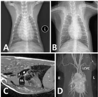
Data for 2 dogs with PLCVC were PRAA and PDA, respectively. First case, a 1-year-6-month-old female, 6.12 kg Cocker Spaniel was admitted to veterinary medical center with chronic vomiting. Clinical problem was onset 4 months ago. At the time of admittance hospital, body temperature and blood pressure were normal range, but respiratory sounds were honking sound and panting. After thoracic and abdomi- nal radiography were conducted, radiography showed sign of mega-esophagus caused by obstruction. After barium con- trast study was performed, obstruction site was clearly found
*Corresponding author
Tel: +82-43-261-3171, Fax: +82-43-261-3224 E-mail: ghkim@cbu.ac.kr
†