Introduction
Gene targeting is commonly referred to as the process in which a cell’s recombination machin- ery integrates a piece of foreign DNA into its own genome at a target locus homologous to the for- eign donor. The process of gene targeting has sev- eral common features in different organisms.
Mechanistically, gene targeting is based on the conserved mechanisms of recombination and DNA repair. Gene targeting enables the tremendous
ability of modifying essentially any endogenous gene at will. Hence, since its invention in yeast over twenty years ago, intense efforts have been devoted into developing this technology in orga- nisms of both research and commercial value.
The breakthrough was brought about by recent successes in mammalian nuclear cloning (Dinnyes et al., 2002). Cloning eliminates the requirement of a stem cell culture because ‘targeted’ animals can be cloned from the nuclei of cultured somatic cells that have been selected for gene targeting events. By this route, ‘targeted’ adult sheep and pigs were produced for the first time (McCreath
─
─ 116 ──
Gene Targeting Study by Homologous Recombination in Cultured Adult Somatic Cells for Targeted
Zebrafish (Danio rerio) Cloning
Ki-Young Lee
Cellular Reprogramming Laboratory, Department of Animal Science, Michigan State University, MI 48824
Forward--genetic screens have generated thousands of mutations, and antisense-- based methods have been used to transiently knockdown gene expression during embryogenesis. Although these methods have made the zebraf ish (Danio rerio) a val- uable system for the identification and functional characterization of developmenta- lly important genes, one deficiency of the zebrafish model is the absence of methods to introduce targeted mutations to generate knockout lines offish. Application of gene-- targeting methods has been limited in traditional transgenic methods using injections of fertilized fish showingrandom genome integration with some mosaicism and episomic expression. In this study, targeted insertion of vector DNA by homologous recombination was demonstrated in the somatic cells derived from zebrafish tail tissues. A targeting construct has been designed to replace the exon 9 and 19 containing these two critical amino acids S222 and K561 for FANCD2 function with a GFP--NEO cassette. To generate a “knock--out” targeting vector, PCR was performed to amplify a 2.8 kb fragment containing sequences from exon1 to exon9, which was inserted upstream of the GFP--NEO cassette. The 3.0 kb fragment downstream of exon 19 was amplified and put downstream of the GFP--NEO cassette. Somatic cells der- ived from tail tissue were transfected with an elecrtoporation expressing the green fluorescent protein (GFP) gene, indicating that targeting construct is introduced into the cultured fibroblast cells.
Key words : Gene Targeting, homologous recombination, cell culture, somatic cell, animal cloning
*Corresponding author: [email protected]
et al., 2000; Denning et al., 2001; Lai et al., 2002).
Compared to mammals, fishes offer easier tra- nsgenic technology because each female produ- ces hundreds of eggs, the manipulated embryos do not need to be incubated inside the mother.
Traditional transgenic methods using injections of fertilized fish have resulted in the generation of many transgenic fish species; however, they sho- wed random genome integration with some mosai- cism and episomic expression. The use of indu- cible gene systems that control temporal and tis- sue expression and of gene-targeting method- ologies based on homologous recombination is desirable to control the expression, efficiency of insertion, and locus of incorporation of transgenes into fish genomes. A variety of systems developed for mammals are now applied in fishes. The use of such systems would require also further develop- ment of stem cell or nuclear transplant technolo- gies in fish.
The availability of gene knockout technique in mouse has greatly advanced our ability to anal- yze gene function in a vertebrate animal. Zebra- fish (Danio rerio), as a superb model for various biological studies, lacks a gene knockout appro- ach to create null mutations by homologous reco- mbination for studying functions of a specific gene.
Since Lee et al. (2002) have shown that long- term cultured cells are still able to promote nor- mal zebrafish development after nuclear transfer, It is commonly believed there is a high probabi- lity that ‘knockout’ zebrafish through nuclear transfer could be obtained.
In order to create some useful disease models of zebrafish, as a result of technology development, any knockout fish generated can also be used by as many investigators as possible. It has focused on cancer, which has the widest impact on human health. One of first candidate is the FANCD2 gene. FANCD2 is responsible for Fanconi anemia (FA), a rare heterogeneous autosomal recessive disorder affecting about 1/100,000 people. This disease is characterized by progressive bone marrow failure, developmental malignancies and typical birth defects including short stature, microcephaly and microphthalmia (D’Andrea and Grompe, 1997; Grompe and D’Andrea, 2001).
The average age of patients who develop cancers is 15 years for leukemia, 16 years for liver tum- ors, and 23 years for other tumors (D’Andrea and Grompe, 1997). Seven FA genes have been clo- ned (A, C, D1/BRCA2, D2, E, F, G) and the enc-
oded proteins cooperate in a common pathway, resulting in the activation of the downstream FANCD2 protein through the monoubiquitination at the lysine 561 (K561) of FANCD2 short form (FANCD2-S) protein after exposure to DNA dam- aging agents. The activated FANCD2 protein is tr- anslocated to the nuclear super complex called BRCA1-associated complex (BASC), which was considered to be a genome damage “sensor”, reco- gnizing and repairing the damaged DNA (Wang et al., 2000; Garcia-Higuera et al., 2001; Grompe, 2002)
The main goal of this study is to develop tech- niques for targeted mutagenesis in zebrafish. This involves designing targeting constructs and sele- cting cultured zebrafish somatic cells carrying ho- mologous recombination events, and then in fut- ure, cloning zebrafish using these cells. It will be anticipated the approach will have a profound im- pact on zebrafish functional genomics, therefore further enhance the utility of zebrafish as a model system for vertebrate development and human diseases.
Meterials and Methods
Zebrafish Strain
Wild-type tubingen strain, zebrafish (Danio rerio), were maintained on 14 h/10 h light-dark cycle and with a rich supply of flake food and live brine shrimp according to standard conditions (Westerfield, 1994).
Caudal Fin Amputation and Cell Culture Fish were anesthetized in 0.02% tricaine pH 7 (3-aminobenzonic acid ethylester, Sigma). Cau- dal fins from the fully anesthetized fish were carefully amputated using sharp scissors (Wes- terfield, 1994). The amputated fins were washed several times with 0.9X PBS supplemented with 100 units/mL penicillin and 100 µ g/mL strepto- mycin (GIBCO). These washed fins were plated into 24 well tissue culture plates (Corning).
For culturing zebrafish cells, the DMEM (Dulb- ecco’s modification of Eagles’s medium) was used as the basic medium. It contains 3 mM L-gluta- mine, 4.5 gm/L glucose, and bFGF (Sigma, 20~
50 ng/mL), but lacks sodium pyruvate (Gibco BRL, Rockville, MD and Cellgro, Mediatech, Inc.). The medium was supplemented with 15%
heat-inactivated fetal bovine serum (vol/vol, Gibco
BRL, Rockville, MD). Cellswere cultured in DM- EM based medium at 28~29� C with 5% CO
2.
Targeting Vector Construct
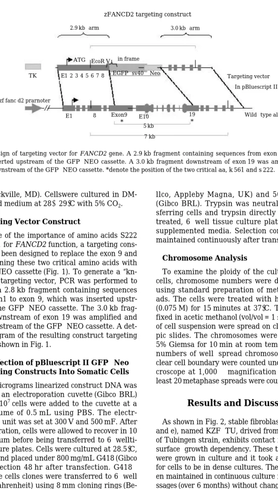
Because of the importance of amino acids S222 and K561 for FANCD2 function, a targeting cons- truct has been designed to replace the exon 9 and 19 containing these two critical amino acids with a GFP-NEO cassette (Fig. 1). To generate a “kn- ock-out” targeting vector, PCR was performed to amplify a 2.8 kb fragment containing sequences from exon1 to exon 9, which was inserted upstr- eam of the GFP-NEO cassette. The 3.0 kb frag- ment downstream of exon 19 was amplified and put downstream of the GFP-NEO cassette. A det- ailed diagram of the resulting construct targeting vector is shown in Fig. 1.
Transfection of pBluescript II GFP- -Neo Targeting Constructs Into Somatic Cells Fifty micrograms linearized construct DNA was added to an electroporation cuvette (Gibco BRL) and 45×10
7cells were added to the cuvette at a final volume of 0.5 mL using PBS. The electr- oporation unit was set at 300 V and 500 mF. After electroporation, cells were allowed to recover in 10 mL medium before being transferred to 6-wellti- ssue culture plates. Cells were cultured at 28.5� C, 5% CO
2and placed under 800 mg/mL G418 (Gibco BRL) selection 48 hr after transfection. G418- resistance cells clones were transferred to 6-well plates (Fahrenheit) using 8 mm cloning rings (Be-
llco, Appleby Magna, UK) and 50 mL trypsin (Gibco BRL). Trypsin was neutralized by tran- sferring cells and trypsin directly into gelatin- treated, 6-well tissue culture plates containing supplemented media. Selection conditions were maintained continuously after transfection.
Chromosome Analysis
To examine the ploidy of the cultured somatic cells, chromosome numbers were determined by using standard preparation of metaphase spre- ads. The cells were treated with hypotonic KCl (0.075 M) for 15 minutes at 37� C. The cells were fixed in acetic methanol (vol/vol = 1 : 3), and drops of cell suspension were spread on clean microsco- pic slides. The chromosomes were stained with 5% Giemsa for 10 min at room temperature. The numbers of well-spread chromosomes within a clear cell boundary were counted under a light mi- croscope at 1,000× magnification under oil. At least 20 metaphase spreads were counted.
Results and Discussion
As shown in Fig. 2, stable fibroblast lines (a, b, c, and e), named KZF-TU, drived from an tail tissue of Tubingen strain, exhibits contact inhibition and surface-growth dependency. These tail tissue cells were grown in culture and it took about 3 weeks for cells to be in dense cultures. The cells have be- en maintained in continuous culture for over 20 pa- ssages (over 6 months) without change in characte-
Fig. 1. Design of targeting vector for FANCD2 gene. A 2.9 kb fragment containing sequences from exon 1 to exon 9 was inserted upstream of the GFP-NEO cassette. A 3.0 kb fragment downstream of exon 19 was amplified and put downstream of the GFP-NEO cassette. *denote the position of the two critical aa, k 561 and s 222.
zFANCD2 targeting construct 2.9 kb-arm
TK E1 2 3 4 5 6 7 8 EGFP-sv40 Neo
E1 8 E10 19
5 kb *
7 kb Exon9
* ATG EcoR V in frame
Targeting vector In pBluescript II
Wild-type allele zf fanc d2 prarnoter
3.0 kb-arm
ristics. A karyotype analysis of these cells showed eudiploid chromosome count (50 chromosomes/di- ploid cell, Fig. 2-d).
FANCD2 protein was also a target of ATM kin- ase. Phosphorylation of serine 222 (S222) of FAN- CD2 by ATM kinase affects the ability of cells to arrest DNA synthesis in response to ionizing radia- tion and prevents mutation from accumulating in daughter cells (Taniguchi et al., 2002). Thus, the FANCD2 protein plays two independent roles in the response to genome insults, first by receiving the signal from both FA and ATM pathways, and second by activating the S phase checkpoint of mi- tosis and initiating a DNA repair reaction (Gro- mpe, 2002).
In the cases of FANCD2 knockouts, to enhance the homologous recombination efficiency in cult- ured cells, a promoter-trap approach was adop- ted in the construction of targeting vectors. Basic- ally, the neomycin resistant gene was flanked by genomic sequences of FANCD2 (Fig. 1) in a way that the neo gene will not be expressed unless it picks up the gene promoters by homologous reco- mbination. This approach has proven very effici- ent in increasing the frequency of identifying ho- mologous recombinants in mouse ES cells and has been used in the production of the first gene- targeted sheep by nuclear transfer from cultured
somatic cells (McCreath et al., 2000). This trap is applicable for FANCD2 because this, as genes involved in general cell cycle and proliferation, are expressed in cultured cells, which have been confirmed by Western blot and mRNA Northern blot analyses (data not shown).
In Fig. 2-e, It shows the GFP expression on the cells after selecting with neomycin indicating that targeting construct is introduced into the cultured fibroblast cells.
These targeted cells can be selected and used to produce cloned zebrafish in future. Once heteroz- ygous fish have been established, they will be mat- ed to generate clutches of offspring with 25% of embryos homozygous for the mutation induced by homologous recombination, 50% heterozygotes and 25% wide-type embryos. The heterozygous embryos are anticipated to have normal development and fertility and will be grown up for line maintenance.
Most important, the fancd2 homozygous embryos are also anticipated to be viable, so that they can be grown up and monitored for the onset of malignant disorders, due to the inability to repair spontaneous DNA damages and resultant genome instability.
The malignant disorders that zfancd2 homologous fish should be predisposed to acquire include leuk- emia and solid tumors arising from different organs.
Once the zfancd2 homologous knockout fish are
Fig. 2. Characterization of cultured adult fibroblast cells (KZF-TU). (a) wild-type primary cells at day 5, 5×, arrow is tail tissue (b) wild-type fibroblast cell lines at day 30 at high density, 5×; (c) wild-type fibroblast cell lines at day 90 at low density, 20×; (d) chromosome smear showing metaphase chromosomes of targeted cells, 10×; (e) a f luorescence image of targeted cells.
a b
d e
c
established, chemical and genetic modifier scre- ens will be performed to search for suppressors or enhancers of the Fanconi phenotype, which will provide a platform for the identification of novel drug targets, as well as the molecular investigat- ion of genome instability-related cancers. If succe- ssful, zebrafish will have all the genetic tools avail- able to the mouse system, and fully realize its pot- ential to study vertebrate gene function and to mo- del human diseases.
References
D’Andrea, A.D. and M. Grompe. 1997. Molecular biology of Fanconi anemia: implications for diag- nosis and therapy. Blood, 90 : 1725 ~1736.
Denning, C., P. Dickinson, S. Burl, D. Wylie, J.
Fletcher and A.J. Clark. 2001. Gene targeting in primary fetal fibroblasts from sheep and pig.
Cloning Stem Cells, 3(4) : 221 ~231.
Dinnyes, A., P. De Sousa, T. King and I. Wilmut.
2002. Somatic cell nuclear transfer: recent prog- ress and challenges. Cloning Stem Cells, 4 : 81~
90.
Garcia -Higuera, I., T. Taniguchi, S. Ganesan, M.S.
Meyn, C. Timmers, J. Hejna, M. Grompe and A.D. D’Andrea. 2001. Interaction of the Fanconi anemia proteins and BRCA1 in a common path-
way. Mol Cell., 7 : 249 ~262
Grompe, M. 2002. FANCD2: a branch -point in DNA damage response. Nat Med., 8 : 555 ~556.
Lai, L., D. Kolber-Simonds, K.W. Park, H.T. Che- ong, J.L. Greenstein, G.S. Im, M. Samuel, A.
Bonk, A. Rieke and B.N. Day. 2002. Production of {alpha}-1, 3-Galactosyltransferase Knockout Pigs by Nuclear Transfer Cloning. Science, 295 : 1089 ~1092
Lee, K.Y., H. Huang, B. Ju, Z. Yang and S. Lin.
2002. Cloned zebrafish by nuclear transfer from long -term-cultured cells. Nature Biotechnology, 20 : 795~799.
McCreath, K.J., J. Howcroft, K.H. Campbell, A.
Colman, A.E. Schnieke and A.J. Kind. 2000. Pro- duction of gene -targeted sheep by nuclear tran- sfer from cultured somatic cells. Nature, 405 : 1066 ~1069.
Taniguchi, T., I. Garcia -Higuera, B. Xu, P.R. Andr- eassen, R.C. Gregory, S.T. Kim, W.S. Lane, M.B.
Kastan and A.D. D’Andrea. 2002. Convergence of the fanconi anemia and taxia telangiectasia signaling pathways. Cell, 17109(4) : 459 ~472.
Wang, Y., D. Cortez, P. Yazdi, N. Neff, S.J. Elledge and J. Qin. 2000. BASC, a super complex of BR- CA1 -associated proteins involved in the reco- gnition and repair of aberrant DNA structures.
Genes Dev., 15; 14(8) : 927 ~939.
Westerfield, M. 1994. In “Zebrafish Book” 2
ndedi- tion. University of Oregon Press, Eugene, OR.
Zebrafish ( Danio rerio )
성체배양세포 수준에서의 유전자적중에 관한 연구 이 기 영미시간대학교 동물자원과학과
전통적인 유전자이식법으로는 유전자적중법을 적용하는데 한계가 있으며 최근 몇 년동안 동물 복제방법에 의한 유전자적중 복제동물들이 생산되고 있다
.따라서 본 연구는 척추동물의 모델로 매우 유용하고 중요시 다뤄지고 있는
Zebrafish (Danio rerio)를 대상으로 유전자적중방법을 응용 하고자 실험을 진행하였다
.본 실험은
Zebrafish의 꼬리지느러미로부터 체세포를 배양한 다음 유 전자적중 벡터를 전이시켜 발현유무를 확인하였다
.대상유전자로는
FANCD2 gene을 선정하였다
. FANCD2기능에 있어 아주 중요한 아미노산
S222와
K561을 포함하고 있는
exon 9과
exon 19를
GFP-Neo cassette로 교체하여 유전자적중 벡터를 제작하였다
.제작된 벡터는 전기충격법에 의해
3개월 이상 배양된 체세포
(KZF-TU라 명명
)에 전이시켰다
.생존한 세포들을
G418으로
2주 동안 선별하여 형광현미경으로 관찰한 결과 정상적으로 유전자가 발현됨이 확인되었다
.이는 체 세포에 정상적으로 유전자적중 유전자가 삽입되었음을 의미하며
,따라서 이들 발현 세포들을 이 용한 유전자적중
Zebrafish의 복제가 가능할 것으로 기대된다
.Received : May 15, 2004 Accepted : June 12, 2004