eISSN 2287-1683 pISSN 1738-8767 Journal of Trauma and Injury Vol. 29, No. 4, December, 2016 http://dx.doi.org/10.20408/jti.2016.29.4.161
� Original Article �
� Address for Correspondence : Kang Kook Choi, M.D., Ph.D.
Department of Trauma Surgery, Gachon University Gil Medical Center, 21, 774beon-gil, Namdong-daero, Namdong-gu, Incheon 21565, Korea
Tel : 82-32-460-3213, Fax : 82-32-461-3214, E-mail : choikangkook@gilhospital.com Submitted : December 7, 2016 Revised : December 10, 2016 Accepted : January 31, 2017 The article was presented at the 3rd Pan-Pacific Trauma Congress, June 4-6, 2015 in Seoul, Korea.
Blush on Computed Tomography and Transcatheter Arterial Embolization in Pelvic Fracture
Jihun Gwak, M.D., Yong-Cheol Yoon, M.D., Min A Lee, M.D., Byungchul Yu, M.D., Myung Jin Jang, M.D., Kang Kook Choi, M.D.
Department of Trauma Surgery, Gachon University, Gil Medical Center, Incheon, Korea
Purpose: Bleeding is the primary cause of death after severe pelvic fracture. Transcatheter arterial embolization (TAE) is the mainstay of treatment for arterial bleeding. This study aimed to determine the frequency of bleeding by angiography of blush-positive pelvic fractures on computed tomography (CT) images. The bleeding arteries that were involved were investigated by pelvic angiography.
Methods: This retrospective cohort study evaluated 83 pelvic fracture patients who were treated in the intensive care unit of the author’s trauma center between January 01, 2013 and April 30, 2015.
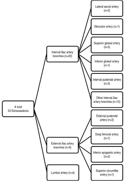
Results: Overall mortality was 9 of 83 patients (10.8%). Blush was observed in 37 patients; blush-positive patients had significantly higher mortality (24.3%) than blush-negative patients (0%). Twenty-four of the 83 patients (28.9%) underwent pelvic angiography. Bleeding was showed in 22 of 24 patients in pelvic angiography. TAE was successfully performed in 21 (95.5%) of the bleeding 22 patients. Angiography was performed in 23 of 37 blush-positive patients, and arterial bleeding was identified in 21 (91.3%). A total 33 bleeding arteries were identified in 22 angiography-posi- tive patients. The most frequent origin of bleeding was internal iliac artery (69.7%) followed by the external iliac artery (18.2%) and lumbar arteries (12.1%).
Conclusion: The vascular blush observed in CT scans indicates sites of ongoing bleeding in pelvic angiography. TAE is an excellent therapeutic option for arterial bleeding and has a high success rate with few complications. [ J Trauma Inj 2016; 29: 161-166 ]
Key Words: Pelvic bones, Fractures, Bone, Hemorrhage, Embolization, Therapeutic
Table 1. The relationship between the mortality and blush on CT scan and posterior pelvic instability
Mortality rate p value
Blush < 0.01
Positive 9/37 (24.3)
Negative 0/46 (0)00.
Posterior pelvic instability 0 0.08
Intact arch 1/21 (04.8)
Incomplete disruption 6/56 (10.7) Complete disruption 02/6 (33.3)
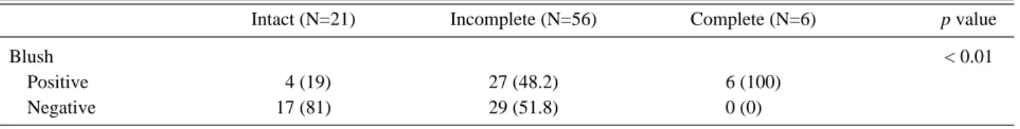
Table 2. The relationship between the posterior pelvic instability and blush in CT scan
Intact (N=21) Incomplete (N=56) Complete (N=6) p value
Blush < 0.01
Positive 04 (19) 27 (48.2) 6 (100)
Negative 17 (81) 29 (51.8) 0 (0)00
Jihun Gwak, et al. Transcatheter Arterial Embolization in Pelvic Fracture
Table 3. The relationship between hypotension and blush in CT scan
Hypotension (N=22) p value
Blush < 0.01
Positive 18/22 (81.8)
Negative 04/22 (18.2)
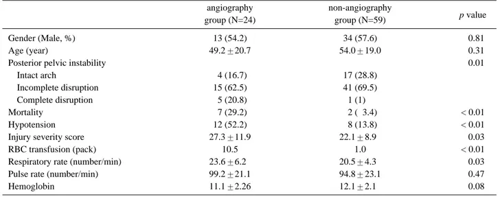
Table 4. The comparison between angiography group and non-angiography group
angiography non-angiography
p value
group (N=24) group (N=59)
Gender (Male, %) 13 (54.2) 34 (57.6) 0 0.81
Age (year) 49.2 20.7 54.0 19.0 0 0.31
Posterior pelvic instability 0 0.01
Intact arch 04 (16.7) 17 (28.8)
Incomplete disruption 15 (62.5) 41 (69.5)
Complete disruption 05 (20.8) 1 (1)0.
Mortality 07 (29.2) 02 (03.4) < 0.01
Hypotension 12 (52.2) 08 (13.8) < 0.01
Injury severity score 27.3 11.9 22.1 8.90 0 0.03
RBC transfusion (pack) 10.5 1.0 < 0.01
Respiratory rate (number/min) 23.6 6.20 20.5 4.30 0 0.03
Pulse rate (number/min) 99.2 21.1 94.8 23.1 0 0.47
Hemoglobin 11.1 2.26 12.1 2.10 0 0.08
Fig. 1. Angiography was performed for 23 patients of 37 blush-positive patients, and arterial bleeding was identified in 21 patients (91.3%) in the angiography.
Fig. 2. The distribution of bleeding arteries in pelvic angiography.
Jihun Gwak, et al. Transcatheter Arterial Embolization in Pelvic Fracture
REFERENCES
01) Starr AJ, Griffin DR, Reinert CM, Frawley WH, Walker J, Whitlock SN, et al. Pelvic ring disruptions: prediction of asso- ciated injuries, transfusion requirement, pelvic arteriography, complications, and mortality. J Orthop Trauma 2002; 16: 553-61.
02) Suzuki T, Smith WR, Moore EE. Pelvic packing or angiogra- phy: competitive or complementary? Injury 2009; 40: 343-53.
03) Osborn PM, Smith WR, Moore EE, Cothren CC, Morgan SJ, Williams AE, et al. Direct retroperitoneal pelvic packing ver- sus pelvic angiography: A comparison of two management protocols for haemodynamically unstable pelvic fractures.
Injury 2009; 40: 54-60.
04) Marzi I, Lustenberger T. Management of Bleeding Pelvic Fractures. Scand J Surg 2014; 103: 104-11.
05) Eastridge BJ, Starr A, Minei JP, O’Keefe GE, Scalea TM. The importance of fracture pattern in guiding therapeutic decision- making in patients with hemorrhagic shock and pelvic ring disruptions. J Trauma 2002; 53: 446-50; discussion 50-1.
06) Agolini SF, Shah K, Jaffe J, Newcomb J, Rhodes M, Reed JF, 3rd. Arterial embolization is a rapid and effective technique for controlling pelvic fracture hemorrhage. J Trauma 1997;
43: 395-9.
07) Cullinane DC, Schiller HJ, Zielinski MD, Bilaniuk JW, Collier
tion with pelvic fractures. Am Surg 2012; 78: 825-30.
09) Anandakumar V, Hussein FK, Varuun B, Zhu R. Predictive parameters for angiography and embolization in the bleeding pelvic fracture. J Clin Orthop Trauma 2013; 4: 70-4.
10) Papakostidis C, Kanakaris N, Dimitriou R, Giannoudis PV.
The role of arterial embolization in controlling pelvic fracture haemorrhage: a systematic review of the literature. Eur J Radiol 2012; 81: 897-904.
11) Brasel KJ, Pham K, Yang H, Christensen R, Weigelt JA.
Significance of contrast extravasation in patients with pelvic fracture. J Trauma 2007; 62: 1149-52.
12) Diamond IR, Hamilton PA, Garber AB, Tien HC, Chughtai T, Rizoli SB, et al. Extravasation of intravenous computed tomog- raphy scan contrast in blunt abdominal and pelvic trauma. J Trauma 2009; 66: 1102-7.
13) Verbeek DO, Zijlstra IA, van der Leij C, Ponsen KJ, van Delden OM, Goslings JC. Management of pelvic ring fracture patients with a pelvic “blush” on early computed tomography.
The journal of trauma and acute care surgery 2014; 76: 374-9.
14) Costantini TW, Bosarge PL, Fortlage D, Bansal V, Coimbra R. Arterial embolization for pelvic fractures after blunt trau- ma: are we all talk? American journal of surgery 2010; 200:
752-7; discussion 7-8.
15) Ono Y, Ishida T, Iwasaki Y, Kawakami Y, Inokuchi R, Tase C, et al. The off-hour effect on trauma patients requiring sub- specialty intervention at a community hospital in Japan: a ret- rospective cohort study. Scand J Trauma Resusc Emerg Med 2015; 23: 20.
16) Balogh Z, Caldwell E, Heetveld M, D’Amours S, Schlaphoff G, Harris I, et al. Institutional practice guidelines on manage- ment of pelvic fracture-related hemodynamic instability: do they make a difference? J Trauma 2005; 58: 778-82.
17) Manson TT, Nascone JW, Sciadini MF, O’Toole RV. Does fracture pattern predict death with lateral compression type 1 pelvic fractures? J Trauma 2010; 69 : 876-9.
18) Sarin EL, Moore JB, Moore EE, Shannon MR, Ray CE, Morgan SJ, et al. Pelvic fracture pattern does not always pre- dict the need for urgent embolization. J Trauma 2005; 58:
973-7.
19) Wright V, Zelle BA, Prayson M. Bilateral sacroiliac joint dis- location without associated fracture or anterior pelvic ring injuries. J Orthop Trauma 2004; 18: 634-7.
20) DuBose J, Inaba K, Barmparas G, Teixeira PG, Schnuriger B, Talving P, et al. Bilateral internal iliac artery ligation as a dam- age control approach in massive retroperitoneal bleeding after pelvic fracture. J Trauma 2010; 69: 1507-14.