Tuberc Respir Dis 2009;66:274-279
CopyrightⒸ2009. The Korean Academy of Tuberculosis and Respiratory Diseases. All rights reserved.
비소세포폐암 세포주에서 Uteroglobin Transduction이 COX-2 및 IDO의 발 현에 미치는 영향
1
동국대학교 의과대학 동국대학교 일산병원 내과,
2서울대학교 의과대학 내과학교실 및 폐연구소,
3분당서울대학교병원 폐센터, 내과
박근민1, 이상민2, 임재준2, 양석철2, 유철규2, 이춘택3, 한성구2, 심영수2, 김영환2Expression of COX-2 and IDO by Uteroglobin Transduction in NSCLC Cell Lines
Gun Min Park, M.D.
1, Sang-Min Lee, M.D.
2, Jae-Joon Yim, M.D.
2, Seok-Chul Yang, M.D.
2, Chul Gyu Yoo, M.D.
2, Choon-Taek Lee, M.D.
3, Sung Koo Han, M.D.
2, Young-Soo Shim, M.D.
2, Young Whan Kim, M.D.
21
Department of Internal Medicine, Dongguk University Ilsan Hospital, Dongguk University College of Medicine, Gyeongju,
2
Department of Internal Medicine and Lung Institute of Medical Research Center, Seoul National University College of Medicine, Seoul,
3Respiratory Center, Department of Internal Medicine, Seoul National University Bundang Hospital, Seongnam, Korea
Background: Uteroglobin (UG) is a secretary protein that has strong immunomodulatory properties, and which is synthesized in most epithelia including lung tissue. Overexpression of UG is associated with decreased expression of cyclooxygenase (COX)-2 and suppression of cancer cell growth. Indoleamine 2,3-dioxygenase (IDO) catalyzes tryptophan along the kynurenine pathway, and both the reduction in local tryptophan and the production of tryptophan metabolites contribute to the immunosuppressive effects of IDO.Methods: In this study, we investigated the pattern of expression of COX-2 and IDO, and the effect of UG transduction in the expression of COX-2 and IDO in several non-small cell lung cancer cell lines, especially A549.
Results: Both COX-2 and IDO were constitutionally expressed in A549 and H460 cells, and was reduced by UG transduction. In A549 cells, the slightly increased expression of COX-2 and IDO with the instillation of interferon-gamma (IFN-γ) was reduced by UG transduction. However, the reduced expression of COX-2 and IDO by UG transduction was not increased with IFN-γ instillation in A549 cells. In both the A549 COX-2 sense and the A549 COX-2 anti-sense small interfering RNA (siRNA)-transfected cells, IDO was expressed; expression was reduced by UG transduction, irrespective of the expression of COX-2.
Conclusion: The results suggest that the anti-proliferative function of UG may be associated with the immune tolerance pathway of IDO, which is independent of the COX-2 pathway.
Key Words: Uteroglobin, Cyclooxygenase 2, Indoleamine 2,3-dioxygenase, Interferon-gamma, Immune tolerance
Address for correspondence: Young Whan Kim, M.D.
Department of Internal Medicine, Seoul National University College of Medicine, 28, Yeongeon-dong, Jongno-gu, Seoul 110-799, Korea
Phone: 82-2-2072-2856, Fax: 82-2-762-9662 E-mail: [email protected]
Received: Feb. 11, 2009 Accepted: Mar. 18, 2009
서 론
Uteroglobin (UG)은 70개 아미노산으로 구성된 두 개 의 단백으로 이루어져 있고, 자궁, 폐, 유선, 전립선 등
여러 장기의 점막 세포에서 분비되며, 혈액, 뇨, BAL fluid 에서 검출된다. UG는 장기에 따라 많은 이름으로 불려지 고 있는데, 폐의 경우 Clara cell에서 발견되어 Clara Cell 분비 단백질(CCSP)이라고 불려진다1. 그 생물학적인 작용 으로는 면역조절과 항염증작용을 하는 것이 알려져 있다.
최근의 연구에서 비소세포폐암 조직이나 세포주에서 UG 의 발현이 현저하게 감소해 있음이 보고되었고2, 반대로 암세포에서 UG를 과발현 시키면 암의 형질이 소실되고, 암세포 성장이 억제됨이 보고되었다3-5.
Cyclooxygenase (COX)는 아라키돈산을 프로스타노이
드로 바꾸는 과정의 속도제한 효소(rate limiting enzyme) 이다. COX-1과 이성체로 존재하는 COX-2는 알레르기 매 개물질 및 박테리아의 lipopolysaccharide의 자극 등에 의 해 트롬복산이나 프로스타글란딘을 생성하여 염증 반응 에 관여한다고 알려져 있었다. 그러나, 최근 여러 암종에 서 COX-2의 발현이 증가되어 있음이 보고되었고, 비소세 포 폐암 조직의 약 30%, 폐선암의 40%에서 COX-2가 발현 됨이 보고되었다6. COX-2는 metalloproteinase의 합성, 미 세혈관 형성 등에 관여하여 암세포의 침윤 및 성장에 영향 을 미치고, PGE2를 생성하여 면역세포인 림프구와 대식구 에서 IL-10의 합성을 자극함으로써 종양세포에 대한 인체 의 면역반응을 억제함이 보고되었다. 한편, 폐암세포주인 A549에 UG를 과발현 시켰을 때 COX-2의 생성물인 PGE2
의 분비가 감소하며, UG를 과발현 시킨 A549의 배양액으 로 면역세포를 배양했을 때 IL-10, TGF-β의 분비가 감소 함이 보고되었다7.
Indoleamine 2,3-dioxygenase (IDO)라는 효소단백이 악성 종양의 면역관용(immune tolerance)에도 관여함이 최근의 연구에서 알려졌다. IDO는 필수 아미노산인 Tryp- tophan의 이화작용에 관여하는 속도제한 효소로서, 폐, 태반을 비롯한 거의 모든 정상조직에서 발현되어 정상적 으로는 면역관용에 중요한 역할을 한다. 그러나, IDO는 여러 암세포주에서 interferon-γ (IFN-γ) 등의 염증물질 에 의해 유도됨이 알려져 있다8,9. 이러한 IDO는 국소적으 로는 정상세포의 생존에 필수적인 tryptophan을 고갈시키 며, tryptophan 대사로 생성된 kynurenine 등의 대사물질 을 통해 면역 T-세포의 성장을 억제하고 T-세포의 anergy 를 유발함으로써 암세포에 대한 면역관용을 일으켜 오히 려 암세포 성장을 돕게 된다10,11. 한편, IFN-γ 투여로 인 해 IDO의 발현이 증가함은 물론, 대식세포를 비롯한 여러 세포에서 COX-2의 발현도 증가함이 알려져 있다12. 또한, IDO의 과발현이 COX의 발현 및 활성도와 관련성이 있음 이 보고된 바 있다13.
본 연구에서는 먼저, 비소세포폐암 세포주에서 COX-2, IDO의 발현양상 및 UG transduction이 COX-2, IDO의 발현에 미치는 영향을 살펴보았다. 다음으로, COX-2를 항상 발현시 킨 암세포주와 COX-2 발현을 억제한 암세포주에서 UG transduction에 의한 IDO의 발현 양상의 변화를 살펴봄으로 써 UG와 IDO 사이의 직접적인 관련성을 보고자 하였다.
대상 및 방법
1. 세포주
세포주는 비소세포폐암 세포주인 A549, H460, H157을 사용하였다. 각각의 세포주를 37oC, 5% CO2 조건 하에서 10% fetal bovine serum (FBS), penicillin (60 μg/mL), streptomycin (100 μg/mL)이 첨가된 RPMI 1640 배지에 배양하였다.
2. Adenovirus-UG (Ad-UG) transduction
각각의 세포주를 1×10⁶/mL으로 플레이트에 준비한 후 한 플레이트의 세포를 떼어, 살아있는 세포의 수를 측정하 였다. 2일간 배양하여 세포가 well의 80% 정도까지 자라면 Ad-UG의 양성 대조군인 Ad-null 혹은 Ad-UG의 용액을 (20 MOI) RPMI 1640 배지와 섞어서 플레이트에 넣고 37oC 에서 1시간 동안 감염시켰다. PBS로 세척한 후 FBS가 포함 된 RPMI 1640 배지를 넣고 2일간 추가 배양하였다.
3. IFN-γ instillation 및 Ad-UG transduction
비소세포폐암 세포주인 A549를 아무 처리도 하지 않은 대조군, Ad-UG transduction을 한 군, IFN-γ (1,000 U) instillation을 한 군, IFN-γ instillation 12시간 후에 Ad-UG transduction을 한 군, Ad-UG transduction 36시간 후에 IFN-γ instillation 한 군으로 나누어 배양한 후 단백질을 추출하여 Western 분석법을 통해 COX-2 및 IDO의 발현 양상을 분석하였다.
4. 단백질의 추출 및 Western 분석법
충분히 배양된 A549를 비롯한 각각의 세포주 및 Ad-UG transduction 시행 후 추가 배양한 A549 세포주를 whole lysis buffer (0.1% NP-40, 5 mM EDTA, 50 mM Tris [pH 7.5∼8.0], 250 mM NaCl, 50 mM NaF)를 이용하여 총세 포 단백을 추출하였다. 30 μg의 세포 단백질을 10%
SDS-polyacrylamide 젤에서 전기영동 시켰다. 젤 상의 단 백질을 nitrocellulose membrane으로 이동시키고, 이 membrane을 blocking solution (5% skim milk in 1×PBS/
Tween 20)으로 1시간 동안 block시킨 후 IDO와 COX-2, actin에 대한 1차 항체를 12시간 동안 반응시켰다. 세척 후에 2차 항체를 반응시킨 후 면역 신호의 검출은 ECL Western blotting detection system (Amersham Pharmacia Biotech, Uppsala, Sweden)을 이용하였다.
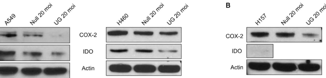
Figure 1. Expression of COX-2 and IDO in NSCLC cell lines transduced with Uteroglobin (UG). (A) Both COX-2 and IDO were expressed in A549 and H460 cells. Transduction of Ad-UG (20 moi) directly to A549 and H460 cells decreased expression of both COX-2 and IDO. (B) COX-2 was expressed in H157 cells. Transduction of Ad-UG (20 moi) directly to H157 cells decreased expression of COX-2. But, IDO was not expressed in H157 cells.
Figure 2. Change of expression of COX-2 and IDO after UG transduction, instillation of IFN-γ, UG transduction af- ter instillation of IFN-γ and instillation of IFN-γ after UG transduction, in A549, non-small cell lung cancer cell line.
5. COX-2 sense 세포주 및 COX-2 anti-sense 세포주 배양
COX-2가 항상 발현하도록 A549에 COX-2를 trans- fection한 A549 COX-2 sense 세포주와 A549에 small in- terfering RNA (siRNA)를 transfection해 COX-2의 발현을 억제시킨 A549 COX-2 anti-sense 세포주를 배양하여 사 용하였다. siRNA transfection은 A549 세포주가 well의 70%가 찰 때까지 키운 후 50 nmol의 COX-2 gene-specif- ic siRNA를 HiPerFect Transfection 시약과 섞으면서 부드 럽게 세포에 주입하여 48∼72시간 동안 감염시키는 과정 으로 진행하였다.
결 과
비소세포폐암 세포주인 A549와 H460에서 COX-2 및 IDO가 발현되었고, UG transduction을 했을 때 COX-2 및 IDO의 발현이 모두 감소하였다(Figure 1A). 또 다른 비소 세포폐암 세포주인 H157에서는 COX-2가 발현되었고, UG transduction에 의해 COX-2의 발현이 감소하였지만, IDO는 발현되지 않았다(Figure 1B).
비소세포폐암 세포주인 A549를 control 군, Ad-UG transduction을 시행한 군, IFN-γ (1,000 U) instillation을 시행한 군, IFN-γ instillation 12시간 후 Ad-UG trans- duction을 시행한 군, Ad-UG transduction 36시간 후 IFN-γ instillation을 시행한 군으로 나누어서 배양한 후 단백질을 추출하고 Western 분석법을 통해 COX-2 및 IDO의 발현 양상을 분석하였다.
A549에 IFN-γ instillation을 한 경우, 처리하지 않은 군 에 비해 COX-2 및 IDO의 발현이 모두 약간씩 증가하는
양상을 보였다. IFN-γ instillation을 먼저 시행한 후 UG transduction을 시행한 군의 경우 IFN-γ instillation만 시 행한 군에 비해 COX-2 및 IDO의 발현이 모두 감소하였 다. 그러나, UG transduction을 먼저 시행한 후 IFN-γ in- stillation을 시행한 군의 경우 UG transduction만 한 군에 비해 COX-2 및 IDO의 발현이 증가하지 않았다(Figure 2).
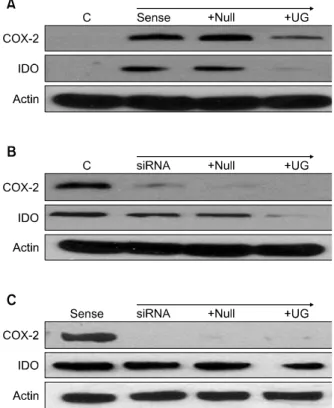
비소세포폐암 세포주인 A549가 COX-2를 항상 발현하 도록 한 COX-2 sense 세포주에서 COX-2와 함께 IDO의 발현도 관찰되었다. 여기에 Ad-UG transduction을 했을 때 COX-2와 IDO의 발현이 모두 감소하였다(Figure 3A).
A549에 siRNA를 transfection해서 COX-2의 발현을 억 제한 COX-2 anti-sense 세포주의 경우 COX-2의 발현은 현저히 감소하였으나, IDO의 발현은 감소하지 않았다. 여 기에 다시 Ad-UG를 transduction 했을 때 COX-2는 거의 변화가 없었으나 IDO의 발현은 감소하였다(Figure 3B).
A549 COX-2 sense 세포에 siRNA를 transfection 시켰을 때도 COX-2의 발현은 현저히 감소하였으나, IDO의 발현 에는 변화가 없었고, 여기에 다시 Ad-UG를 transduction
Figure 3. Expression of COX-2 and IDO in A549 cell line transduced with Uteroglobin (UG) as transfection with COX-2 sense or siRNA. (A) COX-2 and IDO were de- creased when A549 COX-2 sense cell was transduced with Ad-UG (50 moi). (B) Transfection with COX-2 siRNA (50 nM) to A549 cell inhibited not IDO but COX-2. (C) A549 COX-2 sense cell transfected with siRNA (50 nM) had same result B. Uteroglobin suppressed IDO protein level both B and C. It need further study about IDO en- zyme activity test with uteroglobin.
했을 때 COX-2는 변화가 없었고, IDO의 발현은 감소하여 3B와 같은 결과를 보여주었다(Figure 3C).
고 찰
IDO의 유도 면역관용에는 두 가지 기전이 존재한다.
첫 번째는 tryptophan 대사물에 의한 면역저하가 있는데, IDO가 활성화되면서 tryptophan으로부터 Kynurenines 및 Kynurenine 유도체인 3-hydroxyanthranilic acids나 quinolinic acids가 생성되고, 이러한 대사산물이 면역 T 세포(CD4+)의 증식을 억제하고, apoptosis를 유발함으로 써 면역저하에 기여한다14. 두 번째는 IDO 효소활성의 결 과로 면역 T 세포의 증식에 필요한 tryptophan이 국소적 으로 부족하게 되어 T 세포의 세포주기를 정지시키고, T
세포의 anergy를 야기함으로써 면역저하를 유발한다. IDO 에 의한 면역 저하는 종양 주위뿐만 아니라 종양세포가 유출되는 림프절에서도 일어나게 되는데, 종양세포에 접 촉한 IDO를 발현하는 수지상세포(dendritic cells)가 림프 절에 도착하면 직접 종양에 반응하는 면역 T 세포를 억제 하고 무기력화시킬 뿐만 아니라, IDO를 발현하지 않는 수 지상세포와 반응하는 주위의 면역 T 세포의 반응도 억제 시킨다. 또한 regulatory T 세포와의 상호작용을 통해 수 지상세포 내에 IDO의 발현을 유도함이 알려져 있다. 종양 미세환경에서도 종양세포는 스스로 IDO를 발현하거나, 염증 신호에 반응하여 IDO의 발현을 증가시키게 되는데, 특히 IFN-γ가 종양세포의 IDO 발현 증가에 중요하다15. 최근 여러 연구에서 대장암, 유방암, 식도암, 폐암, 그리 고 피부암 등에서 프로스타글란딘(prostaglandin, PG)의 수치가 정상조직에 비해 증가되어 있음이 보고되었다.
COX-2의 산물인 PGE2는 VEGF를 생성하여 암세포 성장 을 촉진하고 면역 체계를 억제하며, bcl-2를 과발현시키고 다양한 matrix metalloproteinase를 과발현시켜 종양의 성 장을 촉진하는 것으로 설명되고 있다. 최근에는 PGE2가 IL-10을 유발하면서 이렇게 유발된 IL-10이 수지상세포에 작용하여 T 세포의 anergy를 유발하여 T 세포의 종양세포 에 대한 면역 활성을 억제함이 보고되었다16. Murine lewis lung carcinoma (3LL) 모델에서 COX-2의 발현을 감소시 키면 IL-10, IL-12가 억제되면서 antitumor reactivity가 활 성화되는데, 여기서 COX-2에 의하여 생성되는 PGE2의 생 성이 감소하고 IL-10의 생성이 억제되면서 면역세포인 림 프구와 대식구의 면역 기능이 활성화되어 종양 생성이 억 제됨이 보고되었다17,18.
이번 연구에서 비소세포폐암 세포주인 A549, H460, H157에서 COX-2와 IDO의 발현 양상을 살펴보았을 때 A549, H460에서는 구조적으로 COX-2와 IDO가 발현되었 고, UG transduction에 의해서 COX-2 및 IDO의 발현이 모두 감소하였다. IDO의 발현이 없었던 H157에서도 COX-2가 발현되었고, UG transduction으로 인해 COX-2 의 발현이 감소하였다. A549, H460에서 UG transduction 으로 인해 COX-2의 발현이 감소하고19, 세포성장이 억제 됨이 보고된 바가 있고, H157에서도 UG transduction으 로 인해 NF-kB의 활성도가 억제되어 COX-2 발현이 감소 함이 보고되었으나20, 본 연구에서처럼 IDO의 발현양상을 함께 살펴본 연구결과는 없었다.
IFN-γ는 A549, THP-1을 비롯한 많은 암세포주에서 IDO의 발현을 유도하는 것으로 알려져 있다8. 또한 IFN-γ
는 수지상세포에서 IDO 발현의 주요 자극제인데, 최근 그 기전으로 Janus kinase-signal transducer and activator (JAK/STAT) 신호체계가 관련이 있음이 보고되었다21. IDO의 발현과 COX-2의 발현의 연관성을 보기 위해 비 소세포폐암 세포주인 A549를 IFN-γ로 자극하여 COX-2, IDO의 발현을 증가시킨 군과 IFN-γ instillation 후 UG transduction을 한 군을 비교했을 때 IFN-γ instillation 후 UG transduction을 한 군에서 COX-2 및 IDO가 모두 감소 되었다. 그러나, UG transduction만 한 군에 비해 UG transduction 후 IFN-γ instillation을 한 군에서 COX-2 및 IDO의 발현이 증가하지는 않았다. 이러한 결과는 UG transduction으로 인한 세포성장 억제효과가 IFN-γ의 효 과보다 더 강력한 것임을 시사한다.
Marshall 등은 murine macrophage cell line RAW 264.7과 murine fibrosarcoma cell line MC57에서 세포 성 장의 증가와 cell adhesion이 증가됨을 관찰하였으며 이러 한 증가는 IDO의 억제 시 감소됨을 관찰하였고, 또한 IDO의 발현은 metalloproteinase 발현 정도와 COX의 활 성도 및 발현 정도에 따라 변화함을 보고하였다13. 최근에 는 COX-2의 생산물인 PGE2가 수지상세포 유도의 잠정적 인 핵심 물질이며, IDO의 발현을 유도하는 물질임이 보고 되었다22. 또한 Basu 등23은 COX-2를 특이적으로 억제한 경우 종양의 미세환경에서 IDO의 발현이 감소함을 보고 하였다. 이렇듯 COX-2와 IDO의 발현 사이에 관련성이 있 음이 제기되고 있으며, 본 연구에서도 A549, H460에서 UG transduction으로 인해 COX-2와 IDO의 발현이 함께 감소하는 결과를 보였다. 그러나, 본 연구에서는 COX-2가 항상 발현되도록 처치한 A549뿐 아니라 siRNA trans- fection을 통해 COX-2의 발현을 억제한 A549에서도 UG transduction으로 인해 IDO의 발현이 감소하는 결과를 보 였다. 이러한 결과는 UG의 세포성장억제 기능이 COX-2 를 통한 기전과는 독립적으로 IDO의 면역관용 기전과 관 련될 가능성이 있음을 시사한다. 향후 IDO의 발현이 없었 던 H157에서 UG transduction이 세포성장에 영향을 미치 지 않음을 MTT assay 등을 통해 확인한다면, UG의 세포 성장억제와 IDO의 관련성을 좀 더 명확히 밝힐 수 있을 것이다.
요 약
연구배경: Uteroglobin (UG)은 폐를 비롯한 우리 몸의 대부분의 표피세포에서 생성되는 면역조절능을 가진 분
비단백이다. UG의 과발현은 cyclooxygenase (COX)-2의 발현의 감소 및 암세포의 성장억제와 관련이 있다.
Indoleamine 2,3-dioxygenase (IDO)는 kynurenine path- way를 통해 tryptophan을 이화시키는 효소로서, 국소적 으로 tryptopha을 고갈시키고 tryptophan 대사물을 생성 함으로써 T 세포의 면역반응을 억제시키는 데 기여한다.
방 법: 본 연구에서는 여러 비소세포폐암 세포주, 특히 A549에서 COX-2와 IDO의 발현양상 및 UG transduction 이 COX-2 및 IDO의 발현에 미치는 영향을 살펴보았다.
결 과: A549와 H460에서 구조적으로 COX-2와 IDO 가 모두 발현되었고, COX-2 및 IDO의 발현은 UG trans- duction에 의해 감소되었다. A549에 IFN-γ를 투여했을 때 COX-2 및 IDO의 발현이 약간 증가하였고, 이는 UG transduction 시행 후 다시 감소하였다. 그러나, A549에 UG transduction 시행하여 감소된 COX-2, IDO의 발현은 IFN-γ 투여 후에도 증가하지 않았다. A549 COX-2 sense 와 A549 COX-2 anti-sense (siRNA 감염) 세포주 모두에서 COX-2의 발현여부와 상관없이 IDO가 발현되었고, UG transduction으로 인해 IDO의 발현이 감소하였다.
결 론: 이러한 결과는 UG의 세포성장억제 기능이 COX-2를 통한 기전과는 독립적으로 IDO의 면역관용 기 전과 관련될 가능성이 있음을 시사한다.
감사의 글
Supported by grant No. 06-2006-1379 from the SNUH Research Fund.
참 고 문 헌
1. Mukherjee AB, Kundu GC, Mantile-Selvaggi G, Yuan CJ, Mandal AK, Chattopadhyay S, et al. Uteroglobin:
a novel cytokine? Cell Mol Life Sci 1999;55:771-87.
2. Linnoila RI, Szabo E, DeMayo F, Witschi H, Sabourin C, Malkinson A. The role of CC10 in pulmonary carci- nogenesis: from a marker to tumor suppression. Ann N Y Acad Sci 2000;923:249-67.
3. Szabo E, Goheer A, Witschi H, Linnoila RI. Overex- pression of CC10 modifies neoplastic potential in lung cancer cells. Cell Growth Differ 1998;9:475-85.
4. Zhang Z, Kundu GC, Panda D, Mandal AK, Mantile- Selvaggi G, Peri A, et al. Loss of transformed pheno- type in cancer cells by overexpression of the uteroglo- bin gene. Proc Natl Acad Sci USA 1999;96:3963-8.
5. Kundu GC, Mandal AK, Zhang Z, Mantile-Selvaggi G, Mukherjee AB. Uteroglobin (UG) suppresses extra- cellular matrix invasion by normal and cancer cells that express the high affinity UG-binding proteins. J Biol Chem 1998;273:22819-24.
6. Hastürk S, Kemp B, Kalapurakal SK, Kurie JM, Hong WK, Lee JS. Expression of cyclooxygenase-1 and cyclo- oxygenase-2 in bronchial epithelium and nonsmall cell lung carcinoma. Cancer 2002;94:1023-31.
7. Yoon JM, Lim JJ, Yoo CG, Lee CT, Han SK, Shim YS, et al. The role of uteroglobin in the immunomodu- lation of nonsmall cell lung cancer cells. Tuberc Respir Dis 2004;57:336-44.
8. Taylor MW, Feng GS. Relationship between interfer- on-gamma, indoleamine 2,3-dioxygenase, and trypto- phan catabolism. FASEB J 1991;5:2516-22.
9. Burke F, Knowles RG, East N, Balkwill FR. The role of indoleamine 2,3-dioxygenase in the anti-tumour ac- tivity of human interferon-gamma in vivo. Int J Cancer 1995;60:115-22.
10. Munn DH, Shafizadeh E, Attwood JT, Bondarev I, Pashine A, Mellor AL. Inhibition of T cell proliferation by macrophage tryptophan catabolism. J Exp Med 1999;189:1363-72.
11. Stone TW, Darlington LG. Endogenous kynurenines as targets for drug discovery and development. Nat Rev Drug Discov 2002;1:609-20.
12. Blanco JC, Contursi C, Salkowski CA, DeWitt DL, Ozato K, Vogel SN. Interferon regulatory factor (IRF)-1 and IRF-2 regulate interferon gamma-dependent cyclo- oxygenase 2 expression. J Exp Med 2000;191:2131-44.
13. Marshall B, Keskin DB, Mellor AL. Regulation of prosta- glandin synthesis and cell adhesion by antrytophan ca- tabolizing enzyme. BMC Biochem 2001;2:5.
14. Fallarino F, Grohmann U, Vacca C, Orabona C, Spreca A, Fioretti MC, et al. T cell apoptosis by kynurenines.
Adv Exp Med Biol 2003;527:183-90.
15. Munn DH, Mellor AL. Indoleamine 2,3-dioxygenase and tumor-induced tolerance. J Clin Invest 2007;117:1147-54.
16. Steinbrink K, Graulich E, Kubsch S, Knop J, Enk AH.
CD4(+) and CD8(+) anergic T cells induced by inter- leukin-10-treated human dendritic cells display antigen- specific suppressor activity. Blood 2002;99:2468-76.
17. Huang M, Stolina M, Sharma S, Mao JT, Zhu L, Miller PW, et al. Non-small cell lung cancer cyclooxygenase- 2-dependent regulation of cytokine balance in lympho- cytes and macrophages: up-regulation of interleukin 10 and down-regulation of interleukin 12 production.
Cancer Res 1998;58:1208-16.
18. Stolina M, Sharma S, Lin Y, Dohadwala M, Gardner B, Luo J, et al. Specific inhibition of cyclooxygenase 2 re- stores antitumor reactivity by altering the balance of IL-10 and IL-12 synthesis. J Immunol 2000;164:361-70.
19. Lee JC, Park KH, Han SJ, Yoo CG, Lee CT, Han SK, et al. Inhibitory effect of adenovirus-uteroglobin trans- duction on the growth of lung cancer cell lines. Cancer Gene Ther 2003;10:287-93.
20. Yoon JM, Lim JJ, Yoo CG, Lee CT, Bang YJ, Han SK, et al. Adenovirus-uteroglobin suppresses COX-2 ex- pression via inhibition of NF-kappaB activity in lung cancer cells. Lung Cancer 2005;48:201-9.
21. Mellor AL, Munn DH. IDO expression by dendritic cells: tolerance and tryptophan catabolism. Nat Rev Immunol 2004;4:762-74.
22. von Bergwelt-Baildon MS, Popov A, Saric T, Chemnitz J, Classen S, Stoffel MS, et al. CD25 and indoleamine 2,3-dioxygenase are up-regulated by prostaglandin E2 and expressed by tumor-associated dendritic cells in vivo: additional mechanisms of T-cell inhibition. Blood 2006;108:228-37.
23. Basu GD, Tinder TL, Bradley JM, Tu T, Hattrup CL, Pockaj BA, et al. Cyclooxygenase-2 inhibitor enhances the efficacy of a breast cancer vaccine: role of IDO.
J Immunol 2006;177:2391-402.