• 검색 결과가 없습니다.

Regulation of Tumor Necrosis Factor-${\alpha}$-induced Airway Mucin Production and Gene Expression by Carbenoxolone, Prunetin, and Silibinin

N/A
N/A
Protected

Academic year: 2021

Share "Regulation of Tumor Necrosis Factor-${\alpha}$-induced Airway Mucin Production and Gene Expression by Carbenoxolone, Prunetin, and Silibinin"

Copied!
6
0
0

로드 중.... (전체 텍스트 보기)

전체 글

(1)

Tuberc Respir Dis 2010;69:348-353

CopyrightⒸ2010. The Korean Academy of Tuberculosis and Respiratory Diseases. All rights reserved.

Regulation of Tumor Necrosis Factor-α-induced Airway Mucin Production and Gene Expression by Carbenoxolone, Prunetin, and Silibinin

Hyun Jae Lee, M.S.

1

*, Su Yel Lee, B.S.

1

*, Byeong Kyou Jeon, Ph.D.

2

, Jae Woo Lee, Ph.D.

3

, Mi Nam Lee, B.S.

1

, Ju-Ock Kim, M.D., Ph.D.

4

, Choong Jae Lee, Ph.D.

1

1

Department of Pharmacology, Chungnam National University School of Medicine, Daejeon,

2

Department of Radiologic Technology, Daegu Health College, Daegu,

3

LG Life Science, Seoul,

4

Pulmonology Section, Department of Internal Medicine, Chungnam National University Hospital, Chungnam National University School of Medicine, Daejeon, Korea

Background: In this study, we tried to investigate whether carbenoxolone, prunetin, and silibinin affect tumor necrosis factor (TNF)-α-induced MUC5AC mucin production and gene expression from human airway epithelial cells.

Methods: Confluent NCI-H292 cells were pretreated with each agent (carbenoxolone, prunetin, and silibinin) for 30 min and then stimulated with TNF-α for 24 hours. The MUC5AC mucin gene expression and mucin protein production were measured by reverse transcription-polymerase chain reaction and enzyme linked immunosorbent assay, respectively.

Results: Carbenoxolone, prunetin and silibinin inhibited the production of MUC5AC mucin protein induced by TNF-α; the 3 compounds also inhibited the expression of MUC5AC mucin gene induced by TNF-α.

Conclusion: This result suggests that carbenoxolone, prunetin and silibinin can inhibit mucin gene expression and production of mucin protein induced by TNF-α, by directly acting on airway epithelial cells.

Key Words: MUC5AC protein, human; Carbenoxolone; Prunetin; Silibinin

Address for correspondence: Choong Jae Lee, Ph.D.

Department of Pharmacology, Chungnam National Universi- ty College of Medicine, 6, Munhwa-dong, Jung-gu, Daejeon 301-747, Korea

Phone: 82-42-580-8255, Fax: 82-42-585-6627 E-mail: [email protected]

*These two authors equally contributed to this work.

Received: Aug. 24, 2010 Accepted: Sep. 14, 2010

Introduction

Mucus in the airway epithelium plays a pivotal role in defensive mechanisms against airborne chemicals, particles and pathogenic microorganisms. The pro- tective function of airway mucus is due mainly to the viscoelastic property of mucous glycoproteins or mucins. However, any abnormality in the quality or quantity of mucins not only cause altered airway physi- ology but may also impair host defenses often leading to serious airway pathology as exemplified in chronic

bronchitis, cystic fibrosis, asthma, and bronchiectasis

1

.

Therefore, we suggest it is valuable to find the possible

activity of controlling (inhibiting) the excess mucin se-

cretion (production) by the components from medicinal

plants that have been used for the management of air-

way diseases. We have tried to investigate the possible

activities of some natural products on mucin secretion

from cultured airway epithelial cells. As a result of our

trial, we previously reported that several natural com-

pounds affected mucin secretion and/or production

from airway epithelial cells

2,3

. According to folk medi-

cine, Glycyrrhiza glabra L. (licorice) has been used for

regulating diverse inflammatory diseases including pul-

monary diseases

4

. Carbenoxolone, a steroid-like com-

pound derived from Glycyrrhiza glabra L. , was reported

to have various biological effects including anti-inflam-

matory effect

5-8

. Prunetin, a flavonoid derived from

Glycyrrhiza glabra L. , showed antioxidant, inhibition of

(2)

phosphodiesterase, inhibition of aldehyde dehydrogen- ase and alcohol dehydrogenase activities

9-12

. Also, ac- cording to a number of reports, flavonoids derived from milk thistle, Carduus marianus L. , showed hepato- protective, antioxidant, anti-inflammatory, anti-cancer and immunomodulatory effects

13-19

. However, to the best of our knowledge, there are no reports about the poten- tial effects of carbenoxolone, prunetin and silibinin on tumor necrosis factor (TNF)-α-induced MUC5AC mucin production and gene expression from human airway ep- ithelial cells. Therefore, in this study, we checked whether carbenoxolone, prunetin and silibinin affect air- way mucin production and gene expression stimulated by TNF-α from NCI-H292 cells, a human pulmonary mucoepidermoid cell line.

Materials and Methods 1. Materials

All the chemicals and reagents used in this experi- ment including carbenoxolone (purity, 98.0%), prunetin (purity, 98.0%) and silibinin (purity, 97.0%) were pur- chased from Sigma (St. Louis, MO, USA) unless other- wise specified.

2. Cell culture

NCI-H292 cells, a human pulmonary mucoepidermoid carcinoma cell line, were purchased from the American Type Culture Collection (ATCC, Manassas, VA, USA) and cultured in RPMI 1640 supplemented with 10% fetal bovine serum (FBS) in the presence of penicillin (100 units/mL), streptomycin (100 μg/mL) and HEPES (25 mM) at 37

o

C in a humidified, 5% CO

2

/95% air, wa- ter-jacketed incubator. For serum deprivation, confluent cells were washed twice with phosphate-buffered saline (PBS) and recultured in RPMI 1640 with 0.2% fetal bo- vine serum for 24 hours.

3. Treatment of cells with carbenoxolone, prunetin, and silibinin

After 24 hours of serum deprivation, cells were pre- treated with carbenoxolone (1, 10, 100 μM), prunetin

(1, 10, 100 μM), and silibinin (1, 10, 100 μM) for 30 minutes and treated with TNF-α (0.2 nM) for 24 hours in serum-free RPMI 1640. After 24 hours, cells were lysed with buffer solution containing 20 mM Tris, 0.5%

NP-40, 250 mM NaCl, 3 mM EDTA, 3 mM EGTA and protease inhibitor cocktail (Roche Diagnostics, Indiana- polis, IN, USA) and collected to measure the production of MUC5AC protein (in 24-well culture plate). The total RNA was extracted for measuring the expression of MUC5AC gene (in 6-well culture plate) by using reverse transcription-polymerase chain reaction (RT-PCR).

4. MUC5AC mucin analysis using ELISA

MUC5AC protein was measured by using enzyme- linked immunosorbent assay (ELISA). Cell lysates were prepared with PBS at 1 : 10 dilution, and 100 μL of each sample was incubated at 42

o

C in a 96-well plate, until dry. Plates were washed three times with PBS and blocked with 2% BSA (fraction V) for 1 hour at room temperature. Plates were again washed three times with PBS and then incubated with 100 μL of 45M1, a mouse monoclonal MUC5AC antibody (NeoMarkers, Freemont, CA, USA) (1 : 200), which was diluted with PBS contain- ing 0.05% Tween 20 and dispensed into each well. After 1 hour, the wells were washed three times with PBS, and 100 μL of horseradish peroxidase-goat anti-mouse IgG conjugate (1 : 3,000) was dispensed into each well.

After 1 hour, plates were washed three times with PBS.

Color reaction was developed with 3,3',5,5'-tetrame- thylbenzidine (TMB) peroxide solution and stopped with 1 N H

2

SO

4

. Absorbance was read at 450 nm.

5. Total RNA isolation and RT-PCR

Total RNA was isolated by using Easy-BLUE Extraction

Kit (iNtRON Biotechnology Inc., Seongnam, Korea) and

reverse transcribed by using AccuPower RT Premix

(BIONEER Co., Daejeon, Korea) according to the manu-

facturer's instructions. 2 μg of total RNA was primed

with 1 μg of oligo (dT) in a final volume of 50 μL

(RT reaction). 2 μL of RT reaction product was PCR

amplified in a 25 μL by using Thermorprime Plus DNA

Polymerase (ABgene, Rochester, NY, USA). Primers for

(3)

Figure 1. Effect of carbenoxolone on tumor necrosis fac- tor (TNF)-α-induced MUC5AC mucin production from NCI-H292 cells. NCI-H292 cells were pretreated with var- ious concentrations of carbenoxolone for 30 minutes and then stimulated with TNF-α (0.2 nM) for 24 hours. Cell lysates were collected for measurement of MUC5AC mu- cin production by enzyme-linked immunosorbent assay.

Means of individual group were converted to percent control and expressed as mean±SEM. The difference between groups was assessed using one-way ANOVA and Student's t-test for unpaired samples. p<0.05 was considered as significantly different. Each bar represents a mean±SEM of 3∼4 culture wells in comparison with that of control set at 100%. con: control; Car: carbenox- olone. *Significantly different from control (p<0.05),

Sig- nificantly different from TNF-α alone (p<0.05).

MUC5AC were (forward) 5'-TGA TCA TCC AGC AGG GCT-3' and (reverse) 5'-CCG AGC TCA GAG GAC ATA TGG G-3'. The size of expected fragment amplified by PCR was 458 bp. As quantitative controls, primers for Rig/S15 rRNA, which encodes a small ribosomal subunit protein, a housekeeping gene that was constitutively ex- pressed, were used. Primers for Rig/S15 were (forward) 5'-TTC CGC AAG TTC ACC TAC C-3' and (reverse) 5'-CGG GCC GGC CAT GCT TTA CG-3'. The size of expected fragment amplified by PCR was 361 bp. The PCR mixture was denatured at 94

o

C for 2 minutes fol- lowed by 40 cycles at 94

o

C for 30 seconds, 60

o

C for 30 seconds and 72

o

C for 45 seconds. After PCR, 5 μL of PCR products were subjected to 1% agarose gel elec- trophoresis and visualized with ethidium bromide under a transilluminator.

6. Statistics

Means of individual group were converted to percent control and expressed as mean±SEM. The difference between groups was assessed using one-way ANOVA and Student's t-test for unpaired samples. p<0.05 was considered as significantly different.

Results

1. Effect of carbenoxolone on TNF-α-induced MUC5AC mucin production from NCI-H292 cells As can be seen in Figure 1, carbenoxolone inhibited TNF-α-induced MUC5AC mucin production. The a- mounts of MUC5AC mucin in the cells of carbenox- olone-treated cultures were 100±10%, 1,250±98%, 1,235±108%, 1,124±76%, and 500±48% for control, TNF-α 0.2 nM only, carbenoxolone 10

-6

M+TNF-α, carbenoxolone 10

-5

M+TNF-α and carbenoxolone 10

-4

M+TNF-α, respectively (Figure 1).

2. Effect of prunetin on TNF-α-induced MUC5AC mucin production from NCI-H292 cells

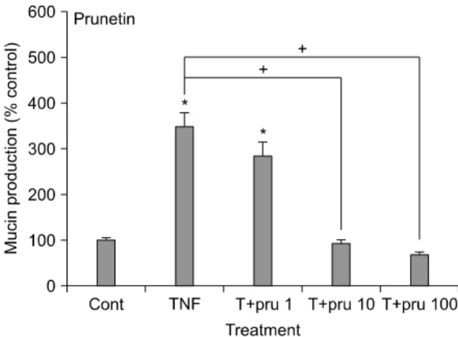
As can be seen in Figure 2, prunetin also inhibited TNF-α-induced MUC5AC mucin production, dose-de- pendently. The amounts of MUC5AC mucin in the cells

of prunetin-treated cultures were 100±6%, 349±29%, 284±30%, 92±9%, and 69±5% for control, TNF-α 0.2 nM only, prunetin 10

-6

M+TNF-α, prunetin 10

-5

M+

TNF-α and prunetin 10

-4

M+TNF-α, respectively (Fig- ure 2).

3. Effect of silibinin on TNF-α-induced MUC5AC mucin production from NCI-H292 cells

As can be seen in Figure 3, silibinin inhibited TNF-α- induced MUC5AC mucin production, dose-dependently.

The amounts of MUC5AC mucin in the cells of silibi- nin-treated cultures were 100±7%, 317±29%, 257±

30%, 198±13%, and 59±5% for control, TNF-α 0.2 nM

only, silibinin 10

-6

M+TNF-α, silibinin 10

-5

M+TNF-α

and silibinin 10

-4

M+TNF-α, respectively (Figure 3).

(4)

Figure 3. Effect of silibinin on tumor necrosis factor (TNF)- α-induced MUC5AC mucin production from NCI-H292 cells. NCI-H292 cells were pretreated with various con- centrations of silibinin for 30 minutes and then stimulated with TNF-α (0.2 nM) for 24 hours. Cell lysates were col- lected for measurement of MUC5AC mucin production by enzyme-linked immunosorbent assay. Means of individual group were converted to percent control and expressed as mean±SEM. The difference between groups was as- sessed using one-way ANOVA and Student's t-test for unpaired samples. p<0.05 was considered as sig- nificantly different. Each bar represents a mean±SEM of 3∼4 culture wells in comparison with that of control set at 100%. cont: control; Sil: silibinin. *Significantly different from control (p<0.05),

Significantly different from TNF- α alone (p<0.05).

Figure 4. Effects of carbenoxolone, prunetin and silibinin on tumor necrosis factor (TNF)-α-induced MUC5AC mucin gene expression from NCI-H292 cells. NCI-H292 cells were pretreated with carbenoxolone, prunetin, and silibinin (100 μM), respectively, for 30 minutes and then stimulated with TNF-α (0.2 nM) for 24 hours. MUC5AC gene expression was measured by reverse transcription-polymerase chain reaction. cont: control; Car: carbenoxolone; Pru: prunetin; Sil:

silibinin.

Figure 2. Effect of prunetin on tumor necrosis factor

(TNF)-α-induced MUC5AC mucin production from NCI-

H292 cells. NCI-H292 cells were pretreated with various

concentrations of prunetin for 30 minutes and then stimu-

lated with TNF-α (0.2 nM) for 24 hours. Cell lysates were

collected for measurement of MUC5AC mucin production

by enzyme-linked immunosorbent assay. Means of in-

dividual group were converted to percent control and ex-

pressed as mean±SEM. The difference between groups

was assessed using one-way ANOVA and Student's t-

test for unpaired samples. p<0.05 was considered as

significantly different. Each bar represents a mean±SEM

of 3∼4 culture wells in comparison with that of control

set at 100%. cont: control, Pru: prunetin. *Significantly

different from control (p<0.05),

Significantly different

from TNF-α alone (p<0.05).

(5)

4. Effects of carbenoxolone, prunetin and silibininon TNF-α-induced MUC5AC gene expression from NCI-H292 cells

As can be seen in Figure 4, carbenoxolone, prunetin and silibinin inhibited TNF-α-induced MUC5AC gene expression at the concentration of 10

-4

M, respectively.

Discussion

MUC5AC was reported to be mainly expressed in goblet cells existing in the airway surface epithelium among the twenty genes coding human mucins (MUC)

20

. Also, TNF-α is a well-known stimulator of secretion and gene expression of airway mucin

21-23

. TNF-α level in spu- tum was reported to be increased, with further increases during exacerbation of diseases

24,25

. TNF-α converting enzyme (TACE) mediated MUC5AC mucin expression in cultured human airway epithelial cells

22

and TNF-α in- duced MUC5AC gene expression in normal human air- way epithelial cells

23

. It also induced mucin secretion from guinea pig tracheal epithelial cells

21

. On the basis of these reports, in this study, we tried to test the possi- ble effects of carbenoxolone, prunetin and silibinin on TNF-α-induced MUC5AC mucin production and gene expression from NCI-H292 cells, a human pulmonary mucoepidermoid cell line, which are frequently used for the purpose of elucidating intracellular signaling path- ways involved in airway mucin production and gene ex- pression

22,26

. As can be seen in results, carbenoxolone, prunetin and silibinin inhibited the production of MUC5AC mucin protein induced by TNF-α. In the re- sult of experiment shown in Figure 1, TNF-α induced MUC5AC mucin production more potently than mucin productions in the other two results of experiments shown in Figures 2 and 3. Actually, based on the pre- liminary experiments performed by our group, the effi- cacy of stimulation of mucin production by TNF-α var- ied depending on the experimental conditions, although the exact cause of this phenomenon is unclear at present. On the other hand, the three compounds in- hibited the expression of MUC5AC mucin gene induced

by TNF-α. This result suggests that carbenoxolone, prunetin and silibinin can regulate mucin gene ex- pression and production of mucin protein, by directly acting on airway epithelial cells. The underlying mecha- nisms of action of these three compounds on MUC5AC production and gene expression are not clear at present, although we are trying to investigate whether these three compounds act as regulators of NF-kB signaling pathway activated by TNF-α in mucin-producing NCI-H292 cells. Taken together, the inhibitory actions of carbenoxolone, prunetin and silibinin on airway mu- cin production and gene expression might explain, at least in part, the traditional use of G. glabra L. as an- ti-inflammatory agent and mucoregulator for airway in- flammatory diseases, in oriental medicine and the folk use of Carduus marianus L. as anti-inflammatory agent.

We suggest it is valuable to find the natural products that have specific inhibitory effects on mucin production and/or gene expression - in view of both basic and clin- ical sciences - and the result from this study suggest a possibility of using carbenoxolone, prunetin and silibi- nin as new efficacious mucoregulators for respiratory diseases, although further studies are essential.

Acknowledgements

This study was partly supported by 2009 grant of Technology Development Program for Agriculture and Forestry, Ministry for Food, Agriculture, Forestry and Fisheries, Republic of Korea.

References

1. Voynow JA, Rubin BK. Mucins, mucus, and sputum.

Chest 2009;135:505-12.

2. Lee CJ, Lee JH, Seok JH, Hur GM, Park YC, Seol IC, et al. Effects of baicalein, berberine, curcumin and hes- peridin on mucin release from airway goblet cells.

Planta Med 2003;69:523-6.

3. Heo HJ, Lee SY, Lee MN, Lee HJ, Seok JH, Lee CJ.

Genistein and curcumin suppress epidermal growth

factor-induced MUC5AC mucin production and gene

expression from human airway epithelial cells. Phyto-

(6)

ther Res 2009;23:1458-61.

4. Research Institute of Natural Products, Seoul National University. Treatise on Asian herbal medicines. Seoul:

Haksulpyeonsugwan; 2003.

5. Tanaka Y, Kikuzaki H, Fukuda S, Nakatani N. Antibac- terial compounds of licorice against upper airway respi- ratory tract pathogens. J Nutr Sci Vitaminol (Tokyo) 2001;47:270-3.

6. Suzuki S, Koyama K, Darnel A, Ishibashi H, Kobayashi S, Kubo H, et al. Dexamethasone upregulates 11be- ta-hydroxysteroid dehydrogenase type 2 in BEAS-2B cells. Am J Respir Crit Care Med 2003;167:1244-9.

7. Suzuki S, Matsuda Y, Sugawara T, Tabata T, Ishibashi H, Hoshikawa Y, et al. Effects of carbenoxolone on al- veolar fluid clearance and lung inflammation in the rat.

Crit Care Med 2004;32:1910-5.

8. Asl MN, Hosseinzadeh H. Review of pharmacological effects of Glycyrrhiza sp. and its bioactive compounds.

Phytother Res 2008;22:709-24.

9. Jung HA, Kim AR, Chung HY, Choi JS. In vitro anti- oxidant activity of some selected Prunus species in Korea. Arch Pharm Res 2002;25:865-72.

10. Ko WC, Shih CM, Lai YH, Chen JH, Huang HL.

Inhibitory effects of flavonoids on phosphodiesterase isozymes from guinea pig and their structure-activity relationships. Biochem Pharmacol 2004;68:2087-94.

11. Sheikh S, Weiner H. Allosteric inhibition of human liver aldehyde dehydrogenase by the isoflavone prunetin.

Biochem Pharmacol 1997;53:471-8.

12. Keung WM. Biochemical studies of a new class of alco- hol dehydrogenase inhibitors from Radix puerariae.

Alcohol Clin Exp Res 1993;17:1254-60.

13. Polyak SJ, Morishima C, Lohmann V, Pal S, Lee DY, Liu Y, et al. Identification of hepatoprotective fla- vonolignans from silymarin. Proc Natl Acad Sci U S A 2010;107:5995-9.

14. Trappoliere M, Caligiuri A, Schmid M, Bertolani C, Failli P, Vizzutti F, et al. Silybin, a component of sylimarin, exerts anti-inflammatory and anti-fibrogenic effects on human hepatic stellate cells. J Hepatol 2009;50:1102-11.

15. Singh RP, Raina K, Sharma G, Agarwal R. Silibinin in- hibits established prostate tumor growth, progression, invasion, and metastasis and suppresses tumor angio- genesis and epithelial-mesenchymal transition in trans- genic adenocarcinoma of the mouse prostate model

mice. Clin Cancer Res 2008;14:7773-80.

16. Li L, Gao Y, Zhang L, Zeng J, He D, Sun Y. Silibinin inhibits cell growth and induces apoptosis by caspase activation, down-regulating survivin and blocking EGFR-ERK activation in renal cell carcinoma. Cancer Lett 2008;272:61-9.

17. Ligeret H, Brault A, Vallerand D, Haddad Y, Haddad PS. Antioxidant and mitochondrial protective effects of silibinin in cold preservation-warm reperfusion liver injury. J Ethnopharmacol 2008;115:507-14.

18. Hogan FS, Krishnegowda NK, Mikhailova M, Kahlen- berg MS. Flavonoid, silibinin, inhibits proliferation and promotes cell-cycle arrest of human colon cancer. J Surg Res 2007;143:58-65.

19. Lee SO, Jeong YJ, Im HG, Kim CH, Chang YC, Lee IS.

Silibinin suppresses PMA-induced MMP-9 expression by blocking the AP-1 activation via MAPK signaling path- ways in MCF-7 human breast carcinoma cells. Biochem Biophys Res Commun 2007;354:165-71.

20. Rogers DF, Barnes PJ. Treatment of airway mucus hy- persecretion. Ann Med 2006;38:116-25.

21. Fischer BM, Rochelle LG, Voynow JA, Akley NJ, Adler KB. Tumor necrosis factor-alpha stimulates mucin se- cretion and cyclic GMP production by guinea pig tra- cheal epithelial cells in vitro. Am J Respir Cell Mol Biol 1999;20:413-22.

22. Shao MX, Ueki IF, Nadel JA. Tumor necrosis factor al- pha-converting enzyme mediates MUC5AC mucin ex- pression in cultured human airway epithelial cells. Proc Natl Acad Sci U S A 2003;100:11618-23.

23. Song KS, Lee WJ, Chung KC, Koo JS, Yang EJ, Choi JY, et al. Interleukin-1 beta and tumor necrosis fac- tor-alpha induce MUC5AC overexpression through a mechanism involving ERK/p38 mitogen-activated pro- tein kinases-MSK1-CREB activation in human airway epithelial cells. J Biol Chem 2003;278:23243-50.

24. Chung KF. Cytokines in chronic obstructive pulmonary disease. Eur Respir J Suppl 2001;34:50s-9s.

25. Cohn L, Whittaker L, Niu N, Homer RJ. Cytokine regu- lation of mucus production in a model of allergic asthma. Novartis Found Symp 2002;248:201-13.

26. Takeyama K, Dabbagh K, Lee HM, Agustí C, Lausier

JA, Ueki IF, et al. Epidermal growth factor system regu-

lates mucin production in airways. Proc Natl Acad Sci

U S A 1999;96:3081-6.

수치

Figure 1. Effect of carbenoxolone on tumor necrosis fac- fac-tor  (TNF)-α-induced  MUC5AC  mucin  production  from  NCI-H292 cells
Figure  4.  Effects  of  carbenoxolone,  prunetin  and  silibinin  on  tumor  necrosis  factor  (TNF)-α-induced  MUC5AC  mucin gene  expression  from  NCI-H292  cells

참조

관련 문서

Abbreviations: TNF-alpha, tumor necrosis factor-alpha; CI, confidence interval; IFX, infliximab; ETA, etanercept; ADA, adalimumab Fig. Tuberculosis risk associated

The development of cutaneous sarcoidosis as a paradoxical adverse event of tumor necrosis factor alpha (TNF-α) block- ers has been reported in the literature; however, an

Methods: This study was designed to define the role of tumor necrosis factor-α (TNF-α) and the vascular endo- thelial growth factor (VEGF) in angiogenesis induced by

Tussilagone suppressed the production and gene expression of MUC5AC mucin via regulating nuclear factor-kappa B signaling pathway in airway epithelial cells.. Byung-Soo Choi 1,#

Abstract – In the present study, we investigated whether quercitrin, quercetin and afzelin derived from Houttuynia cordata affect the production and gene expression of MUC5AC

To ascertain whether these results were attributed to alteration in intracellular TNF-α protein synthesis or TNF-α processing and secretion, the cells also were

The involvement of inducible nitric oxide synthase (iNOS) and tumor necrosis factor alpha (TNF-α), which have diverse roles in the progression of autoimmune

are no reports about the effect of two compounds on airway mucin production from airway epithelial cells in- duced by phorbol 12-myristate 13-acetate (PMA) or tu-