배양된 기도 상피세포에서 종양괴사인자에 의한 IκB의 분해와 NF-κB p65의 핵으로의 이동에 미치는 실리비닌과 레스베라트롤의 영향
박수현*,†·이현재*,†·류지호*·이수열*·신현대*,**·홍장희*·석정호*·이충재*,#
*충남대학교 의과대학 약리학교실, **충남대학교 의과대학 정형외과학교실 (Received September 22, 2013; Revised November 28, 2013; Accepted December 2, 2013)
Effects of Silibinin and Resveratrol on Degradation of I κB and Translocation of NF-κB p65 Induced by Tumor Necrosis Factor- α in Cultured Airway Epithelial Cells
Su Hyun Park*,†, Hyun Jae Lee*,†, Jiho Ryu*, Su Yel Lee*, Hyun-Dae Shin*,**, Jang-Hee Hong*, Jeong Ho Seok* and Choong Jae Lee*,#
*Department of Pharmacology, School of Medicine, Chungnam National University, Daejeon 303-131, Korea
**Department of Orthopedic Surgery, Chungnam National University Hospital, Daejeon 303-131, Korea
Abstract — We examined whether silibinin and resveratrol affect airway mucin production, degradation of IκB and trans- location of NF-κB p65 induced by TNF-α in NCI-H292 cells. Cells were pretreated with each agent for 30 min and then stim- ulated with TNF-α for 24 h or the indicated periods. The two compounds suppressed TNF-α-induced airway mucin production, degradation of IκB and translocation of NF-κB p65. This result suggests that silibinin and resveratrol can reg- ulate the production of mucin induced by TNF-α through the inactivation of NF-κB pathway in airway epithelial cells.
Keywords □ airway mucin, MUC5AC, natural products
기도에 존재하는 점액(airway mucus)은 섬모세포와의 협동작 용을 통해, 호흡기를 통해 유입되는 병원성 미생물, 유해 자극성 기체, 각종 입자성 유해 물질의 제거에 있어서 중요한 역할을 하 는 것으로 알려져 있다. 이러한 기도 점액의 생체방어 작용은 점 액의 생화학적 주 구성요소인 점액성 당단백질, 뮤신의 점탄성 (viscoelasticity)에 기인한다. 그러나, 뮤신의 양과 질의 이상은 기도 생리의 이상뿐 아니라 인체의 방어작용에 영향을 주어 병 리 현상을 유발할 수 있다. 즉, 천식, 만성 기관지염, 폐기종, 기 관지 확장증, 낭포성 섬유증 등의 기도 질환에서 관찰되는 점액 (객담)의 과다 생성 및 분비, 점도의 비정상적 증가 등은 이러한 질환의 예후를 악화시키는 주된 요인으로 알려져 있다.1-4)과다 생성 및 분비된 점액을 기도로부터 제거하는 데는 두 가지 방법
이 있을 수 있다. 첫째, 물리적 방법에 의한 점액의 제거, 즉 점 액의 점도를 낮춘 뒤 흡인해 내는 방법이고, 둘째는, 점액 생성 자체를 억제할 수 있는 약물의 투여이다. 물리적 방법은 기도 내 부의 자극을 유발하고, 반사기전에 의해 점액 분비를 오히려 자 극하게 된다. 마취 하에서 그런 방법이 시도된다고 해도, 점액의 제거는 feedback mechanism을 통해 점액의 생성과 분비를 더욱 더 자극하게 된다. 따라서, 점액에 점성을 부여하는 주 구성요소 인 뮤신의 생성 자체를 조절하거나 혹은 분비를 조절하기 위한 약리학적 접근은 기도질환의 치료에 있어 중요한 방향이 될 수 있다.5)특히, 항염증, 항산화, 항암 효능을 발현하는 것으로 알려 진 다수의 천연물 가운데서 호흡기 염증성 질환에서 관찰되는 기 도 뮤신의 과다분비 혹은 생성을 조절할 가능성이 있는 후보물 질을 찾고자 하는 연구전략은 성공 가능성이 상대적으로 높다고 할 수 있을 것이다. 다수의 연구문헌에 의하면, 과일인 포도 등 에 함유되어 있는 레스베라트롤은 항산화, 항염증, 항천식 효과 등이 보고되어 있고,6-9)이 등10)은 기도 상피세포에서 레스베라트 롤이 기도 뮤신 유전자의 발현, 생성 및 분비를 억제함을 보고하 였다. 또한, 마리아 엉겅퀴(Carduus marianus L.)에 함유된 플라 보노이드성 화합물의 일종인 실리비닌도 항산화, 항염증 효과 및
종설
#Corresponding Author Choong Jae Lee
Department of Pharmacology, School of Medicine, Chungnam National University, Daejeon 303-131, Korea
Tel.: 042-580-8255 Fax.: 042-585-6627 E-mail: [email protected]
†These two authors equally contributed to this work.
간 미토콘드리아 기능 보호효과를 나타냄이 보고되어 있으며,11) 김 등12)은 실리비닌에 의해 호흡기 뮤신 분비, 생성 및 유전자 발현이 조절될 수 있음을 보고하였다. 그러나, 실리비닌과 레스 베라트롤이 인간 기도 상피세포에서 종양괴사인자(tumor necrosis factor-α, TNF-α)에 의해 자극된 뮤신의 생성 및 이와 관련된 전 사인자인 NF-κB p65의 세포질로부터 핵으로의 이동(translocation) 과 그를 가능케 하는 선행 단계인 IκB의 분해(degradation)에 대 해 각각 경시적으로 어떠한 영향을 미치는 지에 대해서는 자세 히 검증된 바가 없었으므로, 본 연구에서는 그러한 검증을 수행 함으로써 효과적인 기도점액 과다생성(분비) 조절 신약의 개발을 위한 기초과학적 정보를 제공하고자 하였다.
실험방법
세포주 및 시약
NCI-H292 세포는 American Type Culture Collection 사 (Manassas, VA, U.S.A.)에서 구입하였다. Protease inhibitor cocktail은 Roche사(Indianapolis, IN, U.S.A.)에서, HRP-Goat Anti-Mouse IgG Conjugate와 mouse anti-MUC5AC clone 45M1은 NeoMarkers사(Freemont, CA, U.S.A.)에서, trypsin- EDTA, tumor necrosis factor-α(TNF-α), Tween 20, bovine serum albumin(BSA), HEPES, dimethyl sulfoxide(DMSO), 3,3',5,5'-tetramethyl-benzidine peroxide solution(TMB), NP- 40, EDTA, EGTA, HEPES, 실리비닌, 레스베라트롤 등은 Sigma 사(St. Louis, Mo., U.S.A.)에서, enhanced chemiluminescence kit(ECL kit)는 Thermo Scientific(PierceECL western blotting substrate, Waltham, MA, U.S.A.)사에서, penicillin-G, strep- tomycin, fetal bovine serum(FBS), RPMI 1640은 GIBCO- BRL사(Grand Island, New York, U.S.A.)에서, Anti-NF-κB p65, anti-IκBα, anti-actin, anti-p84 antibodies는 Santacruz Biotech- nology(Santa Cruz, CA, U.S.A.)에서, secondary antibody conjugated with horseradish peroxidase는 Calbiochem(San Diego, CA, U.S.A.)에서, 기타 제반 시약들은 일급시약 등급 이 상의 것들을 구입하여 사용하였다.
인간 기도상피 세포(NCI-H292) 배양 및 각 약물의 처리 세포는 습도가 충분히 유지되며 95% 공기, 5% CO2를 함유하 는 37oC 배양기 내에서 HEPES(25 mM), penicillin G(100 U/
ml), streptomycin(100 µg/ml), FBS(10%, V/V) 등이 첨가된 RPMI 1640 배양액에서 배양하였는데, 1주에 2회 빈도로 subculture하였고 배양액은 2일마다 1회씩 교체해 주었다. 뮤신 생성에 대한 약물의 작용을 검증하기 위하여, 24 well culture plate 를 기준으로, well 당 2.0×10⁴cells/well의 밀도로 세포를 도포하고 배양하였다. 세포가 각 well의 70~80% 정도를 차지할
정도로 자라면, FBS 의 농도를 0.2%로 감소시킨 배양액을 주고 24시간 동안 배양하고, serum을 첨가하지 않은 배양액(serum- free medium)으로 세포를 세척한 후 약물 1~100 µM을 함유하 는 배양액 200 µl(24 well plate 기준)를 well마다 가하였다. 30 분이 경과한 후 TNF-α 0.2 nM 을 세포에 투여한 후 37oC에서 추가로 24시간 동안 배양하였다.9-11)
인간 기도상피 세포에서 MUC5AC 뮤신 생성량 측정 각 약물의 처리 기간이 종료된 후, 세포 용해용 완충액(20 mM Tris, 0.5% NP-40, 250 mM NaCl, 3 mM EDTA, 3 mM EGTA, protease inhibitor cocktail)을 가하여 세포 내에 존재하 는 MUC5AC 뮤신을 추출한 후 효소연계 면역흡착법(enzyme- linked immunosorbent assay, ELISA)을 이용하여 뮤신의 생성 량을 다음과 같이 측정하였다. 수거된 cell lysate를 PBS로 1/10 배 희석하고 희석된 각 sample을 ELISA 전용의 96-well plate 에 각각 100 µl씩 분포시킨 후 42oC에서 완전히 건조시켰다. 그 후 PBS-Tween 20(0.05%, PBS-T) 용액 200 µl/well을 이용, 각 well 당 3회씩 세척하였다. 세척 후 PBS-T에 용해된 2% BSA 용액 200 µl를 각 well당 가하고 다시 1시간 동안 incubation하 였다. 1시간 후 PBS-T 200 µl로 3회 세척하고MUC5AC에 대한 monoclonal antibody인 mouse anti-MUC5AC clone 45M1을 2% BSA에 1 : 200의 비율로 희석한 후에, 각 well당 100 µl씩 첨 가하고 1시간 동안 incubation하였다. 1시간 후 PBS-T로 3회 세 척하고 2차 항체인 Horse radish peroxidase(HRP)-Goat Anti- Mouse IgG Conjugate를 2% BSA에 1 : 3,000의 비율로 희석한 후, 각 well당 100 µl씩 첨가하고 1시간 동안 incubation하였다.
PBS-T로 다시 3회 세척 후 3,3',5,5'-tetramethyl-benzidine peroxide(TMB) 용액 100 µl를 각 well에 첨가하고 5분 후 1 N H2SO4 50µl를 첨가하여 반응을 정지시켰다. 450 nm에서 각 well 의 흡광도를 측정함으로써 대조군과 약물 처리군의 MUC5AC뮤 신을 정량하였다.9-11)
핵 분획과 세포질 분획의 준비
150 mm culture dish에 배양하여 70~80% 정도 증식된 NCI- H292 cells에, 37oC에서 24시간 동안 실리비닌 50 µM 또는 레 스베라트롤 100 µM을 전 처리하고 제시된 시간대 별로 TNF-α (50 ng/ml)를 처리하였다. 처리가 종료된 후, 세포는 ice-cold PBS 로 2회간 세척하고 3×trypsin-EDTA solution을 처리한 후 세포 만 조심스럽게 긁어 모아 3 ml PBS를 함유하는 15 ml 시험관에 옮겨 두었다. 이 세포 현탁액을 1,200 rpm, 4oC(Hanil centrifuge, MICRO 17 R) 조건에서 3분간 원심분리한 후, 상등액은 제거하 고 침전물(cell pellet)에 PBS 를 가하고 부드럽게 피펫팅하여 세 포를 재현탁시켰다. 이후 NE-PER® nuclear and cytoplasmic extraction reagent(Thermo-Pierce Scientific, Waltham, MA,
U.S.A.)를 제조자의 지시에 따라 적용함으로써 세포질 분획과 핵 분획을 분리 수거하였다. 각 분획은 −20oC 조건에서 보관되었고, 다음 단계의 실험에 사용되기 전에 각 분획 중의 총 단백질량은 Bradford method를 이용하여 측정되었다.
Western blotting을 이용한 IκB의 분해 단계에 미치는 두 약물의 영향 검증
IκB 단백질을 함유하는 세포질 분획(50 µg as protein)을 10%
SDS-polyacrylamide gel을 이용하여 전기영동하였다. Gel 상에서 분리된 단백질들을 PVDF(polyvinylidene difluoride) membrane 에 전기적으로 흡착시켰고 0.05% Tween 20을 함유하는 5%
skim milk in PBS로 blocking한 후, primary antibody against IκBα(1 : 1,000)를 첨가하고 반응을 진행시켰다. 0.05% tween 20 을 함유하는 PBS로 세척한 후 horseradish peroxidase가 부착 된 secondary antibody(1 : 2,000)를 첨가하고 반응을 진행시켰다.
결과로 생성되는 immunoreaction band는 enhanced chemilumine- scence kit(Pierce ECL western blotting substrate, Thermo Scientific, Waltham, MA, U.S.A.)를 제조자의 설명에 따라 적용 함으로써 가시적으로 검출해 내고 대조군, TNF-α 단독군, 실리 비닌 또는 레스베라트롤 처리군 별로 상호 비교하여 실리비닌 또 는 레스베라트롤이 IκB 단백질의 분해(degradation) 단계에 미치 는 경시적 영향을 검증하였다.
Immunoblotting을 이용한 NF-κB p65의 핵으로의 이동 단 계에 미치는 두 약물의 영향 검증
NF-κB p65 단백질을 함유하는 핵 분획(50 µg as protein)을 10% SDS- polyacrylamide gel을 이용하여 전기영동하였다. Gel 상에서 분리된 단백질들을 PVDF(polyvinylidene difluoride) membrane에 전기적으로 흡착시켰고 0.05% Tween 20을 함유하 는 5% skim milk in PBS로 blocking한 후, primary antibody against NF-κB p65(1 : 1,000)를 첨가하고 반응을 진행시켰다.
0.05% tween 20을 함유하는 PBS로 세척한 후 horseradish peroxidase가 부착된 secondary antibody(1 : 2,000)를 첨가하고 반응을 진행시켰다. 결과로 생성되는 immunoreaction band는 enhanced chemiluminescence kit(PierceECL western blotting substrate, Thermo Scientific, Waltham, MA, U.S.A.)를 제조자 의 설명에 따라 적용함으로써 가시적으로 검출해 내고 대조군, TNF-α 단독군, 실리비닌 또는 레스베라트롤 처리군 별로 상호 비교하여 실리비닌 또는 레스베라트롤이 NF-κB p65 단백질의 핵으로의 이동 단계에 미치는 경시적 영향을 검증하였다.
통계처리
모든 측정 결과는 Mean±S.E.M.으로 환산한 후, 약물 처리군 의 측정치는 대조군 측정치의 백분율로 나타냈다. 통계처리는
one-way ANOVA로 하였으며, 사후 검정은 Duncan's Multiple Range test를 실시하였다. p<0.05인 경우 통계적으로 유의성이 있는 것으로 판정하였다.
실험결과
TNF-α로 자극된 MUC5AC 기도뮤신 생성에 대한 실리비닌 과 레스베라트롤의 영향
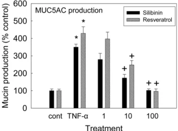
Fig. 1에서 볼 수 있는 것처럼, 실리비닌과 레스베라트롤은 각 각 10, 100 µM 농도에서 0.2 nM의 TNF-α 처리로 증가된 MUC5AC 기도뮤신의 생성을 억제하였다. 실리비닌의 경우, 각 처리 농도별 뮤신의 양은 대조군, TNF 0.2 nM 단독 처리군, 실 리비닌 10−6M+TNF, 실리비닌 10−5M+TNF, 실리비닌 10−4M +TNF 처리군에서 각각 100±10%, 350±17%, 279±35%, 173±21%, 102±9%이었으며, 레스베라트롤의 경우, 대조군, TNF 0.2 nM 단독 처리군, 레스베라트롤 10−6M+TNF, 레스베라트롤 10−5M+TNF, 레스베라트롤 10−4M+TNF 처리군에서 각각 100±9%, 429±38%, 397±39%, 248±25%, 97±13%이었다 (Fig. 1).
실리비닌과 레스베라트롤이 TNF-α로 자극된 IκBα의 분해에 미치는 영향
Fig. 2에서 볼 수 있는 것처럼, NCI-H292 cells에서 TNF-α 처
Fig. 1− Effects of silibinin and resveratrol on TNF-α-induced MUC5AC mucin production from NCI-H292 cells. NCI- H292 cells were pretreated with varying concentrations of silibinin and resveratrol for 30 min and then stimulated with TNF-α (0.2 nM) for 24 h, respectively. Cell lysates were collected for measurement of MUC5AC mucin production by ELISA. Each bar represents a mean±S.E.M.
of 3 culture wells in comparison with that of control set at 100%. * Significantly different from control (p<0.05). + Significantly different from TNF-α alone (p<0.05) (cont:
control, concentration unit is µM).
리에 의해 IκBα의 분해(degradation)가 유발되었으며, 24시간 동안의 실리비닌 50 µM 전처리(pretreatment) 또는 24시간 동 안의 레스베라트롤 100 µM 전처리에 의하여 공히 억제되었다 (Fig. 2).
실리비닌과 레스베라트롤이 TNF-α로 자극된 NF-κB p65의 핵으로의 이동에 미치는 영향
Fig. 3에서 볼 수 있는 것처럼, NCI-H292 cells에서 TNF-α에 의한 NF-κB p65의 세포질로부터 핵으로의 이동(translocation) 이 유발되었으며, 24시간 동안의 실리비닌 50 µM 전처리 (pretreatment) 또는 24시간 동안의 레스베라트롤 100 µM 전처 리에 의하여 공히 억제되었다(Fig. 3).
고 찰
호흡기 질환의 치료 및 조절 과정에서, 과다 생성-분비된 점액 의 효율적 제거를 목적으로 점액용해제 및 거담제 등이 사용되
고 있으나 그 작용 및 작용 기전이 불명확하며 약물 투여 및 점 액의 물리적 제거에 따르는 반사적 과다분비 현상 때문에 점액 과다분비 질환의 효율적 조절은 쉽지 않은 것으로 알려져 있다.13) 현재, 임상적으로 기도점액의 과다 분비를 유의하게 조절할 수 있는 약물은 당질 코르티코이드계 약물로 알려져 있으나 동반되 는 광범위한 부작용이 치료 약물로서의 효용성을 제한하고 있는 실정이다.5)인간의 호흡기에 존재하는 뮤신은 펩티드 골격과 탄 수화물 가지로 이루어진 수백만 dalton의 분자량을 가진 당단백 질(glycoprotein)로서, 뮤신의 펩티드 골격을 coding 하는 유전자 를 MUC로 약칭하는데 현재까지 MUC 1, 2, 3A, 3B, 4, 5AC, 5B, 6, 7, 8, 9, 11, 12, 13, 16, 17, 18, 19, 20 등 20여종 이상의 MUC 유전자가 발견되었으며, 이 중에MUC5AC와 MUC5B 유 전자의 산물인 MUC5AC와 MUC5B 뮤신이 인간의 호흡기에서 발견되는 gel-forming mucin을 구성하고 있다.14)동시에, 인간 기도 뮤신의 생성, 유전자 발현 조절과 연관된 연구모델로 자주 사용되는 인간의 기도 상피세포인 NCI-H292 세포에 대해, TNF- α는 TNF 수용체(tumor necrosis factor receptor)를 매개로 NF- Fig. 2− Effects of silibinin and resveratrol on TNF-α-induced degradation of IκBα. NCI-H292 cells were incubated with 50 µM silibinin or 100µM resveratrol for 24 h at 37oC and treated with TNF-α (50 ng/ml) for the indicated periods. Cytoplasmic extracts were prepared and analyzed by western blot using antibody against anti-IκBα. The results shown are the representative of three independent experiments. Equal protein loading was evaluated by β-actin levels.
Fig. 3− Effects of silibinin and resveratrol on TNF-α-induced nuclear translocation of NF-κB p65. NCI-H292 cells were either untreated or pretreated with 50µM silibinin or 100 µM resveratrol for 24 h at 37oC and then stimulated with TNF-α (50 ng/ml) for the indicated periods. Nuclear protein extracts were prepared and resolved on 10% SDS-PAGE, transferred onto a PVDF membrane, probed with antibody against p65. The results shown are the representative of three independent experiments. To ensure equal protein loading, the membrane was reprobed with anti-p84 antibody.
κB(nuclear factor kappa B)로 이어지는 신호전달 경로를 거쳐, 기도 뮤신인 MUC5AC 뮤신의 생성을 증가(자극)시킬 수 있는 것으로 알려져 있다.15-17)또한, 다수의 연구보고에 의하면 본 연 구에서 사용된 두 약물 모두 호흡기 구성 세포가 아닌 다른 세 포에서도 NF-κB signaling pathway를 조절할 가능성이 있는 것 으로 알려져 있다.18-21)이러한 선행 연구정보에 근거하여 수행 된 본 연구의 결과에서 볼 수 있는 것처럼, 실리비닌과 레스베라 트롤은 TNF-α에 의해 증가된 기도 뮤신의 생성을 억제하는 경 향을 보여주었다(Fig. 1). 두 약물이 MUC5AC 뮤신의 생성을 억 제할 수 있다면 MUC5AC 유전자의 중요 전사조절 인자로 알려 진 NF-κB 신호전달 경로에는 어떠한 영향을 미치는 지 후속 실 험을 통하여 알아보고자 하였다. NF-κB는 세포질 내에서 IκB (inhibitory kappa B)와 결합되어 불활성 상태로 있지만, TNF-α 등에 의한 자극에 의해 비활성 상태의 IKK(inhibitory kappa B kinase)가 활성화됨으로써 이어 IκBα가 인산화되고 이후 분해 (degradation)되면, p65로 대표되는 단백질들이 세포핵 내로 이 동함으로써 MUC5AC 유전자의 전사를 활성화시키는 것으로 알 려져 있다.17,22-24)실험 결과를 통해 알 수 있는 것처럼, 실리비 닌 또는 레스베라트롤의 전 처리(pretreatment) 에 의해 TNF-α- 유도 IκB(inhibitory kappa B)의 분해가 억제(Fig. 2)되었으며, NF-κB p65의 핵 내로의 이동(translocation) 또한 억제(Fig. 3)됨 으로써 결과적으로 TNF-α-유도 NF-κB p65의 활성화 (activation) 가 조절될 가능성을 확인할 수 있었다. 본 연구에서 얻어진 한정 된 연구결과만으로 실리비닌과 레스베라트롤의 분자 수준에서의 작용 기전을 규명했다고는 할 수 없겠지만, 실리비닌과 레스베 라트롤이 배양된 인간 기도 상피세포에서 핵 내 전사 영역으로 의 NF-κB translocation 차단을 통하여, 기도 MUC5AC 뮤신의 생성을 조절할 가능성을 시사하는 결과라고 생각된다. 요약하면, 비록 제한적이지만 본 연구에서 얻어진 이러한 지견들은 호흡기 점액의 과다생성 및 분비를 보이는 천식, 만성 기관지염 등 다양 한 호흡기 염증성 질환의 진행 과정에서 기도 뮤신의 과다한 생 성 억제에 초점을 둔, 점액 조절용 신약후보물질 개발에 대한 기 초과학적 정보를 제공함에 있어서 일부나마 기여할 수 있을 것 으로 판단된다.
감사의 말씀
This research was supported by a grant (12172KFDA989) from Korea Food & Drug Administration in 2012.
ABBREVIATIONS
ELISA : enzyme-linked immunosorbent assay PBS : phosphate-buffered saline
TNF-α : tumor necrosis factor-á
RT-PCR : reverse transcription - polymerase chain reaction NF-κB : Nuclear factor kappa B
IκB : inhibitory kappa B PVDF : polyvinylidene difluoride
SDS-PAGE : sodium dodecyl sulfate polyacrylamide gel electro- phoresis
참고문헌
1) Voynow, J. A. and Rubin, B. K. : Mucins, mucus, and sputum.
Chest 135, 505 (2009).
2) Kim, K. C., Rearick, J. I., Nettesheim, P. and Jetten, A. M. : Biochemical characterization of mucous glycoproteins synthesized and secreted by hamster tracheal epithelial cells in primary culture. J. Biol. Chem. 260, 4021 (1985).
3) Ko, K. H., Lee, C. J., Shin, C. Y., Jo, M.-J. and Kim, K. C. : Inhibition of mucin release from airway goblet cells by polycationic peptides. Am. J. Physiol. 277, L811 (1999).
4) Kim, K. C., Opaskar-Hincman, H. and Bhaskar, K. R. : Secretions from primary hamster tracheal surface epithelial cells in culture: Mucin-like glycoproteins, proteoglycans, and lipids. Exp. Lung Res. 15, 299 (1989).
5) Mutschler, E. and Derendorf, H. : Drug actions. CRC press, Inc., Boca Raton, Florida, 410 (1995).
6) Birrell, M. A., McCluskie, K., Wong, S., Donnelly, L. E., Barnes, P. J. and Belvisi, M. G. : Resveratrol, an extract of red wine, inhibits lipopolysaccharide induced airway neutrophilia and inflammatory mediators through an NF-kappaB-independent mechanism. FASEB J. 19, 840 (2005).
7) Donnelly, L. E., Newton, R., Kennedy, G. E., Fenwick, P. S., Leung, R. H., Ito, K., Russell, R. E. and Barnes, P. J. : Anti- inflammatory effects of resveratrol in lung epithelial cells:
molecular mechanisms. Am. J. Physiol. Lung Cell Mol. Physiol.
287, L774 (2004).
8) Lee, M., Kim, S., Kwon, O. K., Oh, S. R., Lee, H. K. and Ahn, K. : Anti-inflammatory and anti-asthmatic effects of resveratrol, a polyphenolic stilbene, in a mouse model of allergic asthma. Int. Immunopharmacol. 9, 418 (2009).
9) Kode, A., Rajendrasozhan, S., Caito, S., Yang, S. R., Megson, I. L. and Rahman, I. : Resveratrol induces glutathione synthesis by activation of Nrf2 and protects against cigarette smoke-mediated oxidative stress in human lung epithelial cells. Am. J. Physiol. Lung Cell Mol. Physiol. 294, L478 (2008).
10) Lee, S. Y., Lee, H. J., Sikder, M. A., Shin, H. D., Kim, J. H., Chang, G. T., Seok, J. H. and Lee, C. J. : Resveratrol inhibits mucin gene expression, production and secretion from airway epithelial cells. Phytother. Res. 26, 1082 (2012).
11) Ligeret, H., Brault, A., Vallerand, D., Haddad, Y. and Haddad, P. S. : Antioxidant and mitochondrial protective effects of silibinin in cold preservation-warm reperfusion liver injury. J.
Ethnopharmacol. 115, 507 (2008).
12) Kim, K. D., Lee, H. J., Lim, S. P., Sikder, A., Lee, S. Y. and Lee, C. J. : Silibinin regulates gene expression, production and secretion of mucin from cultured airway epithelial cells.
Phytother. Res. 26, 1301 (2012).
13) Ellis, E. F. : Asthma in childhood. J. Allergy. Clin. Immunol. 72 (Suppl), 526 (1985).
14) Rogers, D. F. and Barnes, P. J. : Treatment of airway mucus hypersecretion. Ann. Med. 38, 116 (2006).
15) Takeyama, K., Dabbagh, K., Jeong, Shim J., Dao-Pick, T., Ueki, I. F. and Nadel, J. A. : Oxidative stress causes mucin synthesis via transactivation of epidermal growth factor receptor: role of neutrophils. J. Immunol. 164, 1546 (2000).
16) Takeyama, K., Dabbagh, K., Lee, H., Agusti, C., Lausier, J. A., Ueki, I. F., Grattan, K. M. and Nadel, J. A. : Epidermal growth factor system regulates mucin production in airways. Proc.
Natl. Acad. Sci. USA 6, 3081 (1999).
17) Li, J. D., Dohrman, A. F., Gallup, M., Miyata, S., Gum, J. R., Kim, Y. S., Nadel, J. A., Prince, A. and Basbaum, C. B. : Transcriptional activation of mucin by Pseudomonas aeruginosa lipopolysaccharide in the pathogenesis of cystic fibrosis lung disease. Proc. Natl. Acad. Sci. USA 94, 967 (1997).
18) Bannwart, C. F., Nakaira-Takahagi, E., Golim, M. A., de Medeiros, L. T., Romo, M., Weel, I. C. and Peraoli, M. T. : Downregulation of nuclear factor-kappa B (NF-kappaB) pathway by silibinin in human monocytes challenged with Paracoccidioides brasiliensis. Life Sci. 86, 880 (2010).
19) Liu, P. L., Tsai, J. R., Charles, A. L., Hwang, J. J., Chou, S. H., Ping, Y. H., Lin, F. Y., Chen, Y. L., Hung, C. Y., Chen, W. C., Chen, Y. H. and Chong, I. W. : Resveratrol inhibits human lung adenocarcinoma cell metastasis by suppressing heme oxygenase 1-mediated nuclear factor-kappaB pathway and subsequently downregulating expression of matrix metalloproteinases. Mol.
Nutr. Food Res. 54, S196 (2010).
20) Deng, Y. H., Alex, D., Huang, H. Q., Wang, N., Yu, N., Wang, Y. T., Leung, G. P. and Lee, S. M. : Inhibition of TNF-α- mediated endothelial cell-monocyte cell adhesion and adhesion molecules expression by the resveratrol derivative, trans- 3,5,4'-trimethoxystilbene. Phytother. Res. 25, 451 (2011).
21) Kumar, A. and Sharma, S. S. : NF-kappaB inhibitory action of resveratrol: a probable mechanism of neuroprotection in experimental diabetic neuropathy. Biochem. Biophys. Res.
Commun. 394, 360 (2010).
22) Song, K. S., Lee, W. J., Chung, K. C., Koo, J. S., Yang, E. J., Choi, J. Y. and Yoon, J. H. : IL-1beta and TNF-alpha induced MUC5AC overexpression through a mechanism involving ERK/p38 mitogen-activated protein kinase-MSK1-CREB activation in human airway epithelial cells. J. Biol. Chem. 278, 23243 (2003).
23) Shao, M. X., Ueki, I. F. and Nadel, J. A. : TNF-alpha converting enzyme mediated MUC5AC mucin expression in cultured human airway epithelial cells. Proc. Natl. Acad. Sci. USA 100, 11618 (2003).
24) Ishinaga, H., Takeuchi, K., Kishioka, C., Suzuki, S. and Basbaum, C. : Pranlukast inhibits NF-kappaB activation and MUC2 gene expression in cultured human epithelial cells.
Pharmacol. 73, 89 (2005).