"J. Korean Soc. Radiol., Vol. 14, No. 1, February 2020"
Evaluation of Classification and Accuracy in Chest X-ray Images using Deep Learning with Convolution Neural Network
Ho-Jun Song, Eun-Byeol Lee, Heung-Joon Jo, Se-Young Park, So-Young Kim, Hyeon-Jeong Kim, Joo-Wan Hong
*Department of Radiological Science, Eulji University
Received: January 23, 2020. Revised: February 25, 2020. Accepted: February 28, 2020
ABSTRACT
The purpose of this study was learning about chest X-ray image classification and accuracy research through Deep Learning using big data technology with Convolution Neural Network. Normal 1,583 and Pneumonia 4,289 were used in chest X-ray images. The data were classified as train (88.8%), validation (0.2%) and test (11%).
Constructed as Convolution Layer, Max pooling layer size 2×2, Flatten layer, and Image Data Generator. The number of filters, filter size, drop out, epoch, batch size, and loss function values were set when the Convolution layer were 3 and 4 respectively. The test data verification results showed that the predicted accuracy was 94.67%
when the number of filters was 64-128-128-128, filter size 3×3, drop out 0.25, epoch 5, batch size 15, and loss function RMSprop was 4. In this study, the classification of chest X-ray Normal and Pneumonia was predictable with high accuracy, and it is believed to be of great help not only to chest X-ray images but also to other medical images.
Keywords: Deep Learning, CNN, Pneumonia, Chest X-Ray
Ⅰ. INTRODUCTION
Corresponding to the fourth industrial revolution, accuracy of image analysis using Artificial Intelligence (AI), Deep Learning, is increasing fast in medical image analysis and diagnosis.
[1-3]Deep Learning is a technology that combines Artificial Neural Network (ANN), which is an algorithm based on a mechanism of neural networking, and Big data.
ANN can interpret the input data using perceptron, classification and clustering and can recognize specific pattern of data using this interpretation. Deep Learning uses Convolution Neural Network (CNN), one kind of ANN.
[4,5]CNN has advantages that inputting two-dimensional data and training is easy and there is less parameter. It shows good performance in every part of image and audio.
[6]Chest X-ray was
basic examination for pneumonia and follow up for improvement, and was reported a sensitivity of more than 50% when diagnosed with pneumonia.
[7,8]However, the diagnosis of pneumonia may be misdiagnosed by the doctor, and it takes a long time to Chest X-ray read.
[9]Increasing accuracy of pneumonia diagnosis and reducing reading time would help imaging diagnosis a lot. Therefore, this study aims to evaluate pneumonia classification and accuracy of deep learning using chest X-ray data.
Ⅱ. MATERIAL AND METHODS
1. Chest X-ray image data
This research used 5,872 chest X-ray images provided
by Labeled Optical Coherence Tomography (OCT) and
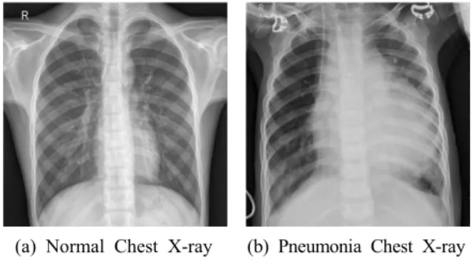
https://doi.org/10.7742/jksr.2019.14.1.39Chest X-ray Images for Classification from Mendeley LTD. 1,583 images were Normal and 4,289 images were Pneumonia as shown in Fig. 1.
(a) Normal Chest X-ray (b) Pneumonia Chest X-ray Fig. 1. Image of Chest PA.
2. Data preprocessing and modeling 2.1. Data classification
Collected chest X-ray image was classified into Training Data about 88.8% (Normal 25.7%, Pneumonia 74.3%), Test Data about 11% (Normal 37.5%, Pneumonia 62.5%) and Validation Data about 0.2%
(Normal 50%, Pneumonia 50%). Size of all image was reduced to 150×150.
2.2. Data modeling
The research used Anaconda (Anaconda Inc, USA, Texas, Ver. 4.7.10), Python (Python Software Foundation, Ver. 3.7.3), Jupyter Notebook (Project Jupyter, Ver. 6.0.0), Keras (Ver. 2.2.4.)
2.2.1 Convolution layer
Among Convolution layer, Conv2D layer was used.
At three layers, filter number was set as 64-128-128 and filter size was set as 2×2 and 3×3. At four layers, filter number was set as 64-128-128-128 and filter size as 3×3.
2.2.2 Max Pooling layer
For Max Pooling layer, pool size was set as 2×2 in order to obtain feature through extracting maximum value from Pixel of output Image through Convolution and resizing.
2.2.3 Flatten layer
To transform two - dimensional data into one - dimensional image in order to convey the characteristics, which is extracted into Convolution layer and Max Pooling layer to Fully-connected layer, Flatten layer was used.
2.2.4 Image Data Generator
It was done to increase performance by reflecting character of Test Dataset fully through overstating Training Dataset which is not good enough with Image Data Generator as shown in Fig. 2.
Fig. 2. Data modeling diagram.
2.3. Data learning
For data learning, an experiment was done under the setting that parameters are Filter number, Filter size, drop out, epoch, batch size and Loss Function when Convolution layer is 3. Secondly, an experiment was done under the setting that parameters are Filter number, Filter size, drop out, epoch, batch size and Loss Function when Convolution layer is 4.
Ⅲ. RESULT
When Convolution layer was 3 and Filter number
was 64-128-128 and set as Filter size 2×2, Drop out
0.25, Epoch 3, Batch size 16, the accuracy was
"J. Korean Soc. Radiol., Vol. 14, No. 1, February 2020"
83.75%. When set as Filter size 3×3, Drop out 0.25, Epoch 3, Batch size 16, the accuracy was 87.5%.
When set as Filter size 3×3, Drop out 0.5, Epoch 3, Batch size 16, the accuracy was 66.25%. When set as Filter size 3×3, Drop out 0.25, Epoch 5, Batch size 16, the accuracy was 90%, as shown in Table 1.
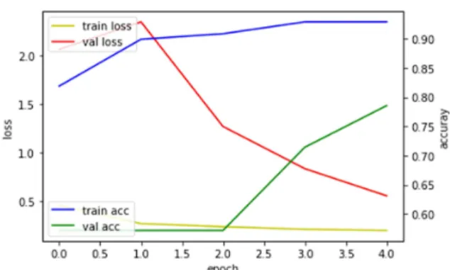
When Convolution layer was 4, filter number was 64-128-128-128 and filter size is 3×3, the accuracy was 82.5% under the setting that Drop out was 0.25, Epoch was 3, Batch size was 16 and loss function was RMSprop. When set as Drop out 0.25, Epoch 5, Batch size 15, loss function as adam, the accuracy was 90.67%, as shown in Table 2. The accuracy was highest as 94.67% when set as Drop out 0.25, Epoch 5, Batch size 15, Loss function rmsprop, as shown in Fig. 3. The progress of learning rate of this model, as shown in Fig. 4.
Table 1. Set value of Convolution layer 3
Setting Value
Layer 3
Filter number 64-128-128
Filter size 2×2 3×3 3×3 3×3
Drop out 0.25 0.25 0.5 0.25
Epoch 3 3 3 5
Batch size 16 16 16 16
Loss function rmsprop
Accuracy 83.75% 87.5% 66.25% 90%
Table 2. Set value of Convolution layer 4
Setting Value
Layer 4
Filter number 64-128-128-128
Filter size 3×3 3×3 3×3
Drop out 0.25 0.25 0.25
Epoch 3 5 5
Batch size 16 15 15
Loss function rmsprop rmsprop adam
Accuracy 82.5% 94.67% 90.67%
Fig. 3. Diagram of convolution layer.
Fig. 4. Learning progress of the highest model.
Ⅳ. DISCUSSION
This research classified into Normal and Pneumonia through Deep Learning. Among the results, minimum was 66.25% and maximum was 94.67%. For the development of program supporting diagnosis of occupational lung disease (pneumoconiosis)
[10], the research classifying Normal and Pneumoconiosis using CNN showed accuracy up to 95%. This research also showed high results with accuracy up to 94.67%. A research about detection of crack in granite X-ray CT image using Deep Learning applied a method increasing image learning data but the accuracy was only 67.91%.
[11]A research classifying lung pattern among interstitial lung disease showed 85.5%
accuracy.
[12]This research showed satisfactory prediction accuracy but there was a few restraints when classifying Chest image using Deep Learning.
Deep Learning is a machine-learning that can obtain more accurate results as data number increases. For medical image, however, it was difficult to obtain enough data for learning due to personal information protection.
[13]Cooperation with database about clinical disorders along with a supporting system for web service or Electronic Medical Record (EMR) system would solve this and make possible to share large volumes of data. Learning with more various and large amount of lung image would show higher accuracy than this research. Also, a phenomenon that perception rate failed to meet expectation would occur
if classify high-resolution medical image with existing algorithm.
[14]Despite these problems, this research developed a method to enable learning at network by dividing large size of image into small size of lattice.
[15]This method would show high accuracy if algorithm for high-resolution image like medical image is developed.
Ⅴ. CONCLUSION
Chest X-ray classification and pneumonia diagnosis using deep learning were high accuracy and it is considered to be a useful basic data for pneumonia diagnosis.
Reference
[1] M. Rastgarpour, J. Shanbehzadeh, "Application of AI Techniques in Medical Image Segmentation and Novel Categorization of Available Methods and Tools," Lecture Notes in Engineering and Computer Science, Vol. 2188, No. 1, pp. 519-523, 2011.
[2] Sarraf, Saman, Ghassem, Tofighi, "Deep learning-based pipeline to recognize Alzheimer's disease using fMRI data," Future Technologies Conference (FTC), IEEE, pp. 816-820, 2016.
[3] U. Niyaz, Abhishek Singh Sambyal, Devanand,
"Advances in Deep Learning Techniques for Medical Image Analysis," 2018 Fifth International Conference on Parallel, Distributed and Grid Computing (PDGC), IEEE, pp. 271-277, 2018.
[4] R. Meng, S. G, Rice, J. Wang, X. Sun, "A Fusion Steganographic Algorithm Based on Faster R-CNN,"
Computers, Materials & Continua, Vol. 55, No. 1, pp. 1-16, 2018.
[5] Schmidhuber, Jürgen. "Deep learning in neural networks: An overview," Neural networks, Vol. 61, pp. 85-117, 2015.
[6] L. Amelio, A. Amelio. "Classification Methods in Image Analysis with a Special Focus on Medical Analytics," Machine Learning Paradigms: Advances in Data Analytics, Vol. 149, pp. 31-69, 2018.
[7] E. J. Park, J. H. Park, M. J. Hong, W. D. Kim, K.
Y. Lee, S. J. Kim, H. J. Kim, K. W. Ha, G. R.
"J. Korean Soc. Radiol., Vol. 14, No. 1, February 2020"
Chon, H. A. Kim, K. H. Yoo. "Usefulness of Vibration Response Imaging(VRI) for Pneumonia Patients," Tuberculosis and Respiratory Diseases, Vol.
71, No. 1, pp. 30-36, 2011.
[8] J. P. Matlay, M. J. Fine, "Testing strategies in the initial management of patients with
community-acquired pneumonia," Annals of Internal Medicine, Vol. 138, No. 2, pp. 109-118, 2003.
[9] M. Tsoumakidou, G. Chrysofakis, I. Tsiligianni, G.
Maltezakis, N. M. Siafakas, N. Tzanakis, "A Prospective Analysis of 184 Hemoptysis Cases – Daignositc Impact of Chest X-Ray, Computed Tomography, Bronchoscopy," Respiration, Vol. 79, No. 6, pp. 808-814, 2006.
[10] J. P. Myung, J. S. Song, S. H. Kang, S. W. Han,
"An artificial intelligence deep learning based diagnostic assistance program for pneumoconiosis,"
Journal of the Korean Academy of Tuberculosis and Respiratory Diseases in the Fall Conference of The Korean Society of Visualization, Vol. 126, pp.
258-258, 2018.
[11] S. H. Hyun, J. S. Lee, S. H. Jeon, Y. J. Kim, K.
Y. Kim, T. S. Yoon, "Pixel-level Crack Detection in X-ray Computed Tomography Image of Granite using Deep Learning," Korean Society for Rock Mechanics and Rock Enginnering in the tunnels and underground spaces, Vol. 29, No. 3, pp. 184-196, 2019.
[12] M. Anthimopoulos, S. Christodoulidis, L. Ebner, A.
Christe, S. Mougiakakou, "Lung pattern classification for interstitial lung diseases using a deep
convolutional neural network," IEEE transactions on medical imaging, Vol. 35, No. 5, pp. 1207-1216, 2016.
[13] Shen, Dinggang, Guorong Wu, Heung-Il Suk, "Deep learning in medical image analysis," Annual review of biomedical engineering, Vol. 19 No. 1, pp.
221-248, 2017.
[14] Y. M. Seo, J. K. Han. "Deep Learning Algorithm to Identify Cancer Pictures," Journal of Broadcast Engineering, Vol. 23, No. 5, pp. 669-681, 2018.
[15] Erickson, Bradley J., et al. "Deep learning in radiology: does one size fit all?," Journal of the American College of Radiology, Vol. 15, No. 3, pp.
521-526, 2018.
컨볼루션 뉴럴 네트워크 기반의 딥러닝을 이용한 흉부 X-ray 영상의 분류 및 정확도 평가
송호준, 이은별, 조흥준, 박세영, 김소영, 김현정, 홍주완
* 을지대학교 보건과학대학 방사선학과본 연구에서는 CNN과 빅데이터 기술을 이용한 Deep Learning을 통해 흉부 X-ray 영상 분류 및 정확성 연 구에 대하여 알아보고자 한다. 총 5,873장의 흉부 X-ray 영상에서 Normal 1,583장, Pneumonia 4,289장을 사 용하였다. 데이터 분류는 train(88.8%), validation(0.2%), test(11%)로 분류하였다. Convolution Layer, Max pooli ng layer pool size 2×2, Flatten layer, Image Data Generator로 구성하였다. Convolution layer가 3일 때와 4일 때 각각 filter 수, filter size, drop out, epoch, batch size, 손실함수 값을 설정하였다. test 데이터로 Convolution layer가 4일 때, filter 수 64-128-128-128, filter size 3×3, drop out 0.25, epoch 5, batch size 15, 손실함수 RMSprop 으로 설정 시 정확도가 94.67%였다. 본 연구를 통해 높은 정확성으로 분류가 가능하였으며, 흉부 X-ray 영 상뿐만 아니라 다른 의료영상에서도 많은 도움이 될 것으로 사료된다.
중심단어: 딥러닝, 합성곱 신경망 네트워크, 폐렴, 흉부 X-ray