의
의
의학
학
학 석
석
석사
사
사학
학
학위
위
위 논
논
논문
문
문
소
소
소아
아
아 상
상
상완
완
완골
골
골 외
외
외과
과
과 골
골절
골
절
절의
의
의 안
안
안정
정적
정
적
적 고
고
고정
정
정을
을
을
위
위
위한
한
한 진
진
진성
성
성 골
골
골절
절
절면
면의
면
의
의 삼
삼
삼차
차
차원
원
원적
적
적 분
분
분석
석
석
아
아
아 주
주
주 대
대
대 학
학
학 교
교
교 대
대
대 학
학
학 원
원
원
의
의
의 학
학
학 과
과
과
김
김
김 준
준
준 용
용
용
소
소
소아
아
아 상
상
상완
완
완골
골
골 외
외
외과
과
과 골
골절
골
절
절의
의
의 안
안
안정
정적
정
적
적 고
고
고정
정
정을
을
을
위
위
위한
한
한 진
진
진성
성
성 골
골
골절
절
절면
면의
면
의
의 삼
삼
삼차
차
차원
원
원적
적
적 분
분
분석
석
석
지
지
지도
도
도교
교
교수
수
수 원
원
원 예
예
예 연
연
연
이
이
이 논
논
논문
문
문을
을
을 의
의학
의
학
학 석
석
석사
사
사학
학
학위
위
위 논
논
논문
문
문으
으
으로
로 제
로
제
제출
출
출함
함
함.
.
.
2
2
20
0
00
0
08
8
8년
년
년 8
8
8월
월
월
아
아
아 주
주
주 대
대
대 학
학
학 교
교
교 대
대
대 학
학
학 원
원
원
의
의
의 학
학
학 과
과
과
김
김
김 준
준
준 용
용
용
김
김
김준
준
준용
용
용의
의 의
의
의
의학
학
학 석
석
석사
사
사학
학
학위
위
위 논
논
논문
문
문을
을 인
을
인
인준
준
준함
함
함.
.
.
심
심
심사
사
사위
위
위원
원
원장
장
장
원
원
원 예
예
예 연
연
연
인
인
인
심
심
심 사
사
사 위
위
위 원
원
원
한
한
한 경
경
경 진
진
진
인
인
인
심
심
심 사
사
사 위
위
위 원
원
원
조
조
조 재
재
재 호
호
호
인
인
인
아
아
아 주
주
주 대
대
대 학
학
학 교
교
교 대
대
대 학
학
학 원
원
원
2
2
20
0
00
0
08
8
8년
년
년 6
6
6월
월
월 2
2
23
3
3일
일
일
국문요약
-소
소
소아
아
아 상
상
상완
완
완골
골 외
골
외
외과
과
과 골
골
골절
절
절의
의
의 안
안
안정
정
정적
적
적 고
고
고정
정
정을
을
을 위
위
위한
한
한
진
진
진성
성
성 골
골
골절
절
절면
면
면의
의
의 삼
삼
삼차
차
차원
원
원적
적
적 분
분
분석
석
석
목 목목 적적적 ::: 한국인 소아 상완골 외과 골절의 분류 및 골절면의 각도를 단순방사선 검사 및 삼차원 컴퓨터 단층 촬영으로 측정하여 생체 역학적으로 가장 안정적인 K-강선 삽입각도를 알아보고자 한다. 연 연연구구구대대대상상상 및및및 방방방법법법 :::2003년에서 2007년까지 본원에 내원한 소아 상완골 외과 골 절 환자 100예와,이 환자들 중 삼차원 컴퓨터 단층 촬영을 한 31예를 대상으로 하였다.100예 환자의 골절을 분류하고,31명의 환자에서 전후면 단순방사선 사 진에서 골절선과 상완골 장축이 이루는 각을 알파각이라고 하고,측면 단순방사 선 사진에서 골절선과 상완골 장축과 이루는 각을 베타각이라 정의하여 측정하 였다.동일한 환자군에서 삼차원 컴퓨터 단층 촬영에서 진성 전후면 사진에서 알 파각을 진성 측면 사진에서 베타각을 다시 측정하였다.이를 토대로 역학적으로 가장 안정된 K-강선 삽입 각을 구하고자 하였다. 결결결 과과과 :::100예의 단순방사선 사진을 분류한 결과 Milch type I은 3명,Milch typeII는 97명,JacobtypeI은 15명,JacobtypeII는 37명,JacobtypeIII는 48, Finnbogason typeA 는 20명,Finnbogason typeB는 31명,Finnbogason type C는 49명이었다.단순방사선 사진으로 측정한 각도는 28명의 평균 알파각은 59 도,베타각은 67도였고 삼차원 컴퓨터 단층 촬영 분석 결과는 28명의 평균 알파 각은 63도,베타각은 65도였다. 결 결결 론론론 :::소아 상완골 외과 골절에 대한 핀 삽입은 삼차원 컴퓨터 단층 촬영 분 석 결과 K-강선이 장축에 대해 전후면상에서 상완골 장축에 대해27도,측면 사 진에서 상완골 장축에 대해 전상방으로 25도로 삽입될 때 골절선의 초기 안정성 을 최대로 얻을 수 있을 것이다.이를 이용하면 상완골 장축과 평행하게 삽입되
는 실수를 줄여 골절 초기 안정성에 기여할 것이다.
핵심어:소아 상완골 외과 골절,삼차원 컴퓨터 단층 촬영, K-강선 고정술,생체 역학
차
차
차 례
례
례
국문요약 ···ⅰ 차례 ···ⅱ 그림 차례 ···ⅲ Ⅰ.서론 ···1 Ⅱ.연구대상 및 방법 ···3 Ⅲ.결과 ···6 Ⅳ.고찰 ···7 Ⅴ.결론 ···9 참고문헌 ···10 ABSTRACT ···12그
그
그림
림
림 차
차
차례
례
례
Fig.1.FractureplaneangleintrueAP view,alphaangle...3 Fig.2.Fractureplaneangleintruelateralview,betaangle...4 Fig.3.Clinicalapplicationoftruefractureplaneduring
I
I
I.
.
.서
서
서 론
론
론
소아에서 상완골 외과 골절(lateralcondylefracture)은 소아 주관절 부위 골 절 중 두 번째로 흔한 형태로 주관절 골절의 약 10-20%에서 발생하며 대개 6 세 전후의 연령에서 호발 한다(Rockwood와 Wilkins,1996;Shirzad등,2001).수 상 기전은 대개 팔을 뻗은 상태에서 상지의 장축에 힘이 가해진 경우,힘의 전달 이 주관절부는 60% 가량이 외측주(lateralcolumn)로 전달되며,이로 인해 내과 골절보다 외과 골절이 많이 발생한다.회내 기전의 경우 요골 두의 전방탈구나 척골 골절의 동반의 가능성이 높아지는 반면 회외 상태의 장축 압박은 외과 골 절을 유발할 가능성을 높인다(Jakob등,1975). 이렇게 발생한 상완골 외과 골절은 과거 소두의 침범 여부에 따라 소두를 침 범한 경우를 MilchtypeI,소두를 침범하지 않은 경우를 MilchtypeII로 분류하 였으며(Milch,1964),이 분류는 예후와 안정성을 예측하는데 일부 도움을 주었으 나,환자의 수술적 방법을 결정하는 데에는 중요한 정보를 주지 못하는 경우가 많았다(Skak등,2001). 최근에 사용되는 분류는 전위된 정도와 회전에 따라 Jacob 분류(Jakob 등, 1975)가 사용된다.TypeI은 전위가 없는 경우이며,전완부를 회외전한 상태에서 주관절을 굴곡하고 장상지 석고 붕대 고정을 시행할 수 있는 형이다(Bast등, 1998).전위가 있으나 원위 골편의 회전 변형이 없는 경우를 typeII라 하고 비관 혈적 정복술 및 K-강선 내고정술을 시행하거나,관혈적 정복술 후 K-강선 내고 정술을 시행하여 치료한다.4mm 이상의 전위나 골편의 회전이 동반된 경우를 typeIII라 분류하는데 이 경우 비관혈적 정복술로 치료하는 경우도 있으나,많은 수에서 관혈적 정복술 및 K-강선 내고정술이 필요하게 된다(Badelon 등,1988; Mintzer등,1994). 골편의 안정성을 평가함에 있어 Finnbogason등(Finnbogason등,1995)은 상 완골 외과 골절선의 외측부의 간극에 의해 평가하기도 하였는데,외측 간극만 조 금 형성하고 원위 상완골 성장판 연골까지 골절선이 침범 안 한 경우를 typeA,
외측 간극이 벌어지며 원위 상완골 성장판까지 간극을 만든 경우를 typeB,외 측 간극과 내측 관절면의 간극이 많이 벌어진 경우를 typeC라고 분류하기도 하 였다.이러한 다양한 골절 분류의 목적은,실제 안정성의 예측과 수술의 방법 결 정,예후의 판정에 도움을 주려는 것이라 할 수 있다. 상완골 외과 골절은 성장판을 관통하는 특성 외에도 관절내 골절 등의 특성 을 지니며,불유합이라는 합병증과 이에 병발되는 외반주,이 후 장시간의 무증 상기를 거친 후 병발하는 지연 척골 신경 마비의 문제점이 있다(Hasler와 Laer, 2001;Jeffery,1958).또한 골절부의 불안정 또는 불유합을 피하기 위해 상완골 과상부 골절 등에 비해 수술 후 장상지 석고 고정을 장기간 유지하려는 경향이 있다(Martina등,2005).과거로부터 상완골 외과 골절은 외과 근처에서 기시하는 많은 신전 회외 근육군으로 인해 전위가 잘 발생하여,수술이 필요한 경우가 많 아 필요 골절(fractureofnecessity)이라고 하였으며,성장판 손상이 동반되므로 정확한 해부학적 정복 및 안정적 고정을 해야 한다. 실제 수술적 치료가 필요한 상완골 외과 골절 환자군에서 관혈적,비관혈적 정복술이 많이 시행되고 있으나,전후면 사진(anteroposteriorview,AP view)에 서의 골절선의 방향은 비교적 이해가 쉬워 K-강선의 삽입 방향에 대한 적절한 삽입이 이뤄지나,측면 사진(lateralview)에서의 골절선의 방향에 대해 생체 역 학적으로 적합한 방향으로 삽입이 이뤄지지 않는 경우가 많다.이는 상완골 장축 을 기준으로 삼는 상완골 과상부 골절의 핀 삽입의 경향이 상완골 외과 골절 시 에도 나타나기 때문이다. 본 연구는 우리나라 소아 상완골 외과 골절을 방사선학적으로 분류하였고 또 한 골절면에 대한 방향성을 단순방사선 사진 및 삼차원 컴퓨터 단층 촬영을 이 용 분석하여,소아 상완골 외과 골절에서 정확한 골절 형태 및 고정을 위한 골절 면 형태에 대하여 이해하고,이를 통해 안정적인 초기 핀 삽입 방향에 대한 정확 한 방향을 확인하고자 한다.
I
I
II
I
I.
.
.연
연
연구
구
구대
대
대상
상
상 및
및
및 방
방
방법
법
법
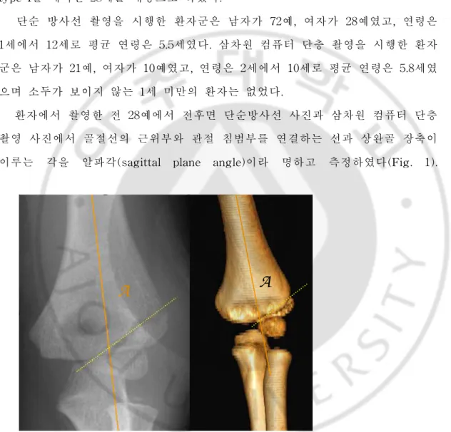
2003년에서 2007년까지 본원에 내원한 소아 상완골 외과 골절 환자 중 초기 수상 단순 방사선 사진이 있는 100예와 이 환자들 중 주관절부 삼차원 컴퓨터 단층 촬영(3-dimensionalcomputerizedtomogram,3-D CT)을 시행한 환자 31예 를 분석한 결과 Milch typeI은 3예,Milch typeII는 28예였으며,희귀한 Milch typeI을 제외한 28예를 대상으로 하였다. 단순 방사선 촬영을 시행한 환자군은 남자가 72예,여자가 28예였고,연령은 1세에서 12세로 평균 연령은 5.5세였다.삼차원 컴퓨터 단층 촬영을 시행한 환자 군은 남자가 21예,여자가 10예였고,연령은 2세에서 10세로 평균 연령은 5.8세였 으며 소두가 보이지 않는 1세 미만의 환자는 없었다. 환자에서 촬영한 전 28예에서 전후면 단순방사선 사진과 삼차원 컴퓨터 단층 촬영 사진에서 골절선의 근위부와 관절 침범부를 연결하는 선과 상완골 장축이 이루는 각을 알파각(sagittal plane angle)이라 명하고 측정하였다(Fig. 1).
A
A
Figure 1. Fracture plane angle in true AP view, alpha angle Fig.1.FFFrrraaaccctttuuurrreeeppplllaaannneeeaaannngggllleeeiiinnntttrrruuueeeAAAPPP vvviiieeewww,,,aaalllppphhhaaaaaannngggllleee
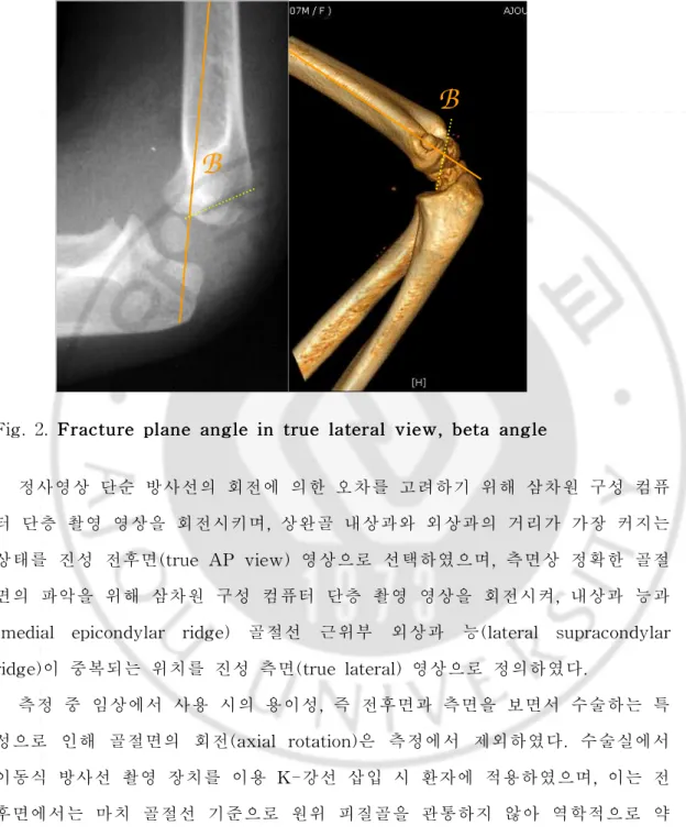
측면 단순방사선 사진과 3D-CT 사진에서 골절선의 골간단 후방 시작점과 소 두 전방의 골절선을 연결하는 선이 상완골 장축과 이루는 각을 베타각(coronal planeangle)이라 명하고 측정하였다(Fig.2).
B
B
Figure 2. Fracture plane angle in true lateral view, beta angle
Fig.2.FFFrrraaaccctttuuurrreeeppplllaananneeeaaannngggllleeeiiinnntttrrruuueeelllaaattteererraaalllvvviiieweeww,,,bbbeeetttaaaaaannngggllleee
정사영상 단순 방사선의 회전에 의한 오차를 고려하기 위해 삼차원 구성 컴퓨 터 단층 촬영 영상을 회전시키며,상완골 내상과와 외상과의 거리가 가장 커지는 상태를 진성 전후면(trueAP view)영상으로 선택하였으며,측면상 정확한 골절 면의 파악을 위해 삼차원 구성 컴퓨터 단층 촬영 영상을 회전시켜,내상과 능과 (medial epicondylar ridge) 골절선 근위부 외상과 능(lateral supracondylar ridge)이 중복되는 위치를 진성 측면(truelateral)영상으로 정의하였다.
측정 중 임상에서 사용 시의 용이성,즉 전후면과 측면을 보면서 수술하는 특 성으로 인해 골절면의 회전(axialrotation)은 측정에서 제외하였다.수술실에서
하게 보이나,측면사진 또는 내회전 45도 사진에서는 골절선에 보다 역학적으로 안정한 위치(수직위)에 가까운 것을 확인할 수 있었다(Fig.3).
Figure3.CCCllliiinnniiiccacaalllaaappppppllliiicccaaatttiiiooonnnoooffftttrrruuueeefffrrraaaccctttuuurrreeepplpllaaannneeeddduuurrriiinnngggooopppeeerraraatttiiiooonnn... C-arm view
I
I
II
I
II
I
I.
.
.결
결
결
과
과
과
100명의 환자 중 Milch typeI은 3명,Milch typeII는 97명,Jacob typeI은 15명,Jacob typeII는 37명,Jacob typeIII는48,Finnbogason typeA 는 20명, Finnbogason typeB는 31명,Finnbogason typeC는 49명이었다.이 중 수술을 받은 환자는 91명이었고 이들의 평균 K-강선 제거일은 수술 후 37일이었다. Jacob type과 Finnbogason type에 따른 K-강선 제거일의 차이는 없었다 (ANOVA test,p<0.05SPSS Koreanversion12.0)
주관절부 삼차원 컴퓨터 단층 촬영을 시행한 환자 31예에서는 Jacob분류상 typeI은 1예,typeII는 15예,typeIII는 12예였으며, FinnbogasontypeA는 2 예,typeB는 13예,typeC는 13예였다. 단순 방사선 촬영을 이용하여 측정한 JacobtypeI1예의 경우 알파각이 65 도,베타각이 75도였다.JacobtypeII15예의 경우 알파각이 63도,베타각이 69도였으며,JacobtypeIII12예의 경우 알파각,베타각이 각각 58도, 60도 였다.28예의 평균 알파각과 베타각은 각각 59도와 67도였다.1세~5세, 5세~10세 두 군으로 분류 시 알파각이 각 군에서 60도, 61도였고 베타각은 각각 64 도, 67도였다. 삼차원 CT를 이용한 경우 JacobtypeI1예의 경우 알파각이 66도,베타각이 75도였다.Jacob type II 15예의 경우 알파각이 69도,베타각이 65도였으며, Jacob typeIII12예의 경우 알파각,베타각이 각각 59도,64도였다.1세~5세, 5 세~10세 두 군으로 분류 시 알파각이 각 군에서 66도,61도였고 베타각은 각각 63도,66도였다.28예의 평균 알파각과 베타각은 각각 63도와 65도였다.
Jacob type과 연령에 따른 알파각과 베타각의 차이는 없었다(paired t-test, p<0.05)
I
I
IV
V
V.
.
.고
고
고
찰
찰
찰
삼차원 CT를 촬영한 환자군에서 희귀하게 발생하는 MilchtypeI은 제외하였 는데 이는 골절선이 분석하고자 하는 대부분의 상완골 외과 골절 양상을 파악하 는데 오류를 초래할 수 있기 때문이었다.삼차원 CT 환자군의 경우 Jacobtype I이나 FinnbogasontypeA가 적었는데 이는 수술보다는 보존적 치료를 하여 삼 차원 CT를 촬영하지 않았기 때문이다. CT를 촬영한 환자군에서는 Milch type I이 전체 31례 중 3례로 상대적으로 많았으며,이는 CT를 촬영 안한 대부분의 JacobtypeI의 환자들이 Milch typeII인 것을 감안하면,그 빈도는 매우적어질 것으로 생각된다. 기존의 치료에서 수술적 치료 시,핀 삽입은 대개 소아 상완골 과상부 골절과 같이 골절선을 관통하는 2개의 K-강선 삽입이 일반적 치료로 받아들여져 왔다. 그러나 그 정확한 삽입 방향에 대한 골절선의 특성을 정확히 연구된 적이 없었 다.따라서 전후면 방사선 사진에서의 골절선에 대한 핀 삽입에 집중하게 되며, 측면 방사선 사진상 골절선에 대한 고정이 상완골 장축에 평행하게 되는 경향이 있었다.그러나 다른 골절부와 마찬가지로 근육의 작용 방향(muscledisplacing force) 을 고려하지 않는다면,골절선과 90도를 이루며 내고정이 되었을 경우 그 기계적 강도가 가장 안정적이라 할 것이다. 전후면 방사선 사진 상의 상완골 외과 골절 선의 방향성은 그 이해가 쉽고 핀 삽입 시 비교적 골절선을 관통하는 강선의 방 향이 바람직하나 측면 골절선의 방향을 삼차원 CT로 정확히 측정한 경우,상완 골 장축에 대한 골절면의 각도인 베타각이 65도이므로,핀 삽입 시 측면에서는 상완골 장축에 평행한 것이 아니라,후하방에서 전상방으로 25도의 각을 주는 것 이 역학적으로 바람직하다 할 수 있을 것이다. 연구 결과 골절선의 전위에 따른 Jacob분류,Finnbogason분류와는 상관없이 모든 환자에서 비교적 일정한 알파각과 베타각을 보였으며,연령에 따른 차이도 거의 없었다(paired t-test,p<0.05).즉 골절면의 각도는 외상의 에너지양에 큰
영향 없이 비교적 유사한 각을 보였고 이를 이용하여 보다 정확한 K-강선 삽입 각도를 결정할 수 있을 것이다.이를 이용 초기 소아 상완골 외과 골절의 안정적 고정으로 장상지 석고 고정 기간을 조절하고,불안정으로 인해 발생하는 불유합, 외반주,지연 척골 신경 마비의 합병증을 방지하기 위해 보다 섬세한 정복과,골 절면의 정확한 입체적 방향을 고려한 치료가 된다면,더 좋은 치료 결과를 유도 할 수 있을 것으로 생각된다. 또한 Jacob분류,Finnbogason분류와 관계없이 모든 환자에서 비슷한 시기에 수술 후 K-강선 제거가 이루어진 것은 골절의 전위 정도 및 이로 인한 골막 손 상정도가 골 유합에 큰 영향을 주지 않는다는 것을 시사하는 것으로 이는 외과 골절시 골유합의 주 기전이 이차 골 치유가 아닌 일차 골 치유에 주로 의존하기 때문인 것으로 생각된다. 알파각과 베타각을 단순방사선 사진과 삼차원 단층 촬영 영상을 이용하여 각 각 측정하였고 두 측정값간의 전체 평균 오차 범위는 5도이내였고 이 중 진성 전후면 및 측면 사진을 얻을 수 있는 삼차원 CT결과를 더 정확한 측정치로 간 주하였다.
V
V
V.
.
.결
결
결 론
론
론
소아 상완골 외과 골절의 골절면의 방향성은 삼차원 CT로 정확히 측정한 경 우 전후면 사진에서 상완골 장축에 대해 63도,측면 사진상 65도의 각을 보이므 로 골절면의 각도를 고려한 적절한 K-강선 삽입각은 전후면 사진에서 상완골 장 축에 대해 27도,측면 사진상 상완골 장축에 대해 25도 정도이다.
참
참
참고
고
고문
문
문헌
헌
헌
1. Badelon O,BensahelH,Mazda K,and VieP:Lateralhumeralcondylar fracturesinchildren:A reportof47cases.J PediatricOrthop,8:31-34, 1988
2. BastS C,Hoffer M Mark,and AvalSoheil:Nonoperative Treatment or Minimally and Nondisplaced LateralHumeralCondyle Fractures in Children.JournalofPediatricOrthopaedics,18(4):448-450,1998
3. Finnbogason T, Karlsson G, and Lindberg L: Non-displaced and minimally displaced fractures of the lateral condyle in children: a prospective radiographic investigation of fracture stability.J.Pediatr. Orthop.,15:422-425,1995
4. Hasler Carol-Claudius and Laer Lutz von: Prevention of Growth DisturbancesafterFracturesoftheLateralHumeralCondyleinChildren. 5. Jakob R, Fowles J V, Rang M, and Kassab TM: Observations
concerning fractures ofthe lateralhumeralcondyle in children.J.Bone JointSurg.Am.57-B(4):430-436,1975
6. Jeffrey C C:Non-union ofthe epiphysis ofthe lateralcondyle ofthe humerus.JBoneJointSurg,40:396-405,1958
displacedfracturesofthelateralhumeralcondylein children.J.Trauma, 58:1202-1207,2005
8. MilchHE:Fracturesandfracture-dislocationsofthehumeralcondyles.J. Trauma,4:592-607,1964
9.Mintzer CM ,Waters PM,Brown DJ,and Kasser JR: Percutaneous pinninginthetreatmentofdisplacedlateralcondylefractures.JPediatric Orthop,14:462-465,1994
10. Rockwood CA and Wilkins KE:Fractures in Children.4th ed.Vol3, Philadelphia,LippincottCo.1996
11. Shirzad Houshian, Bassem Mehdi, and Morten S. Larsen: The epidemiologyofelbow fractureinchildren:analysisof355fractures,with specialreference to supracondylarhumerus fractures.J.Orthop.Sci.,6: 312-315,2001
12. Skak Soren Valdemar, Olsen Soren Due, and Smaabrekke Arvid: Deformity after Fracture of the LateralHumeralCondyle in Children. JournalofPediatricOrthopaedics,10:142-152,2001
ABSTRACT
-3
3
3-
-
-D
D
Di
i
im
me
m
e
en
n
ns
s
si
i
io
o
on
na
n
a
al
l
la
a
an
n
na
a
al
l
ly
y
ys
s
si
i
is
s
so
o
of
ft
f
t
tr
r
ru
u
ue
e
ef
f
fr
r
ra
a
ac
c
ct
t
tu
u
ur
r
re
e
ep
p
pl
l
la
a
an
n
ne
e
ef
f
fo
o
or
r
rt
t
th
h
he
e
e
p
p
pr
r
ro
o
op
p
pe
e
er
r
rf
f
fi
i
ix
x
xa
a
at
t
ti
i
io
o
on
n
no
o
on
n
nl
l
l
a
a
at
t
te
e
er
r
ra
a
al
lh
l
h
hu
u
um
m
me
e
er
r
ra
a
al
l
lc
c
co
o
on
n
nd
d
dy
y
yl
l
l
e
e
ef
f
fr
r
ra
ac
a
c
ct
t
tu
u
ur
r
re
e
e
i
i
in
n
nc
c
ch
h
hi
i
il
l
l
d
d
dr
r
re
e
en
n
n
JoonYongKimDepartmentofMedicalSciences TheGraduateSchool,AjouUniversity (SupervisedbyProfessorYeYeonWon) P
PPuuurrrpppooossesee::: To evaluate the classification and assess the proper angle of Kirschner-wire insertion depending on 3D-CT analysis in pediatric lateral humeralcondylefracture.
M
MMaaattteeerrriiiaaalllaaannndddMMMeeettthhhoododdsss:::100lateralcondylefracturesandthe3D-CT results of31pediatricpatientswerecollectedfrom 2003to2007.Radiographic classificationwasdone.Theangleformedbylongaxisofhumerusandline betweenproximalpointoffracturelineandfracturelineintheelbow jointin anteroposterior3D-CT view andsimpleradiographwasdefinedas"alpha angle",aswellas"betaangle"referringtotheangleformedbylongaxisof humerusandthelinebetweenproximalpointoffractureanddistalpointin lateralview.
R
RReeesssuuullltttsss:::On radiographicclassification of100lateralcondylefractures,Milch typeIwas3,MilchtypeIIwas97.JacobtypeIwas15,JacobtypeIIwas
angles were 59 and 67 degrees on simple radiograph.On 3D-CT,average alphaandbetaangles were63and65degrees.
C
CCooonnncccllluuusssiiiooonnn:::In orderto obtain maximalbiomechanicalstability offracture fixation,Kirschner-wire should be inserted 27 degrees againstlong axis of humerusonAP view and25degreesonlateralview.
This study willcontribute to initialstability of pediatric lateralhumeral condylefracturefixation.
K