Improved Preimplantation Development of Porcine Cloned
Embryos by Flavone Supplement as Antioxidant
Xun Fang1, Ahmad Yar Qamar1, Ki-Young Yoon2 and Jongki Cho1
1College of Veterinary Medicine, Chungnam National University, Daejeon 34134, Republic of
Korea
2Dept. of Companion Animal, Shingu College, Seongnam 13174, Republic of Korea
Abstract
This experiment was conducted to analyse the effects of flavone supplementation on the preimplantation development of in-vitro produced porcine embryos. During in-vitro development, immature oocytes and early embryos were exposed to different concentrations of flavone (0, 1μM, 25μM, 50 μM, and 100 μM respectively). Results showed that 100 µM of flavone significantly reduced the intracellular ROS levels of oocytes accompanied with a significant rise in GSH level. In parthenogenesis, no significant change was observed in the cleavage rates whether flavone was supplemented in IVM or IVC media. In IVM supplemented group, the blastocyst development rate was significantly enhanced by 1 µM concentration than other groups (51.5% vs. 41.3%, 44.0%, 36.3%, 31.7%; P<0.05) respectively. However, in IVC group 1 µM concentration significantly improved the blastocysts production than 50 µM and control groups (50.0% vs. 40.5%, 38.0%; P<0.05) respectively. Following nuclear transfer, the cleavage rate of IVM group was significantly more in 1 µM than 50 µM and 100 µM groups (92.9% vs. 89.7%, 87.8%; P<0.05), followed by similar pattern of cloned blastocysts production being significantly higher in 1 µM group than 50 µM, 100 µM and control groups (16.8% vs. 9.0%, 7.1%, 12.8%; P<0.05) respectively. In IVC group, 1 µM concentration resulted in significantly higher cleavage rate than 25 µM and 50 µM groups (91.7% vs. 87.8%, 88.8%; P<0.05) respectively. However, the blastocysts production was significantly higher in 100 µM group than others (26.2% vs. 13.6%, 14.0%, 18.2%; P<0.05) respectively. The optimal concentrations of flavone significantly enhanced the percentages of ICM:TE than control group (43.8% vs. 37.6%; P<0.05) accompanied with significantly higher expression levels of reprogramming related genes. In conclusion, the optimal concentrations of 1 µM during IVM and 100 µM during IVC can significantly improve the production of porcine
in-vitro embryos.
Received 19 December 2018 Revised 20 December 2018
INTRODUCTION
Somatic cell nuclear transfer (SCNT) plays a vital role in biomedical research and applications, including animal production and biotechnology (Keefer 2015), xenotransplantation (Hwang et al. 2016), and animal disease models (Holm et al. 2016). In addition, SCNT technology holds greater potential for stem cell biology and human therapeutics, such as derivation of patient-specific nuclear transfer embryonic stem cells (Matoba and Zhang 2018). However, the major obstacle associated with the SCNT is the lower overall cloning efficiency (Liu et al. 2015). The reasons held responsible for the lower cloning efficiency, include quality of oocyte, donor cell type, protocol employed for nuclear transfer, culture conditions, aberrant genomic methylation, histone modification, and reprogramming gene expression(Kang et al. 2013; Wrenzycki et al. 2001; Xu et al. 2012).
Abnormal reprogramming, such as chromatin remodeling, histone replacement, transcriptional activation, histone modification and DNA demethylation occur more frequently in cloned embryos than IVF derived embryos (Matoba and Zhang 2018). DNA methylation plays an important role in mammalian embryogenesis (Matoba and Zhang 2018). Inhibition of aberrant DNA re-methylation thought to be one of the critical steps limiting the efficiency of cloning (Gao et al. 2018). Disturbed DNA methylation will lead to declined oocyte quality (Liu et al. 2018). In order to improve the quality of oocytes, there is a need to reduce DNA damage. Antioxidants have a positive effect on the cell survival by reducing the deteriorations caused by reactive oxygen species (ROS) and free radicals. Therefore, antioxidants might theoretically retard spreading of DNA damage and improve cytoplasmic antioxidant cytoprotective systems (Bae et al. 2015). Oocyte damage can reduce the quality, which can affect the development of cloned embryos (Huang and Chan 2017).Flavone is a natural compound which can be extracted from different types of plants. It is widely used in medical research because of its wide range of pharmacological activities. It also possess potent antioxidant characteristics helpful in scavenging reactive nitrogen and oxygen species (Ashaari et al. 2018). The antioxidant properties and neuroprotective effects are found associated with the nuclear factor erythroid 2-related factor 2-(Nrf2) pathway in flavone (Wang et al. 2018a; Wruck et al. 2007). Flavonoid could protect human sperm against the damaging effects of FeSO4/H
2O2 (Huang
et al. 2014). Recently, it has been found that flavone works against cancer growth by direct targeting (Ganai 2017; Sajjadi et al. 2018; Wang et al. 2018b), and inhibits migration through
DLC1/RhoA pathway by decreasing ROS generation in cancer cells (Zhu et al. 2016).
The objective of this experiment was to investigate the improvement in developmental competence of porcine cloned embryos caused by flavone supplementation and explore the relationship with DNA reprogramming and apoptosis. For this purpose, the different parameters including intracellular levels of ROS and GSH, the developmental rates of embryo, and expression levels of genes related with apoptosis & reprograming (BAX, Bcl-2, POU5F1, SOX2 and Nanog) will be investigated.
MATERIALS AND METHODS
1. Chemicals and reagents
The chemicals and reagents utilized were purchased from Sigma-Aldrich company (St. Louis, MO, USA) unless otherwise indicated.
2. Production of embryos
All the experiments were conducted according to our laboratory protocols as previously explained by (Roy et al. 2017). 2.1. Oocytes Collection and IVM
Transportation of ovaries from slaughterhouse to the laboratory was done in a temperature-controlled thermos containing 0.9% saline. After aspiration of the follicular fluid was carried out using 10 mL syringes with the aid of 18 gauges’ needle. Follicles within a diameter range of (3-8 mm) were selected for aspiration. Cellular part was allowed to settle down and washed utilizing HEPES-buffered Tyrode’s (TLH) media, having 0.05% (w/v) polyvinyl alcohol (TLH-PVA) (Bavister et al. 1983). Cumulus-oocyte complexes (COCs) with at least three compact cumulus cells layers were selected. Selected COCs were washed three times with TLH-PVA, and one time in IVM media drop. Selected COCs (50-80) were placed per well of 4-well multi dishes (Nunc, Denmark) containing 500 mL of IVM media including 10 IU/mL PMSG/hCG (Intervet International BV, Holland). Finally, COCs were incubated for 22 h in 5% CO2 humidified atmosphere at
39°C temperature. After 22 h of maturation, transferred COCs in hormone free IVM media and further incubated at same conditions for 18-21 h.
2.2. Parthenogenetic Activation(PA) embryos
After 44 h of IVM, denuding of COCs was performed in IVM medium supplemented with 0.1% (w/v) hyaluronidase with gentle pipetting. After denuding, matured good quality oocytes were activated with 120 V/cm of direct current, 2 pulses for 60 μsec in 280 mM mannitol solution having a concentration of 0.05 mM MgCl2 and0.01 mM CaCl2 using a BTX 2001 Electro-cell
Manipulator (BTX, San Diego, CA, USA) for PA. Post activation of oocytes was carried out in 10 μg/mL cytochalasin B (CB)+ 6’ dimethylaminopurine (DMAP) for 4 h period in humidified atmosphere of 5% CO2 and temperature of 39°C.
2.3. SCNT embryos
Primary cell culture was prepared from Sinclair's kidney. Renal tissue cut into small pieces and centrifuge several times. Finally, cultured until 3/4 passages in CO2 incubator at 38.7 °C
and 5 % CO2. Fibroblasts were cultured till the formation of
complete monolayer cells in 60 mm culture dish using DMEM (Dulbecco’s Modified Eagle Medium) (You et al. 2012) of Sigma-Aldrich containing 10% (v/v) fetal bovine serum (FBS).
Donor cells cycle was synchronized at Go/G1 stage for 48-72 h and a similar number of passages were used in each replicate (3-8 passages). Prior to nuclear transfer, the donor cells pick up by trypsin and cellular suspension was prepared in 0.4% (w/v) BSA with TLH. Denuded oocytes were washed three times in hormone free IVM medium and incubated for 15 min in 5 µg/mL Hoechst 33342 medium of manipulation overlaid by mineral oil. Enucleation of metaphase II and first polar body (PB) was performed with the help of 17 µm beveled glass pipette (Humagen, Charlottesville, VA, USA). Confirmation of enucleation was made by using epifluorescence microscope. Enucleation was followed by the injection of a single cell into the perivitelline space. Electric cell fusion of reconstructed oocytes was done in 280 mM mannitol solution with low Ca2+
concentration (0.001 mM), using 2 DC pulses of 160V/cm for 40 μsec, alternative current of 2V/cm, 2 sec using a BTX 2001 Electro-cell Manipulator (BTX, San Diego, CA, USA). Activation of reconstructed oocytes was performed in 280 mM mannitol solution, using 120 V/cm of direct current, two pulses for 60 μsec. Post activation of reconstructed oocytes was carried out in 10 μ g/mL cytochalasin (CB)+ 6’ dimethylaminopurine (6’DMAP) for 4
Figure 1. Epifluorescence photomicrographic images of in vitro matured porcine oocytes. Oocytes were stained with (a-e) Cell Tracker Blue and (f-j) 2',7'dichlorodihydrofluorescein diacetate (H2DCFDA) to detect intracellular levels of glutathione (GSH) and reactive oxygen species (ROS), respectively whereas (a-f) control and (b-g) Flavone1, (c-h) Flavone25, (d-i) Flavone50 and (e-j) Flavone100 matured oocytes. Effects on intracellular levels on in vitro matured porcine oocytes. GSH samples, N=30; ROS samples, N=30. Experiment was replicated three times.
h period in humidified atmosphere of 5% CO2 and temperature
of 39°C.
2.4. In Vitro Culture
For in-vitro culture, 25 μL droplet of PZM-5 (porcine zygote medium) was used. Prior to culture, embryos were washed three times in PZM-5 medium. Incubation was performed for a period of 6-7 days at 39°C temperature and in humidified atmosphere of 5% CO2, 5% O2, and 90% N2. Designating PA
or SCNT day as day 0, cleavage and blastocyst rates were evaluated on day 2 and 6, respectively.
3. Measurement of intracellular GSH and ROS levels A total of 80 oocytes were used in three independent replicates. After 44 hour of IVM. Briefly, 2ˊ, 7ˊ-dichlorodihydrofluorescein diacetate (H2DCFDA; Invitrogen Corp.) was used to determine the intracellular levels of ROS as green fluorescence. Whereas, the intracellular levels of GSH was determined by using 4-chloromethyl- 6.8-difluoro-7-hydroxycoumarin (CellTracker Blue; CMF2HC; Invitrogen Corp.) as blue fluorescence. Incubated five oocytes per group in TLH-PVA supplemented with 10 μM CellTracker Blue and 10 μM H2DCFDA for 30 min in dark. Dulbecco's phosphate buffered saline (DPBS) (Invitrogen Corporation) having 0.1% (wt/vol) polyvinyl alcohol (PVA) was used to wash oocytes. The evaluation of fluorescence was conducted by an epifluorescence microscope (Leica DM IRB, Wetzlar, Germany) having UV filters (460 nm wavelength for ROS and 370 nm for GSH). Fluorescent images were saved as graphic files in TIFF format for next observations. Finally, ImageJ software (Version 1.41; National Institutes of Health, Bethesda, MD, USA) was utilized to detect the fluorescence intensities of pixels per flavone treated oocytes and control oocytes.
4. Differential staining
Triton X-100 (Sigma-T8532) was diluted in PBS (T solution) to make final con-centration of 0.1% (v/v). Blastocysts were incubated in Hoechst 33342 solution (Sigma B-2261) for 40 min. Blastocyst were incubated in 500 μl of the T solution at room temperature for 1 min. Then blastocysts were inoculated in 500 µL of Propidium iodide (Sigma-P4170) for 35 40 sec followed by PBS washing. Ultimately the blastocysts were transferred onto microscope slides, covered with a drop of glycerol solution and mounted by a cover slip. Blastocysts were observed immediately under UV light (200 400 nm).
5. Gene Expression Analysis by quantitative polymerase chain reaction (qPCR)
5.1. Total RNA extraction and cDNA synthesis
Embryos were harvested at different stages for total RNA transcript analysis of different apoptosis and reprogramming related genes (BAX, Bcl-2, POU5F1, SOX2, Nanog and control gene GAPDH). For homogenization of the sample, 10% volume of TRI REAGENT (MOLECULAR RESEARCH CENTER, Ohio, USA) was used. Homogenate was supplemented with 200 μL of chloroform/1mL TRI REAGENT, followed by vigorous shaking for 15 sec. Incubated the homogenate at room temperature (RT) for 2-3 min and centrifuged at a speed of 12000 rpm for 15 min at 4°C. After centrifugation, 50-60% of upper colorless phase was transferred in a clean eppendorf tube and supplemented with 500 μL of isopropyl alcohol and 20 µg glycogen. Mixed well by vigorous shaking and stored at 4°C overnight. Centrifuged the stored sample at same conditions for 10 min. Discarded the supernatant and washed RNA pellet with 75% of ethanol. Following the manufacturer’s instructions cDNA was synthesized from RNA, (cDNA synthesis kit, iNtRON Bio Inc.).
5.2. Quantitative Polymerase Chain Reaction (qPCR) The analysis to evaluated the gene expression was carried out for reprogramming and apoptosis related genes (BAX, Bcl-2, POU5F1, SOX2, Nanog). Quantitative real-time PCR (CFX connect; BIO-RAD) was performed using 2X Real-Time PCR Pre-Mix (BioFACT) containing specific primers using 1 μ L of the cDNA template. Reactions were conducted till 40 cycles and the cycling parameters were as follow: denaturation at 95 °C for 15 minutes 20 seconds, annealing and extension at 60 °C for 40 seconds. The expression of each target gene was quantified relative to that of the internal control gene (β -actin). Melting curve analysis was used to check PCR specificity. The relative quantification was based on the comparison of threshold cycle (Ct) at constant fluorescence intensity. The relative mRNA expression was calculated using the equation, mRNA expression = 2 - ( Ct sample - Ct control). To determine a normalized
arbitrary value for each gene, every value was normalized to β -actin.
6. Experimental design
In experiment 1, the optimal concentration of flavone required during IVM of porcine oocytes was determined. For this purpose, the intracellular GSH and ROS levels of oocytes were compared among the different concentration groups (0, 1 µM, 25 µM, 50 µM, 100 µM, respectively). The experiment was repeated 8 times. In experiment 2, compared the developmental rates of porcine PA and SCNT embryos derived from oocytes matured in IVM media supplemented with different concentrations of flavone. In addition, also determined the optimal levels of flavone in IVC media that can improve the culture conditions. In experiment 3, the developmental competence of porcine SCNT embryos was compared in terms of cleavage rate, blastocysts rate and percentage of cell number between the most effective concentration of flavone and control group. In experiment 4, analyzed the extent of nuclear reprogramming of porcine SCNT embryos induced by the superior performance group of flavone. So the expression levels of different reprogramming and apoptosis related genes including BAX, Bcl-2, POU5F1, SOX2, NANOG, and control gene GAPDH were compared between optimal concentration of flavone and control group.
7. Statistical Analysis
Each experiment was repeated at least eight times for embryonic development and data was analyzed by SPSS 21.0 software (SPSS Inc., Chicago, USA) with a general linear model by one-way ANOVA. A probability of p-value at <0.05. All experimental data percentage presented as the mean ± SEM (standard error of the mean).
RESULTS
1. GSH and ROS of flavone treated oocytes
Analysis of ROS and GSH levels following flavone treatment were performed by staining the oocytes with DCFHDA and CellTracker Blue CMF2HC, respectively. The intensity of ROS staining ROS was reduced significantly (p<0.05) by 100μ M flavone than others group. (20.8 ± 1.8 vs. 70.0 ± 1.6, 39.2 ± 6.0, 50.7 ± 3.8, 27.4±2.8 respectively). The GSH staining intensity was significantly higher (p<0.05) in 1μM, 50μM and 100μM groups than others (80.3 ± 0.8, 81.8 ± 0.8, 83.2 ± 0.4 VS. 58.8 ± 1.9, 64.2 ± 1.3, respectively).
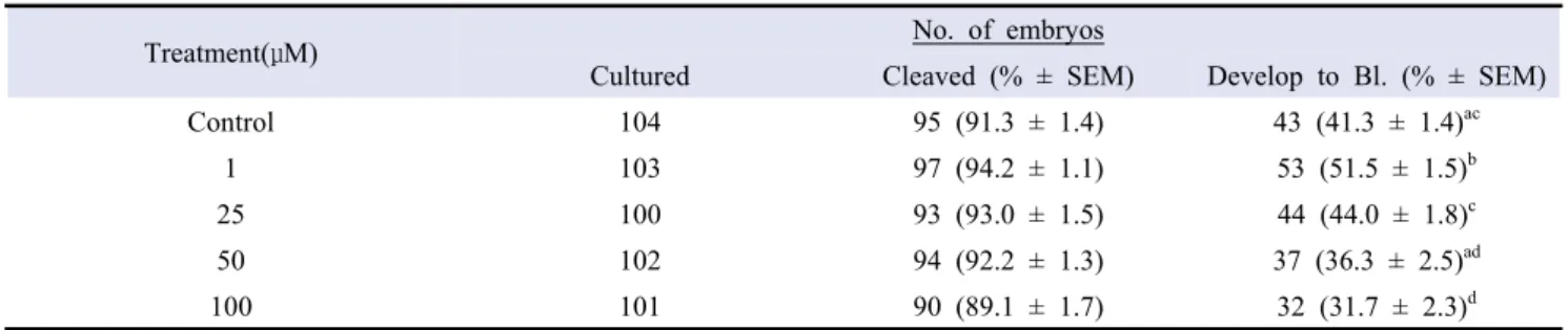
2. In- vitro development of PA embryos after flavone supplementation
The developmental potentials of PA embryos after flavone supplementation were shown in Table 1 and 2, respectively. In the case of supplementation of IVM media, there was no significant variation recorded in the cleavage percentage of oocytes among the groups. However, the blastocysts development rate was significantly raised (p<0.05) in 1 µM group than other groups (51.5 ± 1.5 vs. 44.0 ± 1.8, 36.3 ± 2.5, 31.7 ± 2.3, 41.3 ± 1.4, respectively). In IVC supplemented group, the cleavage rate followed the same trend as found in the case of IVM supplementation. But the percentage of blastocysts produced was enhanced significantly (p<0.05) in 1 μM group than 50 μM and control groups (50.0 ± 3.0 vs. 40.5 ± 2.7, 38.0 ± 3.1, respectively).
Treatment(μM) No. of embryos
Cultured Cleaved (% ± SEM) Develop to Bl. (% ± SEM)
Control 104 95 (91.3 ± 1.4) 43 (41.3 ± 1.4)ac
1 103 97 (94.2 ± 1.1) 53 (51.5 ± 1.5)b
25 100 93 (93.0 ± 1.5) 44 (44.0 ± 1.8)c
50 102 94 (92.2 ± 1.3) 37 (36.3 ± 2.5)ad
100 101 90 (89.1 ± 1.7) 32 (31.7 ± 2.3)d
Means in the same column with different superscripts were significantly different (P<0.05) Values are listed as Mean ± S.E.M.
Table 1. Effect of Flavone supplementation into IVM media with 5 different concentration on preimplantation development of porcine parthenogenetic embryos
3. In- vitro development of SCNT embryos after flavone supplementation
In-vitro developmental rates of porcine SCNT embryos following
flavone supplementation were shown in Table 3 and 4, respectively. Flavone supplementation of IVM media resulted in significantly higher cleavage rate (p<0.05) in 1 µM group as compared to the 50 µM and 100 µM (92.9 ± 0.8 vs. 89.7 ± 1.2, 87.8 ± 1.5%, respectively). Similarly, the blastocyst development rate was also significantly higher (p<0.05) in the 1 μM group than other 50 μM, 100 μM and control groups (16.8 ± 1.6 vs. 12.8 ± 1.1, 14.7
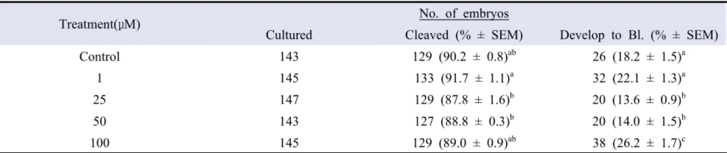
± 1.3, 9.0 ±0.9, 7.1 ± 1.3, respectively). In IVC supplemented groups, the cleavage percentage was significantly more (p<0.05) in 1 μM than 25 μM and 50 μM groups (91.7 ± 1.1 vs. 87.8 ± 1.6, 88.8 ± 0.3, respectively). Whereas, the percentage of blastocysts production was significantly higher (p<0.05) in the 100 μM group than other groups (26.2 ± 1.7 vs. 18.2 ± 1.5, 22.1 ± 1.3, 13.6 ± 0.9, 14.0 ± 1.5, respectively).
4. Differential staining of SCNT blastocysts
The total cell number and percentages of ICM and TE cells in
Treatment(μM) No. of embryos
Cultured Cleaved (% ± SEM) Develop to Bl. (% ± SEM)
Control 158 145 (91.8 ± 1.2) 60 (38.0 ± 3.1)a
1 158 147 (93.0 ± 1.1) 79 (50.0 ± 3.0)b
25 159 143 (89.9 ± 1.1) 69 (43.4 ± 1.2)ab
50 158 144 (91.1 ± 1.0) 64 (40.5 ± 2.7)a
100 158 142 (89.9 ± 1.3) 70 (44.3 ± 3.6)ab
Means in the same column with different superscripts were significantly different (00.05) Values are listed as Mean ± S.E.M.
Table 2. Effect of Flavone supplementation into IVC media with 5 different concentration on preimplantation development of porcine parthenogenetic embryos
Treatment(μM) No. of embryos
Cultured Cleaved (% ± SEM) Develop to Bl. (% ± SEM)
Control 156 144 (92.3 ± 0.8)ab 20 (12.8 ± 1.1)a
1 155 144 (92.9 ± 0.8)a 26 (16.8 ± 1.6)b
25 156 141 (90.4 ± 1.1)ac 23 (14.7 ± 1.3)ab
50 156 140 (89.7 ± 1.2)bc 14 (9.0 ± 0.9)c
100 156 137 (87.8 ± 1.5)c 11 (7.1 ± 1.3)c
Means in the same column with different superscripts were significantly different (P<0.05) Values are listed as Mean ± S.E.M.
Table 3. Effect of Flavone supplementation into IVM media with 5 different concentration on preimplantation development of porcine SCNT embryos
Treatment(μM) No. of embryos
Cultured Cleaved (% ± SEM) Develop to Bl. (% ± SEM)
Control 143 129 (90.2 ± 0.8)ab 26 (18.2 ± 1.5)a
1 145 133 (91.7 ± 1.1)a 32 (22.1 ± 1.3)a
25 147 129 (87.8 ± 1.6)b 20 (13.6 ± 0.9)b
50 143 127 (88.8 ± 0.3)b 20 (14.0 ± 1.5)b
100 145 129 (89.0 ± 0.9)ab 38 (26.2 ± 1.7)c
Means in the same column with different superscripts were significantly different (P<0.05) Values are listed as Mean ± S.E.M.
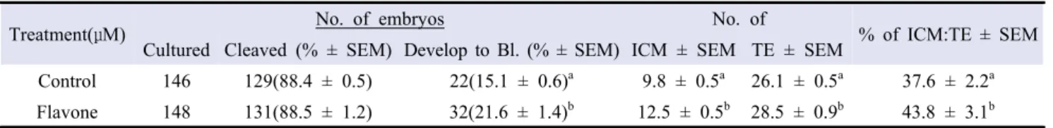
cloned porcine embryos following flavone supplementation were shown in Table 5. The total cell number of flavone supplemented group was significantly more (p<0.05) than control (43.8 ± 3.1 vs.
37.6 ± 2.2). Similarly, the percentages of ICM and TE cells were significantly higher in supplemented groups than control (12.5 ± 0.5 vs. 9.8 ± 0.5; 28.5 ± 0.9 vs. 26.1 ± 0.5, respectively).
Treatment(μM) No. of embryos No. of % of ICM:TE ± SEM
Cultured Cleaved (% ± SEM) Develop to Bl. (% ± SEM) ICM ± SEM TE ± SEM
Control 146 129(88.4 ± 0.5) 22(15.1 ± 0.6)a 9.8 ± 0.5a 26.1 ± 0.5a 37.6 ± 2.2a
Flavone 148 131(88.5 ± 1.2) 32(21.6 ± 1.4)b 12.5 ± 0.5b 28.5 ± 0.9b 43.8 ± 3.1b
Means in the same column with different superscripts were significantly different (P<0.05) Values are listed as Mean ± S.E.M.
Table 5. Effect of Flavone supplementation into IVM and IVC media on preimplantation development of porcine SCNT embryos
A
B
Figure 2. Different staining to compare with control group (A) the Flavone treatment group (B). Staining the ICM and TE by Hoechst 33342 and Propidium iodide. Experiment was replicated eight times.
Figure 3. Relative expression of Bax, Bcl-2, POU5F1, SOX2 and Nanog genes in cloned blastocysts using oocytes supplemented with 1µM of flavone in maturation media and 100 µM of flavone in culture media . a-bWithin the same mRNA
5. Gene expression of flavone treatment embryo
The supplementation of optimal levels of flavone significantly reduced (p<0.05) the expression level of BAX, but Bcl2 gene expression was significantly enhanced (p<0.05). On the other hand, the expression levels of reprogramming related genes POU5F1, SOX2 and NANOG were significantly improved (p<0.05) by flavone supplementation (Figure 3).
DISCUSSION
Damaged caused by increased oxidative stress is one of the main reason behind inefficient development of SCNT embryos. The maturation and culturing environments have higher concentrations of ROS resulting in defective. Flavone has been reported by some of the independent laboratories as a powerful antioxidant, helpful in improving the conditions of cells through a reduced oxidative damage. This study has evaluated the improvements in the developmental rates of porcine embryos derived from flavone supplemented maturation and culturing media. The optimization of its application (concentration) might be the key factor in improving the success rate of maturation and culturing.
The supplementation of IVM media with different inclusion rates of flavone (ranging from 0 to 100 μM) demonstrated that 100 µM group was statistically the most effective in reducing intracellular ROS levels of porcine oocytes. But the intracellular GSH levels were significantly greater in 1 µM, 50 µM and 100 µM than 25 µM and control groups. The reason for raised GSH levels was reduce ROS due to its detoxification, that is helpful both in minimizing the oxidative stress of micro-organelles and also improves the efficiency of enzymes system responsible for GSH production. The results also indicated that the effects of flavone were concentration dependent, might be due to oocyte absorptive capacity.
In parthenogenetic embryos, the cleavage rates remained same among all the groups either flavone was supplemented in IVM or IVC. However, the blastocysts were produced significantly more in 1 µM group than others with no difference in media type. The improved developmental rates of PA embryos might be due to suppressed oxidative damage that not only prevent DNA damage but also maintains the structure and function of different organelles (Zhang et al. 2016).
Insufficient epigenetic reprogramming is not compatible with normal development of cloned embryos (Bohrer et al. 2014).
Because the accumulation of damaged macromolecules, shortened telomeres, and (nuclear donor-derived) age-related DNA damage affects oocyte competency and embryo development (Burgstaller and Brem 2017). Supplementation of the culture conditions with different agents can modulate the gene expressions(Zhou et al. 2008) and ultimately benefits the cloning efficiency. In SCNT embryos, the supplementation of IVM media with 1 µM flavone had significantly better development rates than others. In IVC supplemented group, the development rate was significantly enhanced by 100 µM.
Following determination of optimal levels of flavone, we compared the development rates, quality of blastocysts and gene expression between optimal concentration and control group. The blastocysts rates, total cell number per blastocyst and the percentage of ICM/TE cells were significantly higher in the flavone treatment group than control group. Similarly, the SCNT embryos in flavone treated groups had increased expression levels of reprogramming related genes (POU5F1, SOX2, Nanog). The possible reasons for the improvements after flavone supplementation, may be the development of specific conditions of maturation and culturing with low oxidative stress that are suitable for both reprogramming of genes and integrity of different intra-cellular organelles. Previously, it was observed that reduction in ROS levels by the treatment of antioxidant improved cloned embryo production through better gene expression and reprogramming (Kawasumi et al. 2009).
In conclusion, the flavone supplementation can provide protection to the developing embryos against oxidative stress. In addition, it also improves the nuclear reprogramming, thereby minimizes the deterioration in cloned embryo quality during in vitro culture.
ACKNOWLEDGMENTS
We thank all the members of our laboratory for technical support and helpful discussion. This research was supported by a grant from the Korea Health Technology R & D Project through the Korea Health Industry Development Institute (KHIDI), funded by the Ministry of Health & Welfare, Republic of Korea (grant number: HI13C0954).
REFERENCES
Ashaari Z, Hadjzadeh MA, Hassanzadeh G, Alizamir T, Yousefi B, Keshavarzi Z, Mokhtari T. 2018. The Flavone Luteolin
Improves Central Nervous System Disorders by Different Mechanisms: A Review. Journal of molecular neuroscience : MN 65(4):491-506.
Bae HK, Hwang IS, Kim JY, Lee SY, Park CK, Yang BK, Cheong HT. 2015. Antioxidant treatment during manipulation procedures prevents mitochondrial and DNA damage and enhances nuclear reprogramming of bovine somatic cell nuclear transfer embryos. Reproduction, fertility, and development 27(7):1088-1096.
Bavister BD, Leibfried ML, Lieberman G. 1983. Development of preimplantation embryos of the golden hamster in a defined culture medium. Biol Reprod 28(1):235-247.
Bohrer RC, Duggavathi R, Bordignon V. 2014. Inhibition of histone deacetylases enhances DNA damage repair in SCNT embryos. Cell cycle (Georgetown, Tex) 13(13):2138-2148. Burgstaller JP, Brem G. 2017. Aging of Cloned Animals: A
Mini-Review. Gerontology 63(5):417-425.
Ganai SA. 2017. Plant-derived flavone Apigenin: The small- molecule with promising activity against therapeutically resistant prostate cancer. Biomedicine & pharmacotherapy = Biomedecine & pharmacotherapie 85:47-56.
Gao R, Wang C, Gao Y, Xiu W, Chen J, Kou X, Zhao Y, Liao Y, Bai D, Qiao Z, Yang L, Wang M, Zang R, Liu X, Jia Y, Li Y, Zhang Y, Yin J, Wang H, Wan X, Liu W, Zhang Y, Gao S. 2018. Inhibition of Aberrant DNA Re-methylation Improves Post-implantation Development of Somatic Cell Nuclear Transfer Embryos. Cell stem cell 23(3):426-435.e425. Holm IE, Alstrup AK, Luo Y. 2016. Genetically modified pig
models for neurodegenerative disorders. The Journal of pathology 238(2):267-287.
Huang CH, Chan WH. 2017. Protective Effects of Liquiritigenin against Citrinin-Triggered, Oxidative-Stress-Mediated Apoptosis and Disruption of Embryonic Development in Mouse Blastocysts. International journal of molecular sciences 18(12).
Huang ZS, Xiao HJ, Qi T, Hu ZM, Li H, Chen DL, Xu YL, Chen J. 2014. Antioxidative protective effect of icariin on the FeSO4/H 2O 2-damaged human sperm based on confocal raman micro-spectroscopy. Journal of Huazhong University of Science and Technology Medical sciences = Hua zhong ke ji da xue xue bao Yi xue Ying De wen ban = Huazhong keji daxue xuebao Yixue Yingdewen ban 34(5):755-760. Hwang JH, Kim SE, Gupta MK, Lee H. 2016. Gnotobiotic Miniature
Pig Interbreed Somatic Cell Nuclear Transfer for Xenotransplantation. Cellular reprogramming 18(4):207-213.
Kang JD, Li S, Lu Y, Wang W, Liang S, Liu X, Jin JX, Hong Y,
Yan CG, Yin XJ. 2013. Valproic acid improved in vitro development of pig cloning embryos but did not improve survival of cloned pigs to adulthood. Theriogenology 79(2):306-311.e301. Kawasumi M, Unno Y, Matsuoka T, Nishiwaki M, Anzai M,
Amano T, Mitani T, Kato H, Saeki K, Hosoi Y, Iritani A, Kishigami S, Matsumoto K. 2009. Abnormal DNA methylation of the Oct-4 enhancer region in cloned mouse embryos. Molecular reproduction and development 76(4):342-350.
Keefer CL. 2015. Artificial cloning of domestic animals. Proceedings of the National Academy of Sciences of the United States of America 112(29):8874-8878.
Liu T, Dou H, Xiang X, Li L, Li Y, Lin L, Pang X, Zhang Y, Chen Y, Luan J, Xu Y, Yang Z, Yang W, Liu H, Li F, Wang H, Yang H, Bolund L, Vajta G, Du Y. 2015. Factors Determining the Efficiency of Porcine Somatic Cell Nuclear Transfer: Data Analysis with Over 200,000 Reconstructed Embryos. Cellular reprogramming 17(6):463-471.
Liu X, Nie ZW, Gao YY, Chen L, Yin SY, Zhang X, Hao C, Miao YL. 2018. Sodium fluoride disturbs DNA methylation of NNAT and declines oocyte quality by impairing glucose transport in porcine oocytes. Environmental and molecular mutagenesis 59(3):223-233.
Matoba S, Zhang Y. 2018. Somatic Cell Nuclear Transfer Reprogramming: Mechanisms and Applications. Cell stem cell 23(4):471-485. Roy PK, Fang X, Bahia MSH, Shin ST, Cho JK. 2017. Effects
of Roscovitine on In Vitro Development of Porcine Oocyte Using Brilliant Cresyl Blue. Journal of Embryo Transfer Vol. 32, No. 3:111 122.
Sajjadi SE, Delazari Z, Aghaei M, Ghannadian M. 2018. Flavone constituents of Phlomis bruguieri Desf. with cytotoxic activity against MCF-7 breast cancer cells. Research in pharmaceutical sciences 13(5):422-429.
Wang K, Lv Q, Miao YM, Qiao SM, Dai Y, Wei ZF. 2018a. Cardamonin, a natural flavone, alleviates inflammatory bowel disease by the inhibition of NLRP3 inflammasome activation via an AhR/Nrf2/NQO1 pathway. Biochemical pharmacology 155:494-509.
Wang L, Wang X, Chen H, Zu X, Ma F, Liu K, Bode AM, Dong Z, Kim DJ. 2018b. Gossypin inhibits gastric cancer growth by direct targeting of AURKA and RSK2. Phytotherapy research : PTR.
Wrenzycki C, Wells D, Herrmann D, Miller A, Oliver J, Tervit R, Niemann H. 2001. Nuclear transfer protocol affects messenger RNA expression patterns in cloned bovine blastocysts. Biology of reproduction 65(1):309-317.
Wruck CJ, Claussen M, Fuhrmann G, Romer L, Schulz A, Pufe T, Waetzig V, Peipp M, Herdegen T, Gotz ME. 2007. Luteolin protects rat PC12 and C6 cells against MPP+ induced toxicity via an ERK dependent Keap1-Nrf2-ARE pathway. Journal of neural transmission Supplementum(72):57-67.
Xu W, Wang Y, Li Y, Wang L, Xiong X, Su J, Zhang Y. 2012. Valproic acid improves the in vitro development competence of bovine somatic cell nuclear transfer embryos. Cellular reprogramming 14(2):138-145.
You J, Lee J, Hyun SH, Lee E. 2012. L-carnitine treatment during oocyte maturation improves in vitro development of cloned pig embryos by influencing intracellular glutathione synthesis and embryonic gene expression. Theriogenology 78(2):235-243. Zhang Y, Li W, Ma Y, Wang D, Zhao X, Zeng C, Zhang M,
Zeng X, Meng Q, Zhou G. 2016. Improved development by melatonin treatment after vitrification of mouse metaphase II oocytes. Cryobiology 73(3):335-342.
Zhou W, Xiang T, Walker S, Farrar V, Hwang E, Findeisen B, Sadeghieh S, Arenivas F, Abruzzese RV, Polejaeva I. 2008. Global gene expression analysis of bovine blastocysts produced by multiple methods. Molecular reproduction and development 75(5):744-758.
Zhu W, Ma L, Yang B, Zheng Z, Chai R, Liu T, Liu Z, Song T, Li F, Li G. 2016. Flavone inhibits migration through DLC1/RhoA pathway by decreasing ROS generation in breast cancer cells. In vitro cellular & developmental biology Animal 52(5):589-597.