저작자표시-비영리-변경금지 2.0 대한민국 이용자는 아래의 조건을 따르는 경우에 한하여 자유롭게 l 이 저작물을 복제, 배포, 전송, 전시, 공연 및 방송할 수 있습니다. 다음과 같은 조건을 따라야 합니다: l 귀하는, 이 저작물의 재이용이나 배포의 경우, 이 저작물에 적용된 이용허락조건 을 명확하게 나타내어야 합니다. l 저작권자로부터 별도의 허가를 받으면 이러한 조건들은 적용되지 않습니다. 저작권법에 따른 이용자의 권리는 위의 내용에 의하여 영향을 받지 않습니다. 이것은 이용허락규약(Legal Code)을 이해하기 쉽게 요약한 것입니다. Disclaimer 저작자표시. 귀하는 원저작자를 표시하여야 합니다. 비영리. 귀하는 이 저작물을 영리 목적으로 이용할 수 없습니다. 변경금지. 귀하는 이 저작물을 개작, 변형 또는 가공할 수 없습니다.
Doctoral Thesis in Medicine
Right and Ventral Margins of the
Caudate Lobe: Three-Dimensional
Reconstruction Analysis Using Synapse
3D Software
by
XUEYIN SHEN
Ajou University Graduate School
Major in Medicine
Right and Ventral Margins of the
Caudate Lobe: Three-Dimensional
Reconstruction Analysis Using Synapse
3D Software
Hee Jung Wang,
M.D., Ph.D.
Advisor
I submit this thesis as the
Doctoral thesis in Medicine
August 2020
Ajou University Graduate School
Major in Medicine
Department of Medical Sciences
XUEYIN SHEN
The Doctoral thesis of XUEYIN SHEN in Medicine
is hereby approved.
Thesis Defense Committee President
11111111111111111111
Jin Hong Kim
Member
1111111wook hwan kim
Hee Jung Wang
Member
1S ng uk han
Jae Youn Cheong
Member
111111111111111111111
Hyun Goo Woo
Member
111111111111111111111
Yong Keun Park
Ajou University Graduate School
July 4th, 2020
i
ABSTRACT
Right and Ventral Margins of the Caudate Lobe:
Three-Dimensional Reconstruction Analysis Using Synapse 3D Software
Purpose: Complete removal of the caudate lobe is sometimes necessary, which is accomplished via isolated caudate lobectomy or hepatectomy that includes the caudate lobe. It is impossible, however, to confirm the right and ventral margins of the caudate lobe by preoperative imaging. This study was undertaken to determine whether we could identify the right and ventral margins of the caudate lobe preoperatively using Synapse 3D visualization software.
Methods: Ninety-four preoperative three-dimensional computed tomographic images (1-mm slices) of the liver from candidate donors were examined. The images of the caudate lobe were subjected to a counter-staining method according to Synapse 3D to delineate their dimensions. We first examined whether the right margin of the caudate lobe exceeded the plane formed by the root of the right hepatic vein (RHV) and the right side of the inferior vena cava (IVC). Second, we determined whether the ventral margin of the caudate lobe exceeded the plane formed by the root of the middle hepatic vein (MHV) and the root of the RHV.
Results: For the right margin, 17 cases (18%) exceeded the RHV-IVC plane by a mean of 10.2 mm (range 2.4–27.2 mm). For the ventral margin, 28 cases (30%) exceeded the MHV-RHV plane by a mean of 17.4 mm (range 1.2–49.1 mm).
Conclusion: Evaluating the anatomy of caudate lobe using Synapse 3D preoperatively could be helpful for more precise anatomical resection of the caudate lobe.
Key words: Caudate lobe; Marginal configuration; Paracaval portion; Anatomy of the liver; Anatomical resection.
ii
TABLE OF CONTENTS
ABSTRACT --- i
TABLE OF CONTENTS --- ii
LIST OF FIGURES --- iii
LIST OF TABLES --- iv
ABBREVIATION --- v
I. INTRODUCTION --- - 1 -
II. METHODS --- - 4 -
A. Synapse 3D --- - 5 -
B. Counter-staining technique --- - 5 -
Ⅲ. RESULTS --- - 6 -
Ⅳ. DISCUSSION --- - 13 -
Ⅴ. CONCLUSION --- - 17 -
REFERENCES --- - 19 -
iii
LIST OF FIGURES
Fig. 1. The caudate parenchyma was delineated using a counter-staining technique. Hepatic veins, hepatic arteries, and bile ducts are hidden to identify images more easily. ··· 9
Fig. 2. The methods to define the right and ventral margins are shown. ··· 10
Fig. 3. Right and ventral margins of the caudate lobe are shown. The relations between the caudate lobe and right hepatic vein and inferior vena cava (RHV-IVC) plane and the middle and right hepatic veins (MHV-RHV) plane is shown in Synapse 3D images. ··· 11
iv
LIST OF TABLES
v
ABBREVIATION
RHV: right hepatic vein IVC: inferior vena cava MHV: middle hepatic vein
3D
:three-dimensional
CT
:computed tomography
- 1 -
- 2 -
Because hilar cholangiocarcinomas may send metastases to the caudate lobe via minute bile duct branches [1], total caudate lobectomy, along with hepatic resection, is necessary [2, 3]. Although the importance of the caudate lobe is being increasingly accepted and a copious amount of research carried out, the anatomy of the caudate lobe lacks consensus.
Couinaud suggested a clinically useful anatomy of the liver, but the concept of caudate lobe anatomy has been changing since then [4]. In 1954, Couinaud defined the caudate lobe as segment I according to its portal ramification. This portion of the liver corresponds to the Spiegel lobe in the Kumon classification, which is currently used worldwide. Kumon classified the caudate lobe into three parts: Spiegel’s lobe, the paracaval portion, and the caudate process portion [5]. Many researchers have agreed with the Kumon classification, and it is being used clinically.
For the right margin of the caudate lobe, Kogure et al. advocated that the hepatic vein is the boundary between the caudate process and the right liver [6], but it is more important to find the right margin of the paracaval portion when searching for the right border of the caudate lobe. The paracaval portion is located inside the liver parenchyma, in neighboring segments 7 and 8, making it difficult to identify the margin. Kitakawa, of the Murakami group, initially thought that the right hepatic vein was the most important landmark of the right margin of the paracaval portion. Later, however, he accepted the concept of the right paracaval plane [7]. Recently, Maki et al. found that the paracaval vein of a branch of the right hepatic vein is the boundary between the caudate lobe and segments 7 and 8 [8].
The ventral margin of caudate lobe was believed to correspond to the route of the middle hepatic vein. Couinaud opposed this idea, however, and thought that the margin was situated in the vicinity of the confluence of the middle and right hepatic vein drainage area into the inferior vena cava (IVC) [9]. Later, Kwon et al. reported the existence of the precaudate plane [10].
The current study focuses on identifying the right and ventral margins of the caudate lobe, which is barely recognized in the surgical field. So far, there are no reports on the right and ventral margins of the caudate lobe that can be applied clinically. As the caudate anatomy has been analyzed by Synapse 3D software (Fujifilm, Tokyo, Japan) to visualize the
three-- 3 three--
dimensional (3D) anatomy of the caudate lobe, we set out to delineate the right and ventral margins of the caudate lobe preoperatively using the same system.
- 4 -
II. METHODS
- 5 -
A. Synapse 3D
Contrast-enhanced helical computed tomography (CT) examinations of the liver were conducted. After obtaining abdominal multidetector CT images (1-mm slices), 3D image reconstruction was carried out using Synapse 3D. Digital Imaging and Communication in Medicine file of raw 1-mm CT images were transferred to the Synapse 3D software. After selecting the parenchyma of the whole liver, we traced the portal vein and hepatic vein branches. The caudate lobe was visualized after reconstruction. Although only 15 of 94 cases had portal branches in the caudate lobe, we could delineate the parenchyma of the caudate lobe by electronically clicking on them. Thereafter, we compared it with the parenchyma that remained after staining by clicking on the second-order branches of the portal veins. The directly stained caudate parenchyma was the same as the indirectly stained caudate parenchyma.
B. Counter-staining technique
In 79 subjects in whom there were no portal branches of the caudate lobe visualized, we could delineate the caudate parenchyma by Takayama’s counter-staining technique [11] (Figure 1). The hepatic portal system can be easily identified by hiding the parenchyma of the liver, hepatic veins, hepatic arteries, and bile ducts. When the corresponding parenchyma of the left portal vein, the right anterior portal vein, and the right posterior portal vein is removed, the image of the caudate lobe will be revealed.
We accepted that the right hepatic vein (RHV)-IVC plane is the reliable landmark of the right margin of the caudate lobe, which contains the RHV from the root + 3 cm and the right border of the IVC. For the ventral margin, the MHV-RHV plane could be regarded as the landmark comprising the MHV from the root + 3 cm and RHV from the root + 3 cm. Where the roots of the MHV and RHV overlapped was considered the MHV-RHV plane (Figure 2). The approved number of Institutional Review Board (IRB) was AJIRB-MED-MDB-19-108.
- 6 -
Ⅲ. RESULTS
- 7 -
The characteristics of the caudate lobe in 94 cases are shown in Table 1. For the right margin, 17 subjects (18%) exceeded the RHV-IVC plane by a mean of 10.2 mm (range 2.4–27.2 mm). Among them, 11 were <10 mm, 3 were 10–20 mm, and 3 were >20 mm (Figure 3A, B). For the ventral margin, 28 subjects (30%) exceeded the MHV-RHV plane by a mean of 17.4 mm (range 1.2–49.1 mm). Among them, 12 were <10 mm, 5 were 10– 20 mm, and 11 were >20 mm (Figure 3C, D). In all, 10 subjects were found to exceed both the MHV-RHV and RHV-IVC planes.
- 8 -
Table1. Characteristics of the caudate lobe in 94 cases
Right margin (%) Ventral margin (%) Number of cases exceeding the margin
<10mm 10mm-20mm >20mm 17 (18) 11 (12) 3 (3) 3 (3) 28 (30) 12 (13) 5 (5) 11 (12)
Mean distance exceeding the margin (mm) 10.2 17.4
- 9 -
Fig. 1. The caudate parenchyma was delineated using a counter-staining technique. Hepatic veins, hepatic arteries, and bile ducts are hidden to identify images more easily. A: By hiding the hepatic parenchyma, the portal vein branch of the paracaval portion can be seen (star). B: By clicking on the portal vein branch of the paracaval portion, the parenchyma of caudate lobe is shown (yellow). C: In the same case, we clicked on the left portal vein (square), right anterior portal vein (circle), and right posterior portal vein (diamond), respectively, which then showed the corresponding parenchyma of the liver. D: The shape of the remnant liver parenchyma is the same as in B.
- 10 -
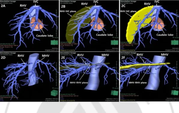
Fig. 2. The methods to define the right and ventral margins are shown. A: IVC and RHV can be identified after hiding the hepatic parenchyma other than the caudate lobe. B: RHV-IVC plane can be seen (translucent brown) by connecting the RHV from the root + 3 cm and the right border of the IVC, C: RHV-IVC plane can be seen (yellow) by connecting the RHV from the root + 3 cm and the right border of the IVC. D: MHV and RHV can be identified after hiding the hepatic parenchyma. E: MHV-RHV plane can be seen (translucent yellow) by connecting the MHV from the root + 3 cm and RHV from the root + 3 cm. F: MHV-RHV plane can be seen (yellow) by connecting the MHV from the root + 3 cm and RHV from the root + 3 cm.
- 11 -
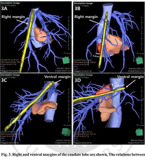
Fig. 3. Right and ventral margins of the caudate lobe are shown. The relations between the caudate lobe and right hepatic vein and inferior vena cava (RHV-IVC) plane and the middle and right hepatic veins (MHV-RHV) plane is shown in Synapse 3D images. A: Caudate lobe is located on the right side of the RHV-IVC plane (yellow). B: The parenchyma of the caudate lobe exceeds the RHV-IVC plane (yellow) is outlined in red. We calculated that the distance exceeds the right margin is 5.6mm in this case. C: Caudate lobe is ventrally located in the MHV-RHV plane (yellow). D: The parenchyma of the caudate lobe exceeds the MHV-RHV plane (yellow) is outlined in
- 12 -
- 13 -
- 14 -
Hilar cholangiocarcinoma is known to invade the parenchyma and bile duct of the caudate lobe rapidly during an early stage of the disease. In the literature, microscopic tumor involvement in caudate lobe branches was found in 42% of the patients at the time of the surgery [1]. Thus, caudate anatomy is of great importance when performing total caudate lobectomy in patients with hilar cholangiocarcinoma. The prognosis reportedly improved in cholangiocarcinoma patients when caudate lobectomy was performed [12]. In the case of a caudate lobe tumor, isolated caudate lobectomy can be performed, as introduced by Lerut et al. in 1990. The surgery included partial or total caudate lobectomy [13]. Partial caudate lobectomy was performed by resecting Spiegel’s lobe or the caudate process only, each of which was easily identified intraoperatively [14]. In other, previous research, total isolated caudate lobectomy was performed by approaching the caudate lobe from the right side of the liver, using the right posterior Glisson pedicle as the anterior border plane of the caudate lobe and reaching the dorsal plane by transecting the parenchyma between the MHV and RHV. There was no mention of the right margin of the caudate lobe [15, 16]. Although it is known that the anatomy of the caudate lobe is important when performing total caudate lobectomy, the exact locations of the right and ventral margins remain obscure.
It is difficult for us to define the right margin of the caudate lobe of the liver because the paracaval portion is located anterior to the IVC, to the right of the ligamentum venosum, and cranial to the hilar plate. The right and ventral margins were not identifiable from the exterior as their locations vary [17]. Couinaud suggested that the right portion of the caudate lobe was a neighbor of segments 7 and 8, lying adjacent to the end point of the right hepatic vein [18]. Kitakawa et al. first thought that the right hepatic vein is the most important landmark of the right margin of the paracaval portion. Later, however, they suggested the concept of the right paracaval plane [7]. These investigators suggested that the right paracaval plane may serve as the imaginary right margin of the caudate lobe. In fact, the right margin of the paracaval portion was located on the left side of the right paracaval plane in 65.4% (36/55) of cases. Therefore, if hepatic resection was carried out in this plane, total caudate lobectomy was complete with 60% anatomical reliability; if carried out at 10 mm to the right of this plane, 80% reliability was guaranteed; and if
- 15 -
performed at 30 mm to the right of this plane, there was 100% reliability. This report was a milestone in the search for a definition of the right margin of the caudate lobe—which is barely visualized from the exterior. Nevertheless, the concept had little effect on identifying the right margin during surgery. Recently, Maki et al. identified the paracaval vein as the landmark for establishing the boundary between the caudate lobe and segments 7 and 8. The paracaval vein, however, cannot be found by intraoperative ultrasonography [8]. In the current study, 17 cases (18%) exceeded the RHV-IVC plane. As landmarks, the RHV and IVC are easily found in the surgical field as well as with intraoperative ultrasonography.
The need to identify the individualized right margin of the caudate lobe is indisputable. The use of Synapse 3D is more likely to enable surgeons to perform total caudate lobectomy based on the patient’s own liver anatomy.
Regarding the ventral margin of the caudate lobe, in 2002 Kwon et al. reported the existence of a precaudate plane [10]. This plane, which they described as flat or slightly curved, was a continuum of the ligamentum venosum and the ventral margin of the hilar plate. According to this report, the ventral margin of the caudate lobe was restricted within the dorsal side of the precaudate line in 63.2% (48/76) of the liver specimens. In 36.8% of the cases, where ventral extension of the caudate lobe was found, the ventral margin was extended to the dome-like area under the terminals of the RHV, MHV, and the left hepatic vein. The authors suggested that the precaudate plane could serve as the imaginary ventral margin of the caudate lobe, which was a milestone in the definition of the ventral margin of the caudate lobe. However, it is an illusory plane that could not be used as the foundation on which to base surgical decisions. In the current study, we used the MHV-RHV plane to facilitate identifying the ventral margin of the caudate lobe. The RHV and MHV could be easily identified during the surgery because their locations were relatively fixed, allowing them to serve as landmarks. Traditionally, the caudate lobe was thought to be confined within the RHV and MHV. Based on this belief, surgeons performed total caudate lobectomy. Our results showed, however, that 28 cases (30%) exceeded the MHV-RHV plane. Kumon noted that the paracaval portion reached the liver surface underneath the diaphragm in 53% of corrosion liver casts [5]. Kwon et al. found the same results in 50% of cadaveric studies [10], and Maki et al. reported the same result in 30% of images of
- 16 -
living patients [8]. In this type of caudate lobe, it is impossible to perform total caudate lobectomy from the dorsal side of the liver.
Although our research has limitations—e.g., that small branches (<1 mm diameter) could not be easily identified on CT images—we learned that preoperative analysis of caudate lobe using Synapse 3D is necessary to evaluate the anatomy of the caudate lobe. Synapse 3D provides the margins of the caudate lobe that two-dimensional CT cannot.
- 17 -
- 18 -
In conclusion, regarding the right margin, 18% exceeded the RHV-IVC plane, whereas 30% exceeded the MHV-RHV plane for the ventral margin.Evaluating the anatomy of the caudate lobe using Synapse 3D preoperatively could be helpful in achieving more precise anatomical resection of the caudate lobe.
- 19 -
REFERENCES
1. Mizumoto R, Kawarada Y, Suzuki M. Surgical management of hilar carcinoma of the bile duct: Surg Gynecol Obstet. 1985;162:153-8.
2. Malhi H, Gores GJ. Cholangiocarcinoma: modern advances in understanding a deadly old disease. J Hepatol 2006;45:856-67.
3. Baton O, Azoulay D, Adam DV, Castaing D. Major hepatectomy for hilar cholangiocarcinoma type 3 and 4: prognostic factors and longterm outcomes. Am Coll Surg 2007;204:250-60.
4. Couinaud C. [Liver lobes and segments: notes on the anatomical architecture and surgery of the liver.] Presse Med 1954;62:709-12 (in French).
5. Kumon M. Anatomy of the caudate lobe with special reference to portal vein and bile duct. Acta Hepatol Jpn 1985;26:1193–9.
6. Kogure K, Kuwano H, Yorifuji H, Ishikawa H, Takata K, Makuuchi M. The caudate processus hepatic vein: a boundary hepatic vein between the caudate lobe and the right liver. Ann Surg 2008;247:288–93.
7. Kitagawa S, Murakami G, Hata F, Hirata K. Configuration of the right portion of the caudate lobe with special reference to the identification of its right margin. Clin Anat 2000;13:321- 40.
8. Maki H, Sakamoto Y, Kawaguchi Y, Akamatsu N, Kaneko J, Arita J, et al. Anatomical
boundary between the caudate lobe of the liver and adjacent segments based on three-dimensional analysis for precise resections. J Gastrointest Surg 2018:22:1709-14.
9. Couinaud C, editor. [Surgical anatomy of the liver revisited.] Paris, Personal edition. 1989 (in French).
- 20 -
10. Kwon D, Murakami G, Hata F, Wang HJ, Chung MS, Hirata K. Location of the ventral margin of the paracaval portion of the caudate lobe of the human liver with special reference to the configuration of hepatic portal vein branches. Clin Anat 2002;15:387-401.
11. Takayama T, Tanaka T, Higaki T, Katou K, Teshima Y, Makuuchi M. High dorsal resection of the liver. J Am Coll Surg 1994;179:72–5.
12. Nimura Y, Hayakawa N, Kamiya J, Kondo S, Shionoya S. Hepatic segmentectomy with caudate lobe resection for bile duct carcinoma of the hepatic hilus. World J Surg. 1990;14:535-44.
13. Lerut J, Gruwez JA, Blumgart LH. Resection of the caudate lobe of the liver. Surg Gynecol Obstet 1990;171:160–2.
14. Cheung TT. Technical notes on pure laparoscopic isolated caudate lobectomy for patient with liver cancer. Transl Gastroenterol Hepatol 2016;1:56.
15. Oh D, Kwon CH, Na BG, Lee KW, Cho WT, Lee SH, et al. Surgical techniques for totally laparoscopic caudate lobectomy. J Laparoendosc Ad Surg Tech A 2016;26:689-692. 16. Lee W, Han HS, Yoon YS, Cho JY, Choi Y, Shin HK, et al. Laparoscopic resection of
hilar cholangiocarcinoma. Ann Surg Treat Res. 2015;89(4):228–232.
17. Murakami G, Hata F. Human liver caudate lobe and liver segment. Anat Sci Int 2002;77:211-224.