Do we have enough evidence for expanding the indications
of ESD for EGC?
Hang Lak Lee, Chang Hwan Choi, Dae Young Cheung Hang Lak Lee, Department of Internal Medicine, Hanyang University College of Medicine, Seoul 133-792, South Korea Chang Hwan Choi, Department of Internal Medicine, Chung-Ang University College of Medicine, Seoul 156-755, South Korea Dae Young Cheung, Division of Gastroenterology, Department of Internal Medicine, Yeouido St. Mary’s Hospital, the Catho-lic University of Korea College of Medicine, Seoul 150-173, South Korea
Author contributions: Lee HL and Choi CH are contributed equally as a first author to this work; Lee HL and Choi CH con-tributed to the design frame work and wrote the paper; Cheung DY contributed to the appraisal of the literature and wrote the paper. Correspondence to: Dae Young Cheung, MD, Division of Gastroenterology, Department of Internal Medicine, Yeouido St. Mary’s Hospital, the Catholic University of Korea College of Medicine, Seoul 150-173, South Korea. [email protected] Telephone: +82-2-37791328 Fax: +82-2-37791331 Received: June 25, 2010 Revised: September 20, 2010 Accepted: September 27, 2010
Published online: June 7, 2011
Abstract
Endoscopic submucosal dissection (ESD) is the most advanced and representative technique in the field of therapeutic endoscopy and has been used for the treat-ment of gastrointestinal neoplasms, including early gas-tric cancer. The major difference and advantage of ESD compared to existing endoscopic resection techniques, such as endoscopic mucosal resection (EMR) and polyp-ectomy, are the width and depth of the resection. Newly developed cutting devices, distal attachable endoscopic accessories, and an advanced electrosurgical unit have helped to overcome the limitations of therapeutic endos-copy in terms of lesion size, location, presence of fibrotic scarring, and accompanying ulcers. As a result, the indications for ESD have been expanded from the clas-sical indication for EMR and polypectomy, and there is now support for a further expansion of ESD indications. At present, the most critical factor to consider in the decision of whether to perform ESD is the probability of
unexpected lymph node metastasis. The guidelines for ESD are continually being updated and debated. In this review, we discuss the strengths and weaknesses of the expanded guidelines, based on evidence found in the literature.
© 2011 Baishideng. All rights reserved.
Key words: Endoscopic submucosal dissection;
Endo-scopic mucosal resection; Early gastric cancer; Indica-tions
Peer reviewers: Damian Casadesus Rodriguez, MD, PhD, Calixto Garcia University Hospital, J and University, Vedado, Havana City, Cuba; Atsushi Nakajima, Professor, Division of Gastroenterology, Yokohama City University Graduate School of Medicine, 3-9 Fuku-ura, Kanazawa-ku, Yokohama 236-0004, Japan; Dr. Noriko Suzuki, MD, PhD, Honorary Consultant, Wolf-son Unit for Endopscopy, St Mark’s Hospital, Watford Road, Harrow, Middlesex, HA1 3UJ, United Kingdom
Lee HL, Choi CH, Cheung DY. Do we have enough evidence for expanding the indications of ESD for EGC? World J
Gas-troenterol 2011; 17(21): 2597-2601 Available from: URL:
http://www.wjgnet.com/1007-9327/full/v17/i21/2597.htm DOI: http://dx.doi.org/10.3748/wjg.v17.i21.2597
INTRODUCTION
Early gastric cancer (EGC) is defined as a gastric cancer that is confined to the mucosa or submucosa, irrespective of the presence of regional lymph node metastasis[1,2].
Endoscopic submucosal dissection (ESD) is a novel endoscopic treatment that enables a clinician to resect a target lesion en bloc. For the last ten years, ESD has been performed in Korea for the management of early gastric cancer[3,4]. ESD for EGC is comparable to conventional
surgery in many aspects, and it has the advantage of be-ing less invasive and more economical. In this article, we
© 2011 Baishideng. All rights reserved. doi:10.3748/wjg.v17.i21.2597
TOPIC HIGHLIGHT
introduce the absolute and expanded indications of ESD and discuss their usefulness, safety, and limitations.
AbsOlUTe AND expANDeD
INDICATIONs Of esD fOR eGC
The curability of EGC depends on the complete removal of the cancerous lesion and its metastatic lymph nodes. Fortunately, because a significant proportion of EGC has no lymph node metastasis, limited surgery, such as ESD, can be legitimately performed in many countries.
Traditionally accepted indications for endoscopic re-section of EGC are small intramucosal EGCs of intestinal histology type. The rationale for this recommendation is based on the knowledge that larger lesions or diffuse histology lesions are more likely to extend into the sub-mucosal layer and thus have a higher risk of lymph node metastasis. In addition, resection of a large lesion was not technically feasible until the ESD procedure was devel-oped. Therefore, at present, the accepted indications for endoscopic mucosal resection (EMR) according to the gastric cancer treatment guidelines published in 2001 by the Japanese Gastric Cancer Association are: (1) well-dif-ferentiated elevated cancers less than 2 cm in diameter; and (2) small (< 1 cm) depressed lesions without ulceration. These lesions must also be moderately or well-differentiat-ed cancers confinwell-differentiat-ed to the mucosa, and have no lymphatic or vascular involvement[5,6]. However, it has been clinically
observed that currently accepted indications for EMR may be too strict, leading to unnecessary surgery[7].
Further studies by Gotoda et al[8] have defined new
criteria to expand the indications for endoscopic treat-ment of gastric cancer. The ESD method has been developed to dissect directly along the submucosal layer using specialized devices. Preliminary studies have been published on the advantage of ESD over conventional EMR for the removal of larger or ulcerated EGC lesions en bloc. Thus, ESD allows the precise histological assess-ment of the resected specimen and may prevent residual disease and local recurrence. Gotoda et al[8] analyzed 5265
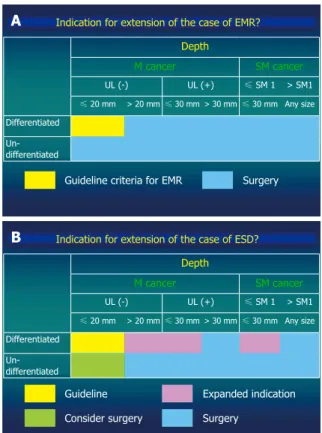
EGC patients who underwent gastrectomy with lymph node dissection. They provided important information on the risks of lymph node metastasis, wherein the dif-ferentiated gastric cancer with a nominal risk of lymph node metastasis was defined. They proposed expanded criteria for endoscopic resection: (1) mucosal cancer with-out ulcer findings, irrespective of tumor size; (2) mucosal cancer with an ulcer ≤ 3 cm in diameter; and (3) minimal (≤ 500 μm from the muscularis mucosa) submucosal invasive cancer ≤ 3 cm in size (Figure 1)[8,9]. However, extending the indications for ESD remains controversial because the long-term outcomes of these procedures have not been fully documented.
ReCeNT eVIDeNCe fOR expANDeD
INDICATIONs Of esD fOR eGC
Over the past 20 years, intraluminal endoscopic surgery,
referred to as EMR, has been advanced in Japan and Ko-rea. Recently, EMR and ESD have been widely used to treat EGC without lymph node metastasis. EMR is indi-cated when the risk of lymph node metastasis is minimal and when the tumor can be removed en bloc with a loop snare. Therefore, in the guidelines issued by the Japanese Gastric Cancer Association (JGCA), differentiated mu-cosal cancers measuring less than 2 cm in diameter best fit the above criteria. However, endoscopic resection has also been used for larger lesions without lymph node me-tastasis. In addition, improved techniques featuring ESD, which involve incision of the mucosa around the lesion followed by direct dissection of the submucosal layer, can provide en bloc resection, regardless of the tumor size. Therefore, provided that there is no metastatic lymph node, and the cancer is confined to mucosa and upper submucosal layer, endoscopic resection featuring ESD can be advocated as a proper management strategy for EGC.
To establish a safe and confident criteria and indica-tion for ESD, we have to classify EGC with and without metastatic lymph nodes and to analyze long-term follow-up data after EMR and ESD. In a recent study of patients who had undergone radical gastrectomy for EGC, none of the 1230 well differentiated mucosa-confined cancers smaller than 3 cm in diameter had associated lymph node metastasis, regardless of the presence of ulceration[8].
Regarding the presence of ulceration in all EGC patients, the probability of lymph node involvement significantly
Depth M cancer SM cancer UL (-) UL (+) ≤ SM 1 > SM1 ≤ 20 mm > 20 mm ≤ 30 mm > 30 mm ≤ 30 mm Any size Differentiated Un-differentiated
Indication for extension of the case of EMR?
Guideline criteria for EMR Surgery A Depth M cancer SM cancer UL (-) UL (+) ≤ SM 1 > SM1 ≤ 20 mm > 20 mm ≤ 30 mm > 30 mm ≤ 30 mm Any size Differentiated Un-differentiated
Indication for extension of the case of ESD?
Guideline B
Consider surgery
Expanded indication Surgery
Figure 1 Guideline criteria for endoscopic mucosal resection (A) and ex-panded criteria for endoscopic submucosal dissection proposed by Gotoda
et al[8] and Soetikno et al[9] (B). EMR: Endoscopic mucosal resection; ESD:
increases in EGC containing an ulcer (3.4%) compared to EGC without an ulcer (0.5%)[8]. In subgroup analysis,
cancer that is confined to the mucosa without an ulcer has no lymph node involvement, regardless of size (95% CI, 0%-0.4%). Mucosal cancer with an ulcer showed a size limitation up to 30 mm to be free of lymph node involve-ment (95% CI, 0%-0.3%). Thus we can conclude that presence of an ulcer and size are factors for indication of ESD for EGC. However, such characterization of EGC based on morphological feature poses certain problems. First, we should consider the life cycle of a malignant ulcer[10]. About one-third of EGCs with depressed
mor-phology can change over time[11]. An EGC with ulceration
on initial EGD can change into non-ulcerative EGC with an antisecretory agent. In addition, EGCs with ulceration, which had not appeared ulcerous on previous examina-tion, are sometimes encountered. Nevertheless, there is no evidence in the literature regarding the outcome of ESD for EGC with healing ulcers and fibrotic scarring. In practice, the decision of ESD for EGC with a shallow ulcer often depends on the timing of the diagnosis and on the willingness of the operator. Regarding the division of ulcerative and non-ulcerative as a criterion for ESD[8],
there has not been sufficient research to allow a definite decision. Second, there can be inter-observer variation in defining an ulcer in EGC. By definition, ulcers measure 5 mm or larger in diameter and are on exposed submu-cosa. However, in real endoscopic examination, the dif-ferentiation between ulcer and erosion is not always clear. Third, the size of a lesion can be different according to the method of measurement. An endoscopic ruler or a standard size disc patch can be applicable but, in most cases, a lesion is measured by eye in comparing to opened grasp of forceps. Thus, a standard reliable measurement method is required.
Another factor for expanded indication of ESD con-cerns the invasion depth of EGC. Although the absolute indication for EMR is applicable to only mucosal cancer, there have been some studies of ESD in submucosal can-cer. Of the 145 well-differentiated tumors that had invad-ed less than 500 μm into the submucosa, and were smaller than 30 mm in diameter, none showed evidence of lymph node metastasis, provided that there was no lymphatic or venous invasion. Based on these findings, it was suggested that the criteria for ESD for EGC could be expanded[12-16].
From the surgical literature, the risk of lymph node metastasis appears to depend on the presence of ulcer rather than on the depth of invasion of EGC. A retro-spective analysis of patients who underwent surgery for EGC in Korea reported that, among 129 cases of mu-cosal cancer compatible with expanded indications for EMR or ESD, three patients (2.3%) had lymph node me-tastasis and, among 52 submucosal cancer cases that met the expanded indications for EMR or ESD, two patients (4%) had lymph node metastasis[17]. The authors suggest
that if EMR or ESD had been performed in these pa-tients, it would not have been curative. However, even in this report, differentiated mucosal cancers without ulcers did not have lymph node metastasis, irrespective of size.
Thus, these data suggest that a well-differentiated muco-sal cancer of any size without ulcer may be considered as an expanded indication for ESD.
Even in the expanded criteria, undifferentiated cancer an indication for surgery. However, studies on feasibility of ESD on undifferentiated EGC have been continuously performed and reported. Ye et al[18] reported that EGC with
undifferentiated histology has no lymph node involvement, provided that the cancer is smaller than 25 mm, is confined to the mucosa or upper third of the submucosa, and has no lymphatic involvement. A similar study for signet ring cell carcinoma was reported by Park et al[19]. EGC with signet
ring cell histology is a high risk for nodal and organ me-tastases, while smaller cancers of less than 25 mm that are confined to the SM2 layer and have no lymphatic-vascular involvement have no lymph node involvement. With regard to poorly differentiated EGC, a Korean study reported a somewhat lower risk of lymph node metastasis than ex-pected[20]. On this retrospective analysis of 234 patients with
poorly differentiated EGC who underwent radical gastrec-tomy with D2 lymph node dissection, half of the cases (n =
116) showed submucosal invasion in the resection specimen and 25.9% (30/116) of those were limited to the upper third (SM1). Lymph node metastasis was found in 3.4% (4/118)
of mucosa-confined cancer. For patients with minor sub-mucosal infiltration (SM1), the lymph node metastasis rate
was non-existent (0/30). However, with SM2/3 invasion,
the lymph node metastasis rate increased sharply to around 30%. Another Korean study[21] focusing on endoscopic
re-section for undifferentiated-type cancer, such as poorly dif-ferentiated adenocarcinoma and signet ring cell carcinoma, showed interesting results. In this study, 58 lesions with un-differentiated EGC (17 poorly un-differentiated; 41 signet-ring cell) were treated by endoscopic resection. The en bloc and complete resection rates in poorly differentiated cases were 82.4% and 58.8%, respectively, whereas those in signet ring cell were 85.4% and 70.7%. The recurrence rate was 5.1% in complete resection during the follow-up period. There-fore, the authors suggested that endoscopic resection might be a feasible local treatment for undifferentiated EGC if complete resection can be achieved. Although the studies so far are still insufficient to form a conclusion, we could take poorly differentiated cancer with mucosa or minimal sub-mucosal invasion into consideration as a possible candidate for ESD in high-risk surgical patients.
A prospective comparative study was reported in Ja-pan[6] concerning the clinical outcomes of absolute and
expanded indication of EMR and ESD. A total of 589 EGC lesions were divided into the guideline group and the expanded group. En bloc, complete, and curative resections were achieved in 98.6 and 93.0, 95.1 and 88.5, and 97.1 and 91.1% of the guideline and expanded criteria lesions, re-spectively, and the differences between the two groups were significant. The complication risks, such as procedure as-sociated perforation and bleeding, were significantly higher and the completeness of resection was statistically superior in the expanded indication group. However, the overall sur-vival was equally adequate in both groups, and the disease-specific survival rates were 100% in both groups.
lIMITATIONs Of esD
Lymph node micrometastasis and delayed cancer dissemi-nation after ESD is one of the concerns about endoscopic resection including ESD. Walter et al[22] reported a fulminant
case in a 67-year-old male patient with EGC of 12 mm and moderate differentiation. The depth of submucosal invasion was 2.3 mm and the cancer free margin could not be established. The patient underwent gastrectomy and the postoperative stage was pT1 (sm3), pNO (0/58), cM0, L0, V0, G2 (UICC stage Ia). Three months later, an ultrasound revealed a new mass in the liver, and biopsy showed a rapidly growing metastasis of the gastric adenocarcinoma. This case highlights the risk of affected lymph nodes in early gastric cancer and the consequent risk of metastasis, which increases with greater depth of infiltration into the submucosa. Micrometastasis can be a reason for cancer recurrence even after curative surgery[23-27]. According to
Cai et al[25], tumor size, macroscopic type, accompanying
ulcers, and depth of invasion are strongly associated with micrometastasis in lymph nodes. Therefore, tumors with suspected submucosal invasion, large size, accompanying ulcers, and undifferentiated histology might have a risk of recurrence owing to micrometastasis, which would be con-traindicated for EMR or ESD.
lONG-TeRM fOllOW Up DATA
Long-term follow up data are needed for the clinical ap-plication of the expanded criteria of ESD. One Japanese study[28] from the National Cancer Center Hospital
in-volving 1955 EGC patients enrolled from January 1999 to December 2005 showed that there were no significant differences in the overall five-year survival rates between the curative resection group, as defined by the expanded indication, and the non-curative resection group, following additional surgery. This data suggest that ESD using the ex-panded criteria can show an excellent long-term outcome.
CONClUsION
ESD makes it possible to perform complete resection for lesions larger than 20 mm, as well as those with ulcer-ation, regardless of location. Many clinical data suggest that ESD might be adequate for lesions that fit both the current guidelines and the expanded criteria. In the near future, when long-term follow-up data accumulate and newer technology is available, endoscopic resection, in-cluding ESD, will be employed in the treatment of EGC with more expanded indications. However, we must keep in mind that accurate diagnosis, characterization of the lesion, and proper appreciation of technical aspects are most essential in therapeutic endoscopy.
RefeReNCes
1 Carter KJ, Schaffer HA, Ritchie WP Jr. Early gastric cancer.
Ann Surg 1984; 199: 604-609
2 Everett SM, Axon AT. Early gastric cancer in Europe. Gut
1997; 41: 142-50
3 Ono H, Kondo H, Gotoda T, Shirao K, Yamaguchi H, Saito D,
Hosokawa K, Shimoda T, Yoshida S. Endoscopic mucosal resection for treatment of early gastric cancer. Gut 2001; 48: 225-229
4 Gotoda T, Kondo H, Ono H, Saito Y, Yamaguchi H, Saito D,
Yokota T. A new endoscopic mucosal resection procedure using an insulation-tipped electrosurgical knife for rectal flat lesions: report of two cases. Gastrointest Endosc 1999; 50: 560-563
5 Japanese Gastric Cancer Association. Japanese
Classifica-tion of Gastric Carcinoma - 2nd English EdiClassifica-tion. Gastric
Cancer 1998; 1: 10-24
6 Yamaguchi N, Isomoto H, Fukuda E, Ikeda K, Nishiyama
H, Akiyama M, Ozawa E, Ohnita K, Hayashi T, Nakao K, Kohno S, Shikuwa S. Clinical outcomes of endoscopic sub-mucosal dissection for early gastric cancer by indication criteria. Digestion 2009; 80: 173-181
7 Gotoda T. Endoscopic resection for premalignant and
malig-nant lesions of the gastrointestinal tract from the esophagus to the colon. Gastrointest Endosc Clin N Am 2008; 18: 435-450, viii
8 Gotoda T, Yanagisawa A, Sasako M, Ono H, Nakanishi Y,
Shimoda T, Kato Y. Incidence of lymph node metastasis from early gastric cancer: estimation with a large number of cases at two large centers. Gastric Cancer 2000; 3: 219-225
9 Soetikno R, Kaltenbach T, Yeh R, Gotoda T. Endoscopic
mucosal resection for early cancers of the upper gastrointes-tinal tract. J Clin Oncol 2005; 23: 4490-4498
10 Sakita T, Oguro Y, Takasu S, Fukutomi H, Miwa T. Obser-vations on the healing of ulcerations in early gastric cancer. The life cycle of the malignant ulcer. Gastroenterology 1971;
60: 835-839 passim
11 Im JP, Kim SG, Kim JS, Jung HC, Song IS. Time-dependent morphologic change in depressed-type early gastric cancer.
Surg Endosc 2009; 23: 2509-2514
12 Yamao T, Shirao K, Ono H, Kondo H, Saito D, Yamaguchi H, Sasako M, Sano T, Ochiai A, Yoshida S. Risk factors for lymph node metastasis from intramucosal gastric carci-noma. Cancer 1996; 77: 602-606
13 Yasuda K, Shiraishi N, Suematsu T, Yamaguchi K, Adachi Y, Kitano S. Rate of detection of lymph node metastasis is cor-related with the depth of submucosal invasion in early stage gastric carcinoma. Cancer 1999; 85: 2119-2123
14 Gotoda T, Sasako M, Ono H, Katai H, Sano T, Shimoda T. Evaluation of the necessity for gastrectomy with lymph node dissection for patients with submucosal invasive gas-tric cancer. Br J Surg 2001; 88: 444-449
15 Kim JJ, Lee JH, Jung HY, Lee GH, Cho JY, Ryu CB, Chun HJ, Park JJ, Lee WS, Kim HS, Chung MG, Moon JS, Choi SR, Song GA, Jeong HY, Jee SR, Seol SY, Yoon YB. EMR for early gastric cancer in Korea: a multicenter retrospective study. Gastrointest Endosc 2007; 66: 693-700
16 Oizumi H, Matsuda T, Fukase K, Furukawa A, Mito S, Taka-hashi K. Endoscopic resection for early gastric cancer: the ac-crual procedure and clinical evaluation. Stomach and Intestine 1991; 26: 289-300
17 Jee YS, Hwang SH, Rao J, Park DJ, Kim HH, Lee HJ, Yang HK, Lee KU. Safety of extended endoscopic mucosal resec-tion and endoscopic submucosal dissecresec-tion following the Japanese Gastric Cancer Association treatment guidelines.
Br J Surg 2009; 96: 1157-1161
18 Ye BD, Kim SG, Lee JY, Kim JS, Yang HK, Kim WH, Jung HC, Lee KU, Song IS. Predictive factors for lymph node me-tastasis and endoscopic treatment strategies for undifferenti-ated early gastric cancer. J Gastroenterol Hepatol 2008; 23: 46-50 19 Park JM, Kim SW, Nam KW, Cho YK, Lee IS, Choi MG,
Chung IS, Song KY, Park CH, Jung CK. Is it reasonable to treat early gastric cancer with signet ring cell histology by en-doscopic resection? Analysis of factors related to lymph-node metastasis. Eur J Gastroenterol Hepatol 2009; 21: 1132-1135 20 Park YD, Chung YJ, Chung HY, Yu W, Bae HI, Jeon SW,
Cho CM, Tak WY, Kweon YO. Factors related to lymph node metastasis and the feasibility of endoscopic mucosal resec-tion for treating poorly differentiated adenocarcinoma of the stomach. Endoscopy 2008; 40: 7-10
21 Kim JH, Lee YC, Kim H, Song KH, Lee SK, Cheon JH, Kim H, Hyung WJ, Noh SH, Kim CB, Chung JB. Endoscopic re-section for undifferentiated early gastric cancer. Gastrointest
Endosc 2009; 69: e1-e9
22 Walter B, Probst A, Märkl B, Wagner T, Anthuber M, Mess-mann H. Fulminant metastatic spread in a patient with an early gastric cancer. Endoscopy 2009; 41: 907-909
23 Maehara Y, Oshiro T, Endo K, Baba H, Oda S, Ichiyoshi Y, Kohnoe S, Sugimachi K. Clinical significance of occult mi-crometastasis lymph nodes from patients with early gastric cancer who died of recurrence. Surgery 1996; 119: 397-402 24 Nakajo A, Natsugoe S, Ishigami S, Matsumoto M,
Nakashi-ma S, Hokita S, Baba M, Takao S, Aikou T. Detection and prediction of micrometastasis in the lymph nodes of patients with pN0 gastric cancer. Ann Surg Oncol 2001; 8: 158-162 25 Cai J, Ikeguchi M, Tsujitani S, Maeta M, Kaibara N.
Micro-metastasis in lymph nodes of mucosal gastric cancer. Gastric
Cancer 2000; 3: 91-96
26 Cai J, Ikeguchi M, Tsujitani S, Maeta M, Liu J, Kaibara N. Significant correlation between micrometastasis in the lymph nodes and reduced expression of E-cadherin in early gastric cancer. Gastric Cancer 2001; 4: 66-74
27 Harrison LE, Choe JK, Goldstein M, Meridian A, Kim SH, Clarke K. Prognostic significance of immunohistochemical micrometastases in node negative gastric cancer patients. J
Surg Oncol 2000; 73: 153-157
28 Kusano C, Gotoda T, Iwasaki M. Long-term outcome of ESD for early gastric cancer. Stom Intes 2008; 43: 73-79