Effects of vimentin protein transduction on
biological function of vascular endothelial cells
Eunsuk Kim
The Graduate School
Yonsei University
Graduate Program in Science for Aging
Molecular Cell Biology
Effects of vimentin protein transduction on
biological function of vascular endothelial cells
A Master’s Thesis Submitted to the Department
of Graduate Program in Science For Aging and
the Graduate School of Yonsei University in
partial fulfillment of the requirements for the
degree of Master in Science for Aging
Eunsuk Kim
This certifies that the master’s thesis of
Eunsuk Kim is approved.
Thesis Supervisor: Yangsoo Jang
Thesis Committee Member: Seok-Min Kang
Thesis Committee Member: Ji Hyung Chung
The Graduate School
Yonsei University
감사의 글
대학졸업을 앞두고 처음 실험실에 들어섰던 설렘을 아직도 잊을 수 가 없습니다. 대학원 석사 과정을 통해서, 작은 것에도 호기심을 갖고 탐구할 수 있는 넓은 시야와 사고를 기를 수 있게 된 것 같아 마음 한 켠이 든든하면서도 아쉽습니다. 지난 학위 과정을 돌이켜보면 많 은 부분 도움을 받고 의지해온 분들이 너무나 많습니다. 넓은 아량과 에너지를 가지고 계신 장양수 교수님, 탁월한 관점으로 현상을 분석하시는 강석민 교수님, 그리고 무궁무진한 아이디어를 가 지고 문제 해결을 위해 스스로 깨우치는 것이 무엇인지 방향을 제시 해 주시는 정지형 교수님. 우리에게는 아버지와 같은 존재이십니다. 실험실 식구들의 작은 것 하나하나에도 관심을 가져주시고 인간적으 로, 학문적으로 많은 감동을 받았습니다. 존경합니다. 석사 과정 동안 가족들보다 더 많은 시간을 함께 보내온 실험실 식 구들을 떠나려니 더 많이 베풀지 못한 것 같아 미안함과 아쉬움이 크 게 남습니다. 우선, 전반적인 실험의 흐름을 잡아주시고 포인트를 찾 아 공부하는 방법을 익힐 수 있게 지도해주시던 이경혜 선생님. 지금은 함께 있지 않지만 언제까지나 보고 싶습니다. 실험실의 분위기를 유쾌하게 이끌어가면서도 잘못된 부분은 유들 있게 집어주시는 김수 혁 선배님, 처음 석사 학위를 시작할 때, 조목조목 자세한 실험설명과 긍정적이고 유머러스한 선배님 덕분에 도움을 많이 받았습니다. 언제 나 에너지 넘치지만 반면에 섬세하고 여린 진태원 선배님, 굳은 일은 먼저 나서서 해결해주시는 진태원 선배님을 보고 적극성과 활력을 배 웠습니다. 친언니처럼 가깝게 의지했던 한 때 룸메이트 은영 언니 덕 분에 동생이자 후배이지만 늘 편하게 대해주고 코드도 잘 맞아 든든 한 대학원 생활을 보냈습니다. 활기차고 붙임성 좋은 착한 현주, 긍정 적이고 유쾌한 현주 덕분에 같은 공간에서 생활하지는 못했지만 그리 운 가족과 같은 느낌으로 언제나 기분 좋은 에너지를 많이 받았습니 다. 그리고 학부생활도 함께 한 수진언니. 여성스럽고 여리지만 한 편 으론 당차고 씩씩한 언니 모습에 많은 것을 배웠습니다. 실험실 식구들 이외에도, 늘 마음의 위안이 되는 친구들 선미, 윤정 이, 주애. 내 생활의 활력소가 되는 비타민 같은 친구들 혜원이(와 정 현님), 서영이, 혜림이. 대학원 생활 동안 바쁘다는 핑계로 자주 약속 을 미뤘지만 웃으며 이해해주고 언제나 먼저 연락해주는 미안한 친구 들 지영이, 아름이, 현정이. 그리고 항상 내 편이 되어 내 말에 귀 기
울여주는 에너지 같은 존재 든든하고 착한 병성이. 다들 너무 사랑합 니다. 무엇보다도 묵묵하게 앞을 보고 달려갈 수 있게 딸들을 적극적 이게 응원해 주시고 믿어주시는 부모님과 나와 외모도 성격도 매우 닮은 동생 현주에게 감사하고 사랑한다고 전하고 싶습니다. 2011.12.17 김은숙 드림
TABLE OF CONTENTS
ABSTRACT ∙∙∙∙∙∙∙∙∙∙∙∙∙∙∙∙∙∙∙∙∙∙∙∙∙∙∙∙∙∙∙∙∙∙∙∙∙∙∙∙∙∙∙∙∙∙∙∙∙∙∙∙∙∙∙∙∙∙∙∙∙∙∙∙∙∙∙∙∙∙∙∙∙∙∙∙∙∙∙∙∙∙∙
1I. INTRODUCTION ∙∙∙∙∙∙∙∙∙∙∙∙∙∙∙∙∙∙∙∙∙∙∙∙∙∙∙∙∙∙∙∙∙∙∙∙∙∙∙∙∙∙∙∙∙∙∙∙∙∙∙∙∙∙∙∙∙∙∙∙∙∙∙∙∙∙∙∙∙
4II. MATERIALS AND METHODS ∙∙∙∙∙∙∙∙∙∙∙∙∙∙∙∙∙∙∙∙∙∙∙∙∙∙∙∙∙∙∙∙∙∙∙∙∙∙∙∙∙∙∙
12 1. Materials ∙∙∙∙∙∙∙∙∙∙∙∙∙∙∙∙∙∙∙∙∙∙∙∙∙∙∙∙∙∙∙∙∙∙∙∙∙∙∙∙∙∙∙∙∙∙∙∙∙∙∙∙∙∙∙∙∙∙∙∙∙∙∙∙∙∙∙∙∙∙∙∙∙∙∙∙∙∙∙∙∙∙∙∙∙∙∙ 12 2. Molecular cloning of human vimentin gene ∙∙∙∙∙∙∙∙∙∙∙∙∙∙∙∙∙∙∙∙∙∙∙∙∙∙∙∙∙∙∙∙∙∙∙∙∙∙∙∙∙ 13 3. Expression and purification of pHis/TAT-vimentin fusion protein ∙∙∙∙∙∙∙∙∙∙∙∙ 13 4. Cell culture ∙∙∙∙∙∙∙∙∙∙∙∙∙∙∙∙∙∙∙∙∙∙∙∙∙∙∙∙∙∙∙∙∙∙∙∙∙∙∙∙∙∙∙∙∙∙∙∙∙∙∙∙∙∙∙∙∙∙∙∙∙∙∙∙∙∙∙∙∙∙∙∙∙∙∙∙∙∙∙∙∙∙∙∙ 14 5. Transduction of pHis/TAT-vimentin fusion protein into cells∙∙∙∙∙∙∙∙∙∙∙∙∙∙∙∙∙∙∙∙ 15 6. Immunoblot analysis ∙∙∙∙∙∙∙∙∙∙∙∙∙∙∙∙∙∙∙∙∙∙∙∙∙∙∙∙∙∙∙∙∙∙∙∙∙∙∙∙∙∙∙∙∙∙∙∙∙∙∙∙∙∙∙∙∙∙∙∙∙∙∙∙∙∙∙∙∙∙∙∙ 15 7. Nitric oxide detection assay ∙∙∙∙∙∙∙∙∙∙∙∙∙∙∙∙∙∙∙∙∙∙∙∙∙∙∙∙∙∙∙∙∙∙∙∙∙∙∙∙∙∙∙∙∙∙∙∙∙∙∙∙∙∙∙∙∙∙∙∙∙∙ 16 8. Tube formation assay ∙∙∙∙∙∙∙∙∙∙∙∙∙∙∙∙∙∙∙∙∙∙∙∙∙∙∙∙∙∙∙∙∙∙∙∙∙∙∙∙∙∙∙∙∙∙∙∙∙∙∙∙∙∙∙∙∙∙∙∙∙∙∙∙∙∙∙∙∙∙∙ 17III. RESULTS
1. Construction and purification of pHis/TAT-vimentin fusion protein ∙∙∙∙∙∙∙∙∙∙ 18
3. Effect of vimentin protein transduction on the tube formation
∙∙∙∙∙∙∙∙∙∙∙∙∙∙∙∙∙ 22
4. Vimentin protein transduction increases VASP phosphorylation ∙∙∙∙∙∙∙∙∙∙∙∙∙∙ 24 5. Effect of PKA inhibitor on vimentin-induced tube formation ∙∙∙∙∙∙∙∙∙∙∙∙∙∙∙∙∙∙ 26 6. Effect of PKA inhibitor on vimentin-induced VASP phosphorylation ∙∙∙∙∙∙∙∙ 28 7. Effect of vimentin protein transduction on adenylate cyclase mediated-PKA activation ∙∙∙∙∙∙∙∙∙∙∙∙∙∙∙∙∙∙∙∙∙∙∙∙∙∙∙∙∙∙∙∙∙∙∙∙∙∙∙∙∙∙∙∙∙∙∙∙∙∙∙∙∙∙∙∙∙∙∙∙∙∙∙∙∙∙∙∙∙∙∙∙∙∙∙∙∙∙∙∙∙∙∙∙∙∙∙ 30 8. Effect of vimentin protein transduction on nitric oxide production ∙∙∙∙∙∙∙∙∙∙∙ 32 9. Expression of junctional protein by vimentin transduction ∙∙∙∙∙∙∙∙∙∙∙∙∙∙∙∙∙∙∙∙∙ 34IV. DISCUSSION ∙∙∙∙∙∙∙∙∙∙∙∙∙∙∙∙∙∙∙∙∙∙∙∙∙∙∙∙∙∙∙∙∙∙∙∙∙∙∙∙∙∙∙∙∙∙∙∙∙∙∙∙∙∙∙∙∙∙∙∙∙∙∙∙∙∙∙∙∙∙∙∙
36V. REFERENCES ∙∙∙∙∙∙∙∙∙∙∙∙∙∙∙∙∙∙∙∙∙∙∙∙∙∙∙∙∙∙∙∙∙∙∙∙∙∙∙∙∙∙∙∙∙∙∙∙∙∙∙∙∙∙∙∙∙∙∙∙∙∙∙∙∙∙∙∙∙∙∙
44LIST OF FIGURES
Fig. 1. The structure of vimentin protein
∙∙∙∙∙∙∙∙∙∙∙∙∙∙∙∙∙∙∙∙∙∙∙∙∙∙∙∙∙∙∙∙∙∙∙∙∙∙∙∙∙∙∙∙∙∙∙ 8
Fig. 2. Expression and purification of pHis/TAT-vimentin protein∙∙∙∙∙∙∙∙∙∙ 19
Fig. 3. Vimentin protein transduction into endothelial cells∙∙∙∙∙∙∙∙∙∙∙∙∙∙∙∙∙∙∙ 21
Fig. 4. Vimentin protein transduction promotes tube formation ofendothelial cells
∙∙∙∙∙∙∙∙∙∙∙∙∙∙∙∙∙∙∙∙∙∙∙∙∙∙∙∙∙∙∙∙∙∙∙∙∙∙∙∙∙∙∙∙∙∙∙∙∙∙∙∙∙∙∙∙∙∙∙∙∙∙∙∙∙∙∙∙∙∙∙∙∙∙∙∙∙ 23
Fig. 5. VASP (Ser157) is phosphorylated by vimentin protein transduction∙∙∙∙∙∙∙∙∙∙∙∙∙∙∙∙∙∙∙∙∙∙∙∙∙∙∙∙∙∙∙∙∙∙∙∙∙∙∙∙∙∙∙∙∙∙∙∙∙∙∙∙∙∙∙∙∙∙∙∙∙∙∙∙∙∙∙∙∙∙∙∙∙∙∙∙∙∙∙∙∙∙∙∙∙∙∙∙∙∙∙∙∙∙∙∙∙∙∙∙∙ 25
Fig. 6. PKA inhibitor suppresses vimentin-induced endothelial cell tube formation
∙∙∙∙∙∙∙∙∙∙∙∙∙∙∙∙∙∙∙∙∙∙∙∙∙∙∙∙∙∙∙∙∙∙∙∙∙∙∙∙∙∙∙∙∙∙∙∙∙∙∙∙∙∙∙∙∙∙∙∙∙∙∙∙∙∙∙∙∙∙∙∙∙∙∙∙∙∙∙∙∙∙∙∙∙∙ 27
Fig. 7. Effect of PKA inhibitor on vimentin-induced VASP phosphorylation∙∙∙∙∙∙∙∙∙∙∙∙∙∙∙∙∙∙∙∙∙∙∙∙∙∙∙∙∙∙∙∙∙∙∙∙∙∙∙∙∙∙∙∙∙∙∙∙∙∙∙∙∙∙∙∙∙∙∙∙∙∙∙∙∙∙∙∙∙∙∙∙∙∙∙∙∙∙∙∙∙∙∙∙∙∙∙∙∙∙∙∙∙∙∙∙∙∙∙∙∙ 29
Fig. 8. Effect of vimentin protein transduction on adenylate cyclase mediated-PKA activation
∙∙∙∙∙∙∙∙∙∙∙∙∙∙∙∙∙∙∙∙∙∙∙∙∙∙∙∙∙∙∙∙∙∙∙∙∙∙∙∙∙∙∙∙∙∙∙∙∙∙∙∙∙∙∙∙∙∙∙∙∙∙∙∙31
Fig. 9. The effect of transduced pHis/TAT-vimentin fusion on NOproduction
∙∙∙∙∙∙∙∙∙∙∙∙∙∙∙∙∙∙∙∙∙∙∙∙∙∙∙∙∙∙∙∙∙∙∙∙∙∙∙∙∙∙∙∙∙∙∙∙∙∙∙∙∙∙∙∙∙∙∙∙∙∙∙∙∙∙∙∙∙∙∙∙∙∙∙∙∙∙∙∙∙∙∙∙∙ 33
Fig. 10. pHis/TAT-vimentin induced ZO-1 expression was decreased byH-89
∙∙∙∙∙∙∙∙∙∙∙∙∙∙∙∙∙∙∙∙∙∙∙∙∙∙∙∙∙∙∙∙∙∙∙∙∙∙∙∙∙∙∙∙∙∙∙∙∙∙∙∙∙∙∙∙∙∙∙∙∙∙∙∙∙∙∙∙∙∙∙∙∙∙∙∙∙∙∙∙∙∙∙∙∙∙∙∙∙∙∙∙∙∙∙∙∙∙ 35
Fig. 11. The structure of vasodilator-stimulated phosphoprotein∙∙∙∙∙∙∙∙∙∙∙∙∙∙38
Fig. 12. The signal of vimentin-induced tube formation∙∙∙∙∙∙∙∙∙∙∙∙∙∙∙∙∙∙∙∙∙∙∙∙42
1
ABSTRACT
Effects of vimentin protein transduction
on biological function of vascular endothelial cells
Eunsuk Kim
Graduate program in Science for Aging The Graduate School, Yonsei University
(Directed by Professor Yangsoo Jang)
The cytoskeleton plays a central role for the integration of biochemical and biomechanical signals across the cell required for complex cellular function. Recent studies indicate that the intermediate filament vimentin is necessary for
2
endothelial function. Vimentin is one of the intermediate filaments that had been considered to be related with cellular integrity and endothelial functions such as cell-cell adhesion, migration and tube formation. It was studied the potential endothelial function of vimentin as a therapeutic protein in HUVECs and its pathways during pHis/TAT-vimentin transduction, using protein delivery system. The results indicated that pHis/TAT-vimentin was effectively transduced in HUVECs and had strong angiogenesis effects though tube formation assay. And it was shown that pHis/TAT-vimentin induced endothelial function is tightly linked to phosphorylation of VASP and activation of protein kinase A (PKA) during tube formation. It acts as stability effects of the cytoskeleton and influences tube formation of endothelial cells.
Previous studies found that vasodilator-stimulated phophoprotein (VASP), actin binding protein, has multiple serind/theronine phosphorylation sites. VASP localizes to endothelial junction complexes and co-localizes with ZO-1. To address the role of phospho-VASP in endothelial function, A phospho-specific VASP antibodies targeting phosphorylation site (Ser 157) were used. The site was preferred by PKA. Transduction of pHisTAT-vimentin induced VASP phosphorylation was attenuated by PKA inhibitors in HUVECs. Overall, pHisTAT-vimentin had strong angiogenesis effects and these effects were inhibited by blocking PKA.
3
Key Words: Vimentin, angiogenesis, Protein transduction domain, vasodilator-stimulated phosphoprotein, protein kinase A.
4
Effects of vimentin protein transduction on biological function
of vascular endothelial cells
Eunsuk Kim
Graduate Program in Science for Aging
The Graduate School, Yonsei University
(Directed by Professor Yangsoo Jang
)I. INTRODUCTION
Endothelial cells play a wide variety of critical roles related with vascular function. The functional properties of the endothelial cells shifted their role from a passive membrane or barrier to a complex tissue with complex functions. Hence, it participates to all aspects of the vascular homeostasis also to physiological or pathological processes like thrombosis, inflammation, or vascular wall remodeling. Endothelial cells participate in role in blood vessel
5
formation, in coagulation in the regulation of vascular tone and in inflammatory reactions and in tumor neoangiogenesis
Because of these characters, endothelial cells are suitable for a diverse range of physiological processes, such as angiogenesis and as the selective blood barrier; and pathophysiological processes, including arterial disease and cancer development. Among endothelial cells, Human Umbilical Vein Endothelial Cells (HUVEC) were used in this study. It was isolated from normal human umbilical vein. They are responsive to cytokine stimulation in the expression of cell adhesion molecules. These cell systems are commonly used for physiological and pharmacological investigations, such as macromolecule transport, blood coagulation, and fibrinolysis.
Many studies make an effort to find angiogenesis pathway to related to regenerate endothelial cells and protects an inner blood vessels. The growth of new capillary blood vessels in the body is an important natural process in vascular healing. It helps regeneration of damaged or lost tissues.4, 5 The somatic maintenance system administrate the balance of angiogenesis modulators. The body controls angiogenesis by producing proper growth and inhibitory factors. In angiogenesis, new blood vessels are formed by vascular endothelial cells. These neo-vessels supply oxygen and nutrients to the ischemic wound region. Fibroblasts grow and form transiently a new extracellular matrix (ECM) during
6
tissue formation. Also, collagen and fibronectin, which are the most critical proteins for the restoration of injured tissue was secreted in angiogenesis.6, 7
Because of these, Angiogenesis-dependent diseases occur when new blood vessels either grow excessively or insufficiently.
Recent studies demonstrate that intermediate filament (IF) proteins play a role in angiogenesis. In many cells and tissue, IF proteins are essential for the integrity of cellular architecture and are protective against mechanical and other kinds of stress. Until recently, intermediate filaments had been considered to be relatively stable. It was proposed solely to provide cellular integrity and resistance against mechanical stresses. The IF proteins consist of greater than 50 members, constituting one of the larger gene families in the human genome. They all share a common predicted domain structure consisting of a central rod domain flanked by head and tail domains
Vimentin is one of the most established actins among the large IF protein family. It is the major IF protein in various cells and frequently used as a developmental marker of cells and tissue. It is a type Ⅲ intermediate filament protein that is expressed frequently in various cells. Vimentin is an important structural feature of endothelial cells. It makes up the cytoskeleton and exists as a dynamic structure. It consists of head, tail and central domain. Central domain and N-terminal domain (Head) plays an important role in intermediate filament
7
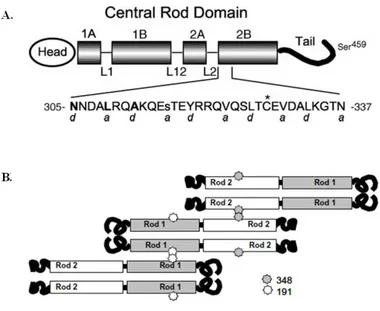
formation and stabilization. The role of the C- terminal domain (Tail) of vimentin is not pronounced. However, the removal of the C-terminal domain alters control of the assembling filament thickness. A structure of the vimentin monomer is presented in Fig. 1A. The amino acid sequence at this site is presented. The position of each amino acid in the heptad repeat pattern is indicated by notations of “a-g". This heptad repeat is thought to impose a coiled-coil assembly of polypeptide chains. It is studied that spin labels along the exterior surface of a dimer, and established in intact filaments that two dimers overlap at residue 348 in rod domain 2 (Fig. 1B). The overlap of two dimers at residue 348 in rod 2 presages an interaction between the free rod 1 domains. It was shown for such an overlap by introducing spin labels along the exterior surface of the dimer in the rod 1 region the broadening centered near position 191 indicates an interaction between dimers or higher level oligomers.
8
Fig. 1. The structure of vimentin protein. (A) The predicted vimentin molecule is shown diagrammatically, with the central rod domain emphasized. alpha-helical rod subdomains 1A, 1B, 2A, and 2B are shown. Hypothesized non-helical linker regions L1, L12, and L2 are drawn as thin lines. The region of rod subdomain 2B is expanded, and the sequence of this region is shown in single-letter amino acid abbreviations. Positions 305, 309, and 312 are bold; position 316 is lowercase. An asterisk marks the site of the single endogenous cysteine. Letters a and d below the amino acid sequence represent positions within individual heptad repeats in this area. The location of Ser459 is abstractly indicated near the end of the vimentin tail. (B) vimentin dimer interacts with rod domain 2 of one dimer (position 348), and rod domain 1 of another dimer (position 191). Both interactions are anti-parallel.
9
Vimentin has key roles in adhesion by regulating integrin functions. Integrins are heterodimer transmembrane cell adhesion receptors that are expressed in all metazoan cell-types, except erythrocytes, and the number of integrins expressed increases with increased complexity of the organism. Its filaments are required for endolysosomal vesicle transport and positioning of endosomes and lysosomes. The vimentin is the major IF protein present in leukocytes. Regarding the role of the vimentin in the transmigration on the endothelial side, it has been suggested that an ICAM-1 and VCAM-1-based “transmigratory cup” would be involved in leukocyte diapedesis through individual vascular endothelial cells. Most likely vimentin-deficient mice and/or phosphomimetic vimentin mutant cells and mice will help dissect the roles of the still poorly characterized transmigratory extravasation route. The protein can also affect the operation of protein complexes on the cell membrane by regulating cytosolic components in a fashion resembling the keratin-mediated modulation of TNFreceptor signaling by sequestering TNF receptor-associated death domain protein (TRADD). The vimentin protein has been demonstrated to function as a potential regulator of transcription, as it is able to interact and sequester transcriptional determinants such as p53. The ability complex with p53 represents another paradigm on the participation of vimentin in regulation of cell death and survival through protein interactions.
10
Many kinases are recognized as substrate for vimentin, including protein kinase C, A, G (PKC, PKA, PKG), cGMP kinase, RhoA kinase and PAK and so on.8 The cores of focal adhesions consist of integrin receptors, hetero-dimers of alpha- and beta-submits which are intracellular surrounded by many adapter proteins as VASP.16, 17, 27 Vasodilator stimulating phosphoprotein (VASP) is focal adhesion proteins that transmit mechanically induced alterations in the intermediate filament network into biochemical information.
In the recent years, people found a kind of protein functional domain, they can guide the bio-macromolecule to permeate the plasma membrane and accumulate in the cells, so they were called protein transduction domain (PTD). A new powerful protein drug as PTD-fusion protein using the PTD system is used. Protein drugs are expected as effective and strong drug, because they can affect specific targets directly, reduce the period of drug development, and have high safety better then drugs using target gene or chemical substances. PTD-fusion proteins entering into cells occurred quickly and affected target protein properties. The molecules carried into the cells hold their native activity. They can do their native work. So PTD is very charming because the character. It is possible for the macromolecule protein or polypeptide to permeate cytoplasmic membrane because of the PTD. PTD accelerates the protein engineering and the exploitation of protein medicine. At present, many kinds of protein have been
11
carried into the different cells or the animals, they hold their native activity. PTD will be applicator to find new therapy method and new function of protein. This study was used to the PTD system for protein delivery. Protein transduction domain (PTD) in a novel therapeutic perspective has been introduced to deliver target protein into cells directly. One of PTDs is the human immunodeficiency virus (HIV-1) transactivated TAT protein. By using TAT protein, numerous target proteins can be delivered into cells. The interaction of PTD with cell surface causes the internalization of TAT-fusion proteins.
As described above, it was established that endogenous vimentin considerably influences migration and proliferation in cells. However, effects on tube formation by endogenous vimentin was unsatisfied. It seems that transduced external vimentin protein support form new blood vessel.
The studies of tube formation functions have not been adequate. Therefore the PTD-fusion vimentin protein is transduced as the possible regulator in tube formation in this study.
12
Ⅱ. Material and Method
1. Materials
Vimentin and ZO-1 antibodies were obtained from abcam (Cambridge, MA, USA). Phospho-VASP (Ser157) was purchased from Cell Signaling Technology (Beverly, MA, USA). Anti-phospho-VASP (Ser239) was obtained from Calbiochem (San diego, CA, USA). KT5823 (PKG inhibitor) was purchased from Tocris Bioscience (Cookson Inc., USA), and H-89 (PKA inhibitor) were obtained from Santa Cruz Biotechnology (Santa Cruz, CA, USA). Nitric oxide Detection Kit obtains from intronbio biotechnology. (Seoul, Korea). Anti-b-actin antibody was purchased from Sigma-Aldrich (St. Louis, MO, USA). In Vitro Angiogenesis Assay Kit was purchased from CHEMICON. (Termecula, CA, USA). Horseradise peroxidase-conjugated secondary antibodies and enhanced cheminescense (ECL) Western blotting detection system were obtained from Santa Cruz Biotechnology (Santa Cruz, CA, USA).
13
2. Molecular cloning of human vimentin gene
To construct pHis/TAT-vimentin fusion protein, the sense primer was 5'- ATC TAC GGA TCC ACC AGG TCC GTG TCC TCG- 3' and the antisense primer was 5'- AGT GTG GTC GAC TTA TTC AAG GTC ATC GTG -3'. The PCR product was restricted with BamHI and SalI, and then subcloned into the BamHI and SalI sites of the pHis/TAT bacterial expression vector. The correct sequence of the pHis/TAT vector was confirmed by DNA sequencing using the universal T7 primer.
3. Expression and purification of pHis/TAT-vimentin fusion protein
Escherochia coli BL21 pLysS (Novagen, Madison, WI, USA) was transformed to the pHis/TAT-vimentin plasmid, and then grown for 24 h at 37 °C in LB broth supplied with 100 μg/ml ampicillin and 34 μg/ml chloramphenicol and while shaking at 200 rpm. Protein expression was induced by the addition of 1 mM β-D-1-thiogalactopyranoside (IPTG) for 4 h while shaking at 37 °C. The pHis/TAT-vimentin fusion protein were then isolated using a urea-denaturing protein purification protocol. The bacterial pallet was isolated by centrifugation at 3000 rpm, resuspended in buffer Z (8 M urea, 100 nM NaCl and 20 mM
14
HEPES, pH 8.0) , and sonicated 6 times with 15 second pulse continually adding 1 mM phenylmethanesulphonylfluoride (PMSF). The sample was then clarified by centrifugation at 14,000 rpm 4 °C for 1 h. The clarified lysate was loaded to Ni-NTA, Ni-IDA and Ni-TED column at 1 ml/min and then the column was washed using buffer A (washing buffer) (50 mM NaH2PO4, 300 mM NaCl) for 1 h at 1 ml/min. The elute vimentin fusion protein, the column was loaded by using buffer B (elution buffer) contained increasing concentrations of imidazole (10-500 mM) at 1 ml/min. Concentration of the protein was quantified by the Bradford assay. (BioRad, Hercules, CA, USA), using bovine serum albumin (BSA) as the standard. The purity of the fusion protein was assessed by Coomassie Brilliant blue staining. The purified fusion proteins were dissolved in PBS 10 % glycerol were aliquot and stored at -80 °C.
4. Cell culture
Primary HUVEC cells (Cambrex Walkersville, MD, USA ) were cultured in endothelial cell growth medium (EGM-2; Lonza, MD, USA) containing 2 % fetal bovine serum (FBS), 0. 4% hydrocortisone, 4 % hFGF-B, 0.1 & VEGF, 0.1 % R3-IGF, 0.1 % ascorbic acid, 0.1 % hEGF, 0.1 % GA-1000 and 0.1 % heparin, according to the manufacturer's instruction. Cells were grown in
15
atmosphere of 95 % air, 5 % CO2 at 37 °C. Sub-cultured HUVECs from passage 2 to 10 were used in these experiments.
5. Transduction of pHis/TAT-vimentin fusion protein into cells.
To transduce pHis/TAT-vimentin fusion protein, cells were grown to confluence in 60 mm dish or rotated in 1.5 ml tube. The culture was replaced with fresh containing 0.04 % FBS and supplement, then treated with various concentrations of pHis/TAT-vimentin fusion protein. The cells were washed by using PBS for 2 times while centrifuged. Cells were prepared for analysis by immunoblot.
6. Immunoblot analysis
At the termination of culture, the lysate were scraped into microcentrifuge tubes and centrifuged at 14,000 rpm for 15 min at 4 °C. The supernatant were washing 2 times with PBS. And then, harvested cells were solubilized in cell lysis buffer containing 1 M HEPES (pH 7.5), 5 M NaCl, 0.5 M EDTA, Triton-X 100, protease inhibitor cocktail 1ea (Roche). Protein concentration was measured using a BSA. The same amounts of proteins from whole cell lysates
16
were loaded to 10 % sodium dodecyl sulfate polyacrylamide gel electrophoresis (SDS-PAGE) and transferred onto PVDF membranes (Millipore Co, Bedford, MA, USA). After blocking the membrane with Tris-buffered saline-Tween 20 (TBS-t 0.1 % tween 20) containing 10 % non-fat skim milk for 1 h at room temperature, washed with TBS-t at 4 times for each 7 min and incubated with primary antibodies for overnight at 4 °C. The membranes were washed 4 times with TBS-T for each 7 min and HRP-conjugated secondary antibodies for 1 h at room temperature. After washing 4 times for each 7 min, the object size of proteins was detected using ECL Kits. (Millipore Co, Bedford, MA, USA and Santa Cruz Biotechnology).
7. Nitric oxide detection assay
NO activity was determined by the NO assay kit. This kit was composed of N1, N2 buffer, Nitric standard and plate. Cells were transduced with pHis/TAT-vimentin. And then, supernatant was collected. When supernatant was collected, Should be careful not to include cell debris. About 100 μl of supernatant was dispensed into wells in lines. Continually, dispense 100 μl of N1 buffer and incubate for 5-10 min at room temperature. After 10 min, final reaction was carried out by adding 50 μl of N2 buffer for 10min in room temperature. Finally,
17
measure the absorbance value between 520-560nm using a plate reader.
8. Tube formation
HUVEC tube formation assay was performed with an In Vitro Angiogenesis Assay Kit. ECMatrix gel solution was thawed at 4 °C overnight, mixed with ECMatrix diluent buffer and then placed in a 96-well plate at 37℃ for 1 h to allow the matrix solution to solidify. HUVECs were harvested with trypsin/ EDTA and 1×10⁴ HUVECs were placed on a matrix solution with EGM-2 medium. It was incubated at 37 °C for 16 h. Tubule formation was inspected under an inverted light microscope (×100).
18
Ⅲ. RESULTS
1. Construction and purification of pHis/TAT-vimentin fusion protein.
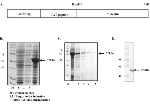
The plasmid which produce artificial vimentin was designed as a cell permeable protein fused with histaq and TAT protein transduction domain. Constructed plasmid was transformed to Competent Escherichia coli BL21 plysS. The bacteria were cultured in LB medium with shaking at 37 °C. 1 mM IPTG was added (O.D = ) and incubated for additional 4 h in order to induce the expression of the recombinant proteins. Recombinant pHis/TAT-vimentin protein was purified by using Ni-NTA, Ni-IDA and Ni-TED affinity column chromatography. Or because pHis/TAT-vimentin was aggregated easily by elution buffer containing imidazole, it was not went through elution step and directly purified. The protein was analyzed by SDS-PAGE gel and stained with Coomassie brilliant blue. The purified protein was confirmed by immunoblot analysis with anti-vimentin antibody.
19
Fig. 2. Expression and purification of pHis/TAT-vimentin protein.
(A) TAT domain and vimentin-coding sequence were fused. (B) Recombinant pHis/TAT-vimentin transformed in BL21 pLysS was induced by 1 mM IPTG for 4 h. (C) pHis/TAT-vimentin was purified using NI-NTA affinity chromatography. Lane from 3 to 5 show continuous elution fraction. However,because pHis/TAT-vimentin was aggregated easily in elution buffer with imidazole, (D) pHisTAT-vimentin which went through elution step was purified directly using Ni-NTA, Ni-IDA and Ni-TED affinity chromatography.
20
2. Transduction of pHis/TAT-vimentin fusion protein into HUVECs
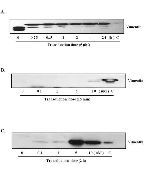
HUVECs were treated with pHisTAT-vimentin for 15 min or 2 h to determine efficient time of transduction. Efficiency of transduction was measured by immnoblot analysis. The immnoblot images presented that pHis/TAT-vimentin was effectively transduced into HUVECs either in short or long expose. However, transduction tendency was fluctuant in short expose. Additionally, vimentin was unstabilized in high concentration which led to apoptosis or hypertrophy of HUVECs. Taken together, the vimentin dosage was appointed at 5 μM to satisfy subjected conditions. Fig. 3C. was shown that pHis/TAT-vimentin fusion protein was greater transduced at 5 μM for 2 h.
21
Fig. 3A. Vimentin protein transduction into endothelial cells.
HUVECs were incubated in EGM-2 with pHis/TAT-vimentin. Immunoblot
analysis was performed using anti-vimentin antibody. (A) pHis/TAT-vimentin (5 μM) was continuously transduced in HUVECs as time-denepent manner. (B) The cells were treated with pHis/TAT-vimentin (0.1, 1, 5, 10 μM) for 15min or 2 h.
22
3. Effects of vimentin protein transduction on the tube formation.
Vimentin has been reported to be effective in endothelial functions such as
adhesion, migration and proliferation. 9, 10, 11 However effect of tube formation
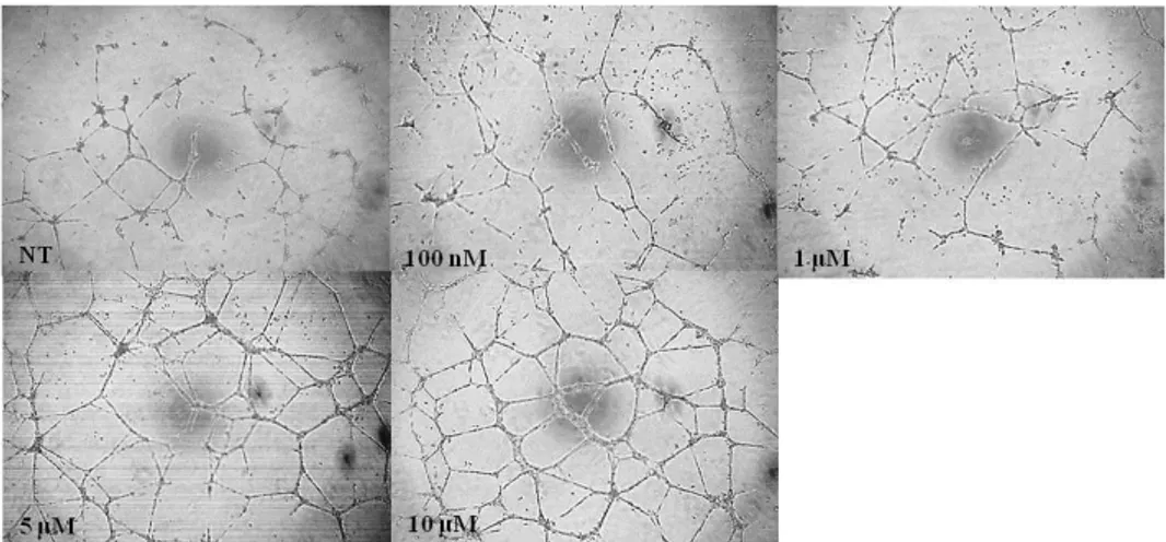
was unsatisfied and there was a lack of research on tube formation effects of the vimentin.HUVECs were incubated with pHis/TAT-vimentin to study further tube formation function of the constructed protein. The vimentin-transduced cells was seeding on ECMatrix gel condition. After 22 h, tube formation patterns by vimentin were measured by optical microscopy (CKX41, OLYMPUS, Tokyo, Japan). It was shown that the pHis/TAT-vimentin increased tube formation as dose-dependent manner in the cells
23
Fig. 4. Vimentin protein transduction promotes tube formation of endothelial cells.
HUVECs were treated with pHis/TAT-vimentin (0.1, 1, 5 and 10 μM ) for 2 h
After 20 h, each well was measured with microscope. pHis/TAT-vimentin increased tube formation on the cells as dose-dependent manner.
24
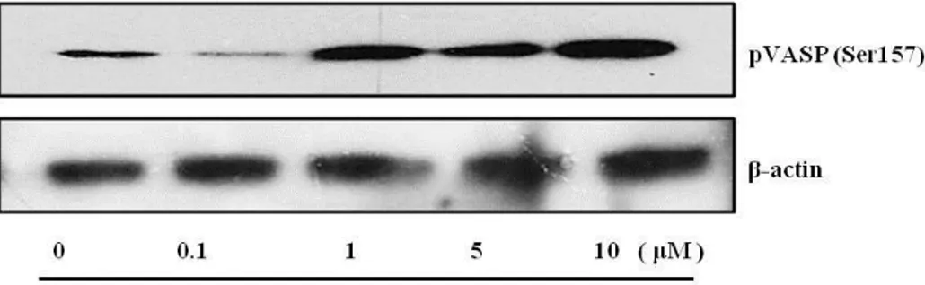
4. Vimentin protein transduction increases VASP phosphorylation.
Previous data have showed that the transduced pHis/TAT-vimentin induced tube formation. Many studies indicate that Vasodilator-stimulated phosphoprotein (VASP) plays a central role in endothelial barrier function. 27, 28, The focal adhesion protein induced alterations in the intermediate filament network into biochemical information 30, 37, 40 The VASP was regulated through Nitric oxide or many pathways in endothelia function. 41 Hence, it was further more studied the relation between transduced pHis/TAT-vimentin and phosphorylation of VASP. An immnoblot analysis was examined to determine whether phosphorylation of VASP was regulated by the pHis/TAT-vimentin.
Vimentin (5 μM) was transduced for 15min because phosphorylation of VASP peaks before 1 h and lasted during 24 h. 30 Compared to control, the protein-transduced cells shown increased phosphorylation of VASP as dose-dependent manner. It was presented that vimentin transduction induces elevation of phosphorylation of VASP.
25
Fig. 5. VASP (Ser157) is phosphorylated by vimentin protein transduction.
The cells were transduced with pHis/TAT-vimentin (5μM) for 15min. Cell lysates were analyzed with anti-phospho VASP (Ser157). And it was quantified by anti- β -actin antibody. Phosphorylation of VASP was increased as dose-dependence compared to control.
26
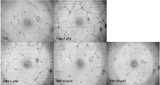
5. Effects of PKA inhibitor on vimentin-induced tube formation.
Many studies indicated that phosphorylation of VASP is related to protein kinase A (PKA) 29, 31, 50 VASP is originally identified as a substrate of PKA which recognize Ser157 site of VASP. The PKA has been reported to involve in endothelial function. 40, 48 H-89 was used as PKA inhibitor to study whether the vimentin affect PKA activity on angiogenesis. Although previous studies used H-89 for 30 min or 1 h, 35 it didn't block PKA activity in this study. Therefore H- 89 was treated for 2 h before treatment of vimentin (5 μM )
Tube formation was reduced as concentration-dependent manner of H-89. (1, 10 and 20 μM). Results were presented that vimentin-induced tube formation was related to PKA activity.
27
Fig. 6. PKA inhibitor suppresses vimentin-induced endothelial cell tube formation.
Cells were treated with PKA inhibitor (1, 10 and 20 μM ) for 2h before the
cells were incubated with pHis/TAT-vimentin (5 μM) and PBS (control) for 2 h Tube formation assay was performed using a ECMatrix gel kit. Each well was photographed under microscope at X100 magnification. pHis/TAT-vimentin-mediated increased tube formation was significantly diminished by PKA inhibitor treatment.
28
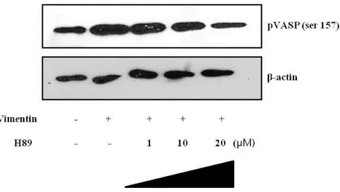
6. Effects of PKA inhibitor on vimentin-induced VASP phosphorylation.
Recent studies reported that VASP was structurally localized at endothelial junction and rapidly phosphorylated (Ser157) by agent which activates PKA. 29, 35, 40, 48
Immunoblot assay was used to elucidate whether the vimentin induced-PKA has an effect on phosphorylation of VASP. Cells were pretreated with PKA inhibitor for 2 h and then of pHis/TAT-vimentin (5 μM) was added for 2 h As tube formation assay, VASP phosphorylation (Ser157) was increased by pHis/TAT-vimentin. The phosphorylation of VASP (Ser157) was decreased after H-89 exposure as dose-dependent manner.
The result shown that pVASP (Ser157) was associated with PKA activity in pHis/TAT-vimentin transduced HUVECs.
29
Fig. 7. Effects of PKA inhibitor on vimentin-induced VASP phosphorylation.
HUVECs were pre-incubated with H-89 (1, 10, 20 μM ) for 2h before it was transduced with pHis/TAT-vimentin 5 μM for 2 h. Lane 2 indicated vimentin-induced pVASP (Ser157) and Lane 3-5 treated with H-89 showed decrease as concentration-dependent manner.
30
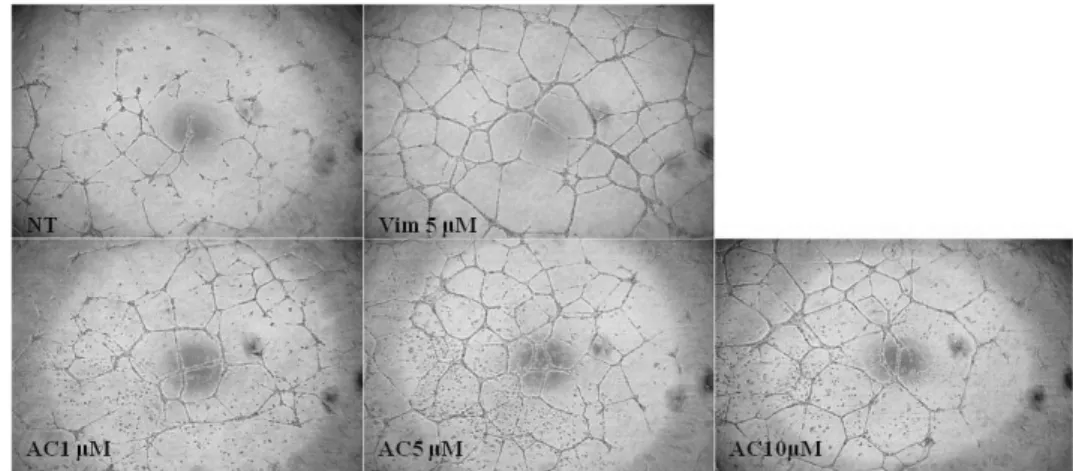
7. Effects Adenylate cyclase inhibitor on vimentin-induced tube formation It is known that adenylate cyclase alter ATP to cAMP that activates PKA.
The cells were pretreated with adenylate cyclase inhibitor (AC inhibitor) for 2 h before pHis/TAT-vimentin was transduced for 2 h to determine the protein induced-adenylate cyclase. After 24 h, tube formation was not reduced regardless of concentration of AC inhibitor. It was shown that PKA induced-tube formation was not affected by adenylate cyclase. Namely pHis/TAT-vimentin directly affects PKA activation.
31
Fig. 8. Adenylate cyclase inhibitor did not affect vimentin-induced tube formation.
HUVECs were incubated with adenylate cyclase (AC) inhibitor (1, 5 and 10 μM) for 2 h. And then pHis/TAT-vimentin (5 μM ) was treated on the cells for 2 h. pHis/TAT-vimentin induced tube formation lasted though AC inhibitor. It indicated that pHis/TAT-vimentin directly influenced PKA activation.
32
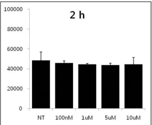
8. Effects of vimentin protein transduction on nitric oxide production.
It is famous that Nitric oxide plays crucial roles in the endothelial function and influence VASP phosphorylation 41, 42 NO production assay was performed using NO production kit to show the effect of pHis/TAT-vimentin on the production. Cells were treated with the protein for 15 min or 2 h as dose-dependent manner. Nitric oxide level in the supermant was measured
Results were shown that the fusion protein didn't influence Nitric oxide production. The phosphorylation of VASP by PKA was regardless of nitric oxide.
33
Fig. 9. The effect of transduced pHis/TAT-vimentin fusion on NO production.
HUVECs were treated with pHisTAT-vimentin (5μM) for 15 min or 2 h. Nitric level in the culture media with vimentin was measured using the NO production Kit. The statistical analysis was performed using the synergy H4.
34
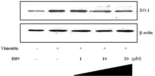
9. Expression of junctional protein by vimentin transduction.
ZO-1 is known as a dominant tight junction-associated phosphoprotein and associate VASP. 40 ZO-1 is associated with actin microfilaments and was thought to influence alterations in actin distribution within endothelial cells as junction markers. PKA inhibitor was pretreated on HUVECs and pHis/TAT-vimentin was added to elucidate expression of ZO-1 by pHis/TAT-vimentin-induced PKA. ZO-1 was expressed in the fusion protein-transduced cells as Fig, 9 shown. The expression by vimentin was decreased by PKA inhibitor. This result indicated that vimentin-induced PKA expressed ZO-1 protein.
35
Fig. 10. pHis/TAT-vimentin induced ZO-1 expression was decreased by H-89. HUVECs were pre-incubated with H-89 (1, 10, and 20 μM) for 2h before
pHisTAT-vimentin (5 μM) was added for 2 h to determine whether PKA expressed 1 in the cells. Results showed that vimentin-induced ZO-expression was decreased by H-89.
36
Ⅳ. DISCUSSION
Intracellular delivery of vimentin fusion protein-fused to a protein transduction domain TAT was performed to induced angiogenesis. The fusion protein was produced as TAT-fusion vimentin protein containing the 11-amino acid transduction domain of TAT. It was constructed by recombinant technology. (Fig.2) Recent studies demonstrate that vimentin has been shown to participate in a number of critical functions and is related to organization of proteins that are involved in adhesion, migration and cell signal. The protein has key roles in angiogenesis by regulating integrin functions. It has also been shown to play a functionally important role in the regulation of cell-cell contacts in endothelial cells.9, 10, 11 . Endogenous vimentin obviously affects migration and proliferation on HUVECs. Presence and absence of vimentin IFs markedly affects the organization and expression of surface molecules critical for adhesion or angiogenesis.17, 20, 21, 22 However tube formation by the vimentin was comparative unsatisfied. Because the ability of endothelial cells to form capillary-like tubule structure is a necessary precursor for angiogenesis, it was studied that whether inserted vimentin increased tube formation in HUVECs.
Fig. 3 showed pHisTAT-vimentin fusion protein transduces into HUVECs for 15 min or 2 h continually. The vimentin protein was unstabilized in high concentration which led to hypertrophy and produce debris of the protein on the
37 cells.
Recent studies found that vimentin increases PKA levels in HUVECs and leads to phosphorylation of VASP protein on serine 157 (Fig. 4) Vasodilator stimulating phosphoprotein (VASP) is a focal adhesion protein that induces alteration in the intermediate filament network into biochemical information. The phosphoprotein associates with focal adhesion and areas of dynamic membrane activity and regulates actin polymerization. Furthermore, it is known to be a regulator of fibroblast migration, protrusion formation and adhesion. In endothelial cells, VASP is involved in tube formation and phosphorylated by vimentin.27, 28, 30, 37 Previous studies show that vimentin is essential for VASP localization to the cell membrane. The VASP is phosphorylated by cAMP dependent kinase (PKA) and cGMP dependent kinase (PKG) in endothelial cells.29, 31,32 VASP contains three phosphorylation sites: Ser239, Ser157 and Thr278. The equivalents of Ser157 and Ser239 but not Thr278 are found in separated by a divergent central proline-rich region, whereas Ser157 is the only site found in EVL(Ena/VASP homology). (Fig.11) Phosphorylation of VASP proteins at the conserved N-terminal (EVH1) serine (Ser157) leads to a major conformational change, which results in an angiogenesis and alterations in protein-protein interactions. PKA regulates filopodia formation in endothelial cells and increases VASP phosphorylation at Ser157.31, 32, 33, 38, 40 In vitro, VASP
38
phosphorylation at Ser239 and Thr278 decreases its anti-capping and filament-bundling activity, and its ability to bind F- and G-actin. Phosphorylation at Thr278 is associated with decreased F-actin content, suggesting that it has a negative role in F-actin assembly, whereas phosphorylation at Ser239 in response to NO treatment which disrupts VASP localization in lamellipodia that leads to loss of lamellipodial protrusions.41, 42, 43, 44, 45, 46 Phosphorylation at Ser157 is correlated with activities such as filopodia formation and regulation of protein-protein interactions. Namely, vimentin scaffold is essential for VASP localization to the cell membrane and VASP phosphorylation by PKG and PKA in vessels and endothelial cells. VASP was originally indicated as substrate of both cAMP and cGMP-dependent kinase and localized at actin-rich sites which regulate actin cytoskeleton dynamics. 27, 29, 31, 32, 40, 48, 50, 51
Fig. 11. The structure of vasodilator-stimulated phosphoprotein. VASP is a
member of a family of proline-rich proteins. VASP family members are organized into three distinct domains. The homologous N-terminal (Ena/VASP
39
homology domains-EVH1) and COOH-terminal domains (EVH2) are separated by a divergent central proline-rich region . 1) The EVH1 region is responsible for VASP binding to proline-rich motif proteins, such as the adhesion proteins. 2) The central VASP domain binds the G-actin regulatory protein profilin that can promote the polymerization of F-actin. 3) The EVH2 region binds and bundles F-actin and localizes to stress fibers. VASP has three phosphorylation sites (Ser157, Ser239, and Thr278). Both PKA and PKG recognize and phosphorylate all three sites, but with different specificities and kinetics. Ser157 is the site preferred by PKA, whereas Ser239 is phosphorylated first by PKG. In this study, VASP is a key regulator of endothelial permeability elicited by PKA activation.40
Vimentin-induced phosphorylation of VASP was inhibited by the PKA-specific inhibitor H-89. (Fig. 6) . It is known that VASP is involved with PKA and cAMP-mediated PKA activation is an important signaling pathway for endothelial barrier stabilization. 50 PKA inhibitor completely blocked vimentin-induced increased pVASP. Inhibition of PKA by H-89 blunted vimentin-mediated tube formation of endothelial cells, providing evidence that this signaling pathway was PKA dependent under this experimental condition.
40
These data underline the significant role of PKA activation for endothelial tube formation and demonstrate that recruitment of VASP and PKA are required for this process. Elevated PKA activation promotes cell-cell, cell-matrix association, intracellular gap gomation, and permeability that are regulated by phosphorylation of complex-associated proteins. Activity of PKA plays a key role for endothelial tube formation. Fig. 5 showed that PKA inhibitor before treatment of vimentin prevented tube formation of HUVECs. Continually, Fig. 6 shown that pHisTAT-vimentin induced phosphorylation of VASP (Ser157) was decreased by H-89. It was demonstrated that increased PKA by vimentin led to strong phosphorylation of VASP (Ser157) in endothelial cells. It has been proposed that VASP phosphorylation at Ser157 by PKA induce tube formation and relaxation of cytoskeletal tension. This phosphorylation of VASP stabilizes endothelial function. 27, 28, 30, 37 It is known that cAMP activates the PKA that is essential to actin-based responses. The cAMP is mediated by adenylase cyclase. The adenylyl cyclase is an enzyme and integral membrane proteins that consist of two bundles of six transmembrane segments. Two catalytic domains extend as loops into the cytoplasm, as depicted in the figure to the right. A soluble (non-membrane bound) form of adenylyl cyclase has recently been characterized in mammalian sperm. This form of the enzyme appears to be activated by bicarbonate ion. When adenylyl cyclase is activated, it catalyses
41
the conversion of adenosine triphosphate (ATP) to cyclic adenosine monophosphate (cAMP), which leads to an increase in intracellular levels of cyclic AMP. However, the fusion vimentin-induced tube formation was not affected by adenylase cyclase inhibitor. It was shown that pHis/TAT-vimentin directly affects PKA activity regardless of adenylase cyclase.(Fig. 7)
Among recent studies, Redistribution of VASP to filopodia was response to a Nitric Oxide donor. NO is recognized as a mediator of angiogenesis, emerged as an important cellular regulator, and plays important roles in diverse physiologic and pathologic processes. 41 Even though nitric oxide (NO) signaling has anti-inflammatory effects in the vasculature, NO production were not increased by vimentin transduction in this study. (Fig. 8). Also, it was known that VASP was localized to endothelial junctional complexes and co-localized with ZO-1, occlidin, and junctional adhesion molecule-1 (JAM-1). 40 Studies whether ZO-1 was expressed by vimentin were performed because PKA-induced phosphorylation of VASP appeared at cell-cell junction where ZO-1 was expressed. 40. PKA inhibitor was pretreated as concentration-dependent manner before vimentin was added. Fig. 9 showed vimentin-induced ZO-1 expression level was decreased by PKA inhibitor. These evidences show how vimentin affects the angiogenesis. 10, 11, 14
42
VASP (ser157) occurred from PKA activation. The fusion vimentin protein directly activates PKA which phosphorylate VASP (Ser157) and continuously expressed ZO-1 at cell-cell junction. This signal led to tube formation on HUVECs. Therefore, discovering a variety role of vimentin and its expressing mechanism or studying agents that express vimentin will be a solution to improve angiogenesis-dependent diseases
Fig. 12. The signal of vimentin-induced tube formation. The pHis/TAT-
43
transduced into HUVECs and it directly activated PKA without other stimulation. The activated PKA phosphorylated VASP and expressed ZO-1. It induced tube formation on HUVECs.
44
V. REFERENCES
1. Moulton KS, Plaque angiogenesis and atherosclerosis, Curr Atheroscler Rep 2001;3:225-233
2. Meltem Avci-Adali, Gerhard Ziemer, Hans P. Wendel, Induction of EPC homing on biofunctionalized vascular grafts for rapid in vivo self-endothelialization – A review of current strategies, Biotechnology Advances 28 (2010) 119-129
3. Khurana R, Simons M, Martin JF, Zachary IC, Role of angiogenesis in cardiovascular disease: a critical appraisal. Circulation 2005;112:1813-1824
4. Min ji Lee, Jumi Kim, Kyung il Lee, Jeong min Shin, Jung il Chae & Hyung min Chung, Enhancement of wound healing by secretory factors of endothelial precursor cells derived from human embryonic stem cells, Cytotherapy, 2011; 13: 165–178
45
5. Anna Zampetaki, Joh,n Paul kirton, and Qingbo Xu, Vascular repair by endothelial progenitor cells, Cirdiovascular Research (2008) 78,413-421
6. Barcat JA, Diapedesis, Between or through the endothelial cells? Medicina (B Aires). 2011; 71(6):581-4.
7. Stubbs SL, Hsiao ST, Peshavariya H, Lim SY, Dusting GJ, Dilley RJ. Hypoxic Preconditioning Enhances Survival of Human Adipose-Derived Stem Cells and Conditions Endothelial Cells in vitro. Stem Cells Dev. 2011 Dec 14.
8. Natalie Lund, Daniel Henrion, Petra Tiede, Marina Ziche, Heribert Schunkert, Wulf D. Ito, Vimentin expression influences flow dependent VASP phosphorylation and regulates cell migration and proliferation, Biocheminal and Biophysical Research Communications 395 (2010) 401-406
46
9. Kai Wang, Lane K. Bekar, Kendra Furber, Wolfgang Walz, Vimentin-expressing proximal reactive astrocytes correlate with migration rather than proliferation following focal brain injury, Brain Research 1024 (2004) 193– 202
10. E. Colucci-Guyon, M.M. Portier, I. Dunia, D. Paulin, S. Pournin, C. Babinet, Mice lacking vimentin develop and reproduce without an obvious phenotype, Cell 79 (1994) 679–694.
11. J.C. Jones, S.B. Hopkinson, L.E. Goldfinger, Structure and assembly of hemidesmosomes, BioEssays 20 (1998) 488–494.
12. M.D. Kottke, E. Delva, A.P. Kowalczyk, The desmosome: cell science lessons from human diseases, J. Cell Sci. 119 (2006) 797–806.
47
13. B. Eckes, D. Dogic, E. Colucci-Guyon, N. Wang, A. Maniotis, D. Ingber, A. Merckling, F. Langa, M. Aumailley, A. Delouvee, V. Koteliansky, C. Babinet, T. Krieg, Impaired mechanical stability, migration and contractile capacity in vimentin-deficient fibroblasts, J. Cell Sci. 111 (Pt 13) (1998)1897–1907.
14. M. Nieminen, T. Henttinen, M. Merinen, F. Marttila-Ichihara, J.E. Eriksson, S. Jalkanen, Vimentin function in lymphocyte adhesion and transcellular migration, Nat. Cell Biol. 8 (2006) 156–162.
15. E. Dejana, The transcellular railway: insights into leukocyte diapedesis, Nat. Cell Biol. 8 (2006) 105–107.
16. D.D. Tang, Y. Bai, S.J. Gunst, Silencing of p21-activated kinase attenuates vimentin phosphorylation on Ser-56 and reorientation of the vimentin network during stimulation of smooth muscle cells by 5-hydroxytryptamine, Biochem. J. 388 (2005) 773–783.
48
17. John E. Eriksson, Tao He, Amy V. Trejo-Skalli, Ann-Sofi Härmälä-Braskén, Jukka Hellman, Ying-Hao Chou5 and Robert D. Goldman, Specific in vivo phosphorylation sites determine the assembly dynamics of vimentin intermediate filaments, Journal of Cell Science 117, 919-932 (2004)
18. Beate Eckes1, Emma Colucci-Guyon2, Hans Smola1, Sue Nodder3, Charles Babinet2, Thomas Krieg1 and Paul Martin, Impaired wound healing in embryonic and adult mice lacking vimentin, Journal of Cell Science 113, 2455-2462 (2000)
19. Beate Eckes, Dagmar Dogic, Emma Colucci-Guyon, Ning Wang, Andrew Maniotis, Donald Ingber, Alexandra Merckling, Francina Langa, Monique Aumailley, Annie Delouvée, Victor Koteliansky6, Charles Babinet and Thomas Krieg, Impaired mechanical stability, migration and contractile capacity in vimentindeficient Fibroblasts, Journal of Cell Science 111, 1897-1907 (1998)
49
20. Hugh Kim, Fumihiko Nakamura, Wilson Lee, Yulia Shifrin, Pamela Arora and Christopher A. McCulloch, Filamin A is required for vimentin-mediated cell adhesion and spreading, Articles in PresS. Am J Physiol Cell Physiol October 28, 2009.
21. Mary J. C. Hendrix, Elisabeth A. Seftor, Richard E. B. Seftor, and Katrina T. Trevort, Experimental Co-Expression of Vimentin and Keratin Intermediate Filaments in Human Breast Cancer Cells Results in Phenotypic Interconversion and Increased Invasive Behavior, Am J Pathol 1997,150:483-495
22. Marie Schoumacher, Robert D. Goldman, Daniel Louvard, and Danijela M. Vignjevic, Actin, microtubules, and vimentin intermediate filaments cooperate for elongation of invadopodia, J. Cell Biol. Published April 26, 2010
50
inhibits carcinoma cell migration and adhesion, Biochemical and Biophysical Research Communications 360 2007, 109–114
24. Miri Yoon, Robert D. Moir, Veena Prahlad, and Robert D. Goldman, Motile Properties of Vimentin Intermediate Filament Networks in Living Cells, The Journal of Cell Biology, Volume 143, Number 1, October 5, 1998 147–157
25. Veena Prahlad, Miri Yoon, Robert D. Moir, Ronald D. Vale, and Robert D. Goldman, Rapid Movements of Vimentin on Microtubule Tracks: Kinesin-dependent Assembly of Intermediate Filament Networks, The Journal of Cell Biology, Volume 143, Number 1, October 5, 1998 159–170
26. Daisuke Tsuruta and Jonathan C. R. Jones, The vimentin cytoskeleton regulates focal contact size and adhesion of endothelial cells subjected to shear stress, Journal of Cell Science 116, 4977-4984 © 2003
51
27. B. Harbeck, S. Huttelmaier, K. Schluter, B.M. Jockusch, S. Illenberger, Phosphorylation of the vasodilator-stimulated phosphoprotein regulates its interaction with actin, J. Biol. Chem. 275 (2000) 30817–30825.
28. L. Wei, S. Muller, J. Ouyang, J.F. Stoltz, X. Wang, Changes of vasodilatorstimulated phosphoprotein (VASP) and its phosphorylation in endothelial cells exposed to laminar flow, Clin. Hemorheol. Microcirc. 28 (2003) 113–120.
29. K. Horstrup, B. Jablonka, P. Honig-Liedl, M. Just, K. Kochsiek, U. Walter, Phosphorylation of focal adhesion vasodilator-stimulated phosphoprotein at Ser157 in intact human platelets correlates with fibrinogen receptor inhibition, Eur. J. Biochem. 225 (1994)
30. Sylvaine Muller, Lei Wei, YungMing Liu, Ke Li, Xiong Wang, Baoha Wang JingPing Ouyang and Jean-François Stoltz, EXPRESSION AND LOCATION OF VASP IN HUMAN ENDOTHELIAL CELLS. EFFECT
52 OF A LAMINAR SHEAR STRESS
31. L.A. MacMillan-Crow, J.E. Murphy-Ullrich, T.M. Lincoln, Identification and possible localization of cGMP-dependent protein kinase in bovine aortic endothelial cells, Biochem. Biophys. Res. Commun. 201 (1994) 531– 537.
32. T.A. Wyatt, T.M. Lincoln, K.B. Pryzwansky, Vimentin is transiently co-localized with and phosphorylated by cyclic GMP-dependent protein kinase in formylpeptide-stimulated neutrophils, J. Biol. Chem. 266 (1991) 21274–21280.
33. A. Smolenski, C. Bachmann, K. Reinhard, P. Honig-Liedl, T. Jarchau, H. Hoschuetzky, U. Walter, Analysis and regulation of vasodilator-stimulated phosphoprotein serine 239 phosphorylation in vitro and in intact cells using a phosphospecific monoclonal antibody, J. Biol. Chem. 273 (1998) 20029– 20035.
53
34. CE´ SAR IBARRA-ALVARADO, JAN GALLE, VOLKER O. MELICHAR, ALEXANDER MAMEGHANI, and HARALD H. H. W. SCHMIDT, Phosphorylation of Blood Vessel Vasodilator-Stimulated Phosphoprotein at Serine 239 as a Functional Biochemical Marker of Endothelial Nitric Oxide/Cyclic GMP Signaling, Mol Pharmacol 61:312– 319, 2002
35. Rachael E. Eckert and Samuel L. Jones, Regulation of VASP serine 157 phosphorylation in human neutrophils after stimulation by a chemoattractant, J. Leukoc. Biol. 82: 1311–1321; 2007.
36. Albert Smolenski, Wolfgang Poller, Ulrich Walter, and Suzanne M. Lohmann, Regulation of Human Endothelial Cell Focal Adhesion Sites and Migration by cGMP-dependent Protein Kinase I, Vol. 275, No. 33, Issue of August 18, pp. 25723–25732, 2000
54
Thompson, and I. Bernard Weinstein2, Vasodilator-stimulated Phosphoprotein (VASP) Phosphorylation Provides a Biomarker for the Action of Exisulind and Related Agents That Activate Protein Kinase G1, Vol. 1, 803–809, August 2002 Molecular Cancer Therapeutics
38. Bachmann, C., Fischer, L., Walter, U., and Reinhard, M. (1999). The EVH2 domain of the vasodilator-stimulated phosphoprotein medi- ates tetramerization, F-actin binding, and actin bundle formation. J. Biol. Chem. 274, 23549–23557.
39. Cecile Lebrand,1,5 Erik W. Dent,1,5 Geraldine A. Strasser,1 Lorene M. Lanier,2 Matthias Krause,1 Tatyana M. Svitkina,3 Gary G. Borisy,4 and Frank B. Gertler1, Critical Role of Ena/VASP Proteins for Filopodia Formation in Neurons and in Function Downstream of Netrin-1, Neuron, Vol. 42, 37–49, April 8, 2004
55
Levi, and Sean P. Colgan, Role of vasodilator-stimulated phosphoprotein in protein kinase A-induced changes in endothelial junctional permeability, The FASEB Journal express article 10.1096. 2002.
41. Sergio Li Calzi, Daniel L. Purich, Kyung Hee Chang, Aqeela Afzal, Takahiko Nakagawa, Julia V. Busik, Anupam Agarwal, Mark S. Segal, and Maria B. Grant1, Carbon Monoxide and Nitric Oxide Mediate Cytoskeletal Reorganization in Microvascular Cells via Vasodilator-Stimulated Phosphoprotein Phosphorylation, Diabetes 57:2488–2494, 2008
42. Xiao-ming Liu a,b, Kelly J. Peyton a,b, Diana Ensenat a, Hong Wang c, Mark Hannink d, Jawed Alam e, William Durante a, Nitric oxide stimulates heme oxygenase-1 gene transcription via the Nrf2/ARE complex to promote vascular smooth muscle cell survival, Cardiovascular Research 75 (2007) 381–389
56
43. Qiong Huang1 and Nader Sheibani, High glucose promotes retinal endothelial cell migration through activation of Src, PI3K/Akt1/eNOS, and ERKs, Am J Physiol Cell Physiol 295: C1647–C1657, 2008.
44. Yung-Hsiang Chen, Shing-Jong Lin, Feng-Yen Lin, Tao-Cheng Wu, Chen-Rong Tsao, Po-Hsun Huang, Po-Len Liu, Yuh-Lien Chen, and Jaw-Wen Chen, High Glucose Impairs Early and Late Endothelial Progenitor Cells by Modifying Nitric Oxide–Related but Not Oxidative Stress– Mediated Mechanisms, DIABETES, VOL. 56, JUNE 2007
45. Prasad V. Gade, MD, Jos& A. Andrades, PhD, Marcel E. Nimni, PhD, Jos6 Becerra, PhD, James Longoria, MD, Nadereh Asemanfar, BA, and Nino Sorgente, PhD, Brooklyn, N.Y.; Los Angeles, Calif.; and Malaga, Spaind, Nitric oxide mediates hyperglycemia,induced defective migration in cultured endothelial cells, J Vasc Surg 1997;26:319-26.
57
Feng-Yen Lin , Hsin-Bang Leu, Tao-Cheng Wu, Shing-Jong Lin , Jaw-Wen Chen, Globular adiponectin improves high glucose-suppressed endothelial progenitor cell function through endothelial nitric oxide synthase dependent mechanisms, Journal of Molecular and Cellular Cardiology 51 (2011) 109–119
47. Mick Burkhardt, Margarita Glazova, Stepan Gambaryan, Tobias Vollkommer, Elke Butt, Benjamin Bader, Katrin Heermeieri, Thomas M. Lincoln, Ulrich Walter, and Alois Palmetshofer, KT5823 Inhibits cGMP-dependent Protein Kinase Activity in Vitro but Not in Intact Human Platelets and Rat Mesangial Cells, THE JOURNAL OF BIOLOGICAL CHEMISTRY Vol. 275, No. 43, Issue of October 27, pp. 33536–33541, 2000.
48. Deling Zhang , Jingping Ouyang , Nian Wang , Yahui Zhang , Jinghua Bie , Yemin Zhang, Promotion of PDGF-induced endothelial cell migration by phosphorylated VASP depends on PKA anchoring via AKAP, Mol Cell Biochem (2010) 335:1–11,
58
49. Nicolas schlegel and Jens Waschke, VASP is involved in cAMP-mediated RAC 1 activation in microvascular endothelial cells, Am J physiol Cell physiol 296: C453-C462, 2009
50. Alan K. Howe, Linda C. Baldor, and Brian P. Hogan, Spatial regulation of the cAMP-dependent protein kinase during chemotactic cell migration, J Cell Physiol. 207:767-774
51. Elke Butt, Kathrin AbelS, Monika KriegerS, Dieter Palmn, Viviane Hoppen, Jurgen Hoppen, and Ulrich Walter8, CAMP- and cGMP-dependent Protein Kinase Phosphorylation Sites of the Focal Adhesion Vasodilator-stimulatedP hosphoprotein (VASP) in Vitro and in Intact H uman Platelets*, The American Society for Biochemistry and Molecular Biology, Vol. 269, No. 20, Issue of May 20, pp. 14509-14517, 1994
59
ABSTRACT (KOREAN)
비멘틴 단백질 전달이 혈관내피세포 기능에 미치는 영향 <지도교수 장양수> 연세대학교 대학원 노화과학협동과정 김은숙 세포골격은 복합적인 세포의 기능을 위하여 생화학적이고 생체역학적인 신호전달의 중요한 역할을 하고 있다. 최근 연구들은 중간미세섬유인 비멘틴 단백질은 혈관내피에 중요한 역할을 한다고 말한다. 세포에 내재되어 있는 비멘틴은 대표적인 중간미세섬유로 세포의 상태와 세포-세포 접합, 이동 그리고 증식과60
같은 내피 기능과 관련이 있다고 알려져 있다. 그러나 혈관형성의 가장 기본적인 특성인 신생혈관형성에 관한 연구는 활발하게 이루어지지 않고 있다.
본 연구에서는 생체 물질 전달 시스템 중의 하나인 단백질 전달 도메인 (protein transduction domain, PTD) 을 이용하여 비멘틴 단백질을 인간 제대 정맥 내피세포로 전달하여 생물학적 효능을 관찰하였다. 인간의 비멘틴 DNA는 단백질 전달 도메인 TAT-PTD에 결합되어 세포 내로 침투가 가능한 재조합 단백질인 TAT-비멘틴으로 제조된다. 본 연구를 통해서 비멘틴 단백질이 인간 제대 정맥 내피세포에 전달 될수록 내재된 비멘틴과 시너지 효과를 이루어 신생혈관이 효과적이게 형성되는 것을 확인하였다. 그리고 신생혈관이 형성되는 동안 프로테인 카이네이즈 A과 프로테인 카이네이즈 G와 같은 프로테인을 활성화 시키는 인자들이 관련되어 있다는 것을 확인하였다. 최근 연구들을 통하면, 액틴과 결합하는 혈관 확장 조절 인단백질은 액틴의 구조과 세포막의 활성을 조절한다고 알려져 있다. 이 것은 세린 157과 세린 239와 같은 다양한 활성화 부위로 이루어져 있는데, 이 부위에 각각 프로테인 카이네이즈 A와 프로테인
61 카이네이즈 G가 결합하여 혈관 확장 조절 인단백질을 활성화 시킨다. 본 실험에서, 인간 제대 정맥 내피세포에 프로테인 카이네이즈 A와 프로테인 카이네이즈 G를 억제하고 비멘틴 단백질을 전달시켜보니, 혈관 확장 조절 인단백질의 활성이 감소하는 것을 관찰하였다. 결론적으로, 이 연구를 통해 단백질 전달 기술을 통한 비멘틴 단백질의 신생혈관형성 효과를 확인함으로써 필요한 단백질을 부가적으로 충족시켜주어 상처치유 가능성을 제시해 주었으며, 이 것이 프로테인 카이네이즈 A, 프로테인 카이네이즈 G에 의해 억제되는 것을 확인함으로써, 세포 내로 전달된 단백질에 의해 형성되는 신생혈관형성을 조절할 수 있는 가능성을 제시 하였다. 핵심 되는 말: 비멘틴 프로테인, 신생혈관형성, 단백질 전달 도메인, 혈관 확장 조절 인단백질, 단백질 효소 A, 단백질 효소 G