Arteriovenous Malformation Infiltrating the
Extensor Hallucis Longus Tendon
A Case Report
By Jae Kwang Kim, MD, PhD, and Jeong Suh Kim, MD
Investigation performed at the Department of Orthopedic Surgery, School of Medicine, Ewha Womans University, Seoul, South Korea
A
rteriovenous malformations are defined as congenitallesions of vascular origin with multiple large feeding arteries. When these lesions become symptomatic, they can adversely affect the surrounding soft tissues and osseous structures1,2
. Surgical resection with or without embolization
therapy is commonly used to treat localized symptomatic ar-teriovenous malformations. Total surgical resection is neces-sary to prevent recurrence but may sometimes produce challenging anatomic defects and expose important structures such as bone, joints, tendons, and neurovascular bundles3
.
Fig. 1-A
T2-weighted sagittal magnetic resonance image showing the localized mass (5.5· 2.5 cm in size) with mixed in-termediate and high signal intensity.
Disclosure: The authors did not receive any outside funding or grants in support of their research for or preparation of this work. Neither they nor a member of their immediate families received payments or other benefits or a commitment or agreement to provide such benefits from a commercial entity.
Here, we describe an arteriovenous malformation that infiltrated the extensor hallucis longus tendon, resulting in a checkrein deformity of the great toe. The segmental defect of the extensor hallucis longus was reconstructed with use of a tendon graft consisting of one-third of the nearby anterior tibial tendon.
The patient was informed that data concerning the case would be submitted for publication, and she consented.
Case Report
A
thirty-three-year-old woman presented with a painful,slow-growing soft-tissue mass on the anterior aspect of the distal third of the right leg of ten years’ duration. In ad-dition, she had a checkrein deformity of the right great toe of approximately two years’ duration. More specifically, she was unable to flex the great toe plantarward when the ankle was in plantar flexion, and the great toe extended during ankle plantar flexion. No previous treatment had been rendered. She reported no recent history of trauma, and the medical history was otherwise unremarkable. Her general health was excellent.
A firm 6 · 4-cm soft-tissue mass was palpable on the anterior aspect of the distal third of the right leg. The mass was slightly mobile and had a nodular, spongy consistency. It was not pulsatile and lacked a bruit or thrill.
Radiographs showed a calcific density just underneath the skin on the anterior aspect of the leg 5 cm proximal to the ankle joint. Magnetic resonance imaging showed a rela-tively well-defined soft-tissue mass of low signal intensity (similar to that of muscle) on T1-weighted images, mixed intermediate and high signal intensity on T2-weighted images (Fig. 1-A), and high signal intensity on gadolinium-enhanced images (Fig. 1-B). Furthermore, the mass was observed to infiltrate the extensor hallucis longus tendon and a portion of the extensor digitorum longus muscle. However, there was no
Fig. 1-B
Gadolinium-enhanced axial magnetic resonance image showing high signal intensity with some signal voids. The extensor hal-lucis longus tendon is not visible because of infiltration by the mass. The arrowhead indicates the extensor digitorum longus tendon, and the arrow indicates the anterior tibial tendon.
Fig. 2
When the ankle was plantar flexed with the patient under general anesthesia, the great toe dorsi-flexed passively.
211
TH EJO U R N A L O FBO N E& JO I N T SU R G E R YdJ B J S.O R G VO L U M E9 2 - AdNU M B E R1dJA N UA R Y2 0 1 0 AR T E R I O V E N O U SMA L F O R M AT I O NIN F I LT R AT I N G T H E EX T E N S O RHA L L U C I SLO N G U S TE N D O Ncortical destruction of bone or bone-marrow change. The mass appeared to be a hemangioma or a vascular malformation.
Surgical resection was attempted to relieve pain and to resolve the checkrein deformity of the great toe because the lesion was relatively well demarcated. When the patient was under general anesthesia, the great toe extended as the ankle was moved plantarward (Fig. 2). A 10-cm-long zigzag incision was made over the anterior aspect of the leg, extending from
the ankle proximally (Fig. 3-A). The superficial peroneal nerve was identified and protected. The mass was observed to lie just beneath the superficial fascia, and it was necessary to transect the superior extensor retinaculum and part of the inferior extensor retinaculum to expose the entire mass (Fig. 3-B). The mass infiltrated the extensor hallucis longus tendon from 2 cm to 8 cm proximal to the ankle joint (Fig. 3-C); however, the anterior tibial and extensor digitorum longus tendons were
Fig. 3-A
The anterior zigzag incision.
Fig. 3-B
The superficial peroneal nerve has been identified and protected. The mass can be seen just beneath the superficial fascial layer. The black arrows indicate the transected superior extensor retinaculum, and the white arrows indicate the partially transected inferior retinaculum.
free of the mass. The lesion was completely excised, including a whole 6-cm-long segment of the extensor hallucis longus tendon and some of the extensor digitorum longus muscle (Fig. 3-D). Multiple veins and arteries were encountered and ligated during the excision, and feeding arteries were consid-ered to be branches of the anterior tibial artery. However, the anterior tibial artery and the deep peroneal nerve were readily separated from the mass. To bridge the segmental defect created in the extensor hallucis longus tendon, we harvested 8 cm of one-third of the width of the nearby anterior tibial
tendon and grafted it end to end to the extensor hallucis longus tendon ends while adjusting tension (Fig. 3-E). The transected retinacula were primarily repaired without difficulty because of the reduced volume of the anterior compartment of the ankle.
The postoperative pathology report described a lesion of innumerable veins and arteries of various sizes and shapes surrounded by fibrovascular and fibroadipose tissues (Fig. 4). Many of the blood vessels, especially the arteries, showed ex-tensive subintimal fibrosis. Furthermore, smooth-muscle
hy-Fig. 3-C
The mass has infiltrated the extensor hallucis longus tendon.
Fig. 3-D
The lesion was excised completely, including a whole 6-cm segment of the extensor hallucis longus tendon and a portion of the extensor digitorum longus muscle.
213
TH EJO U R N A L O FBO N E& JO I N T SU R G E R YdJ B J S.O R G
VO L U M E9 2 - AdNU M B E R1dJA N UA R Y2 0 1 0
AR T E R I O V E N O U SMA L F O R M AT I O NIN F I LT R AT I N G T H E
pertrophy was evident in the walls of many of the vascular structures. These findings are consistent with the diagnosis of an arteriovenous malformation.
The postoperative recovery was uneventful. A short leg splint with neutral dorsiflexion of the ankle and great toe was applied. The closed suction drain was removed at three days
postoperatively, and passive dorsiflexion of the great toe was started. The incision healed without complications. The su-tures were removed at three weeks postoperatively, and a short leg cast was applied. At six weeks postoperatively, the short leg cast was removed and active motion of the great toe and ankle was permitted. At the one-year follow-up examination,
Fig. 3-E
Eight centimeters of one-third of the width of the nearby anterior tibial tendon was harvested and used as a graft to bridge the segmental defect of the extensor hallucis longus tendon.
Fig. 4
Photomicrograph of the mass, demonstrating a venous structure filled with blood. Small-to-medium arterial vessels, which appear thickened because of substantial subintimal fibrosis, were found to be scattered throughout fibrovascular and fibroadipose tissues (hematoxylin and eosin,· 100).
there was no evidence of tumor recurrence and the great toe showed a nearly normal active range of motion (Figs. 5-A and 5-B).
Discussion
V
ascular anomalies are classified as hemangiomas andvas-cular malformations. Hemangiomas are considered to be tumors of infancy, whereas vascular malformations are congenital lesions that are present at birth and do not regress. Vascular malformations are believed to be the result of aber-rant vascular morphogenesis during the fourth to tenth weeks of intrauterine life4
. They are divided into low-flow or high-flow lesions on the basis of their hemodynamic high-flow charac-teristics. Capillary, lymphatic, and venous malformations are classified as low-flow malformations, whereas any malforma-tion with an arterial component—such as an arteriovenous malformation or an arteriovenous fistula—is classified as a high-flow malformation1,4
.
Although the lower extremities are among the most common locations of arteriovenous malformations, arterio-venous malformations are substantially less common than other lesions that affect the foot and ankle, and they are
usually not considered in the differential diagnosis5
. The majority of arteriovenous malformations are isolated anoma-lies in otherwise healthy individuals, and they appear to be activated by factors such as trauma or hormonal stimulation6
. They are often too small to be clinically relevant and may
remain dormant and undetected throughout life1,2
.
Arteriovenous malformations located in the foot and ankle are not believed to be capable of shunting sufficient blood to affect the cardiac system substantially. However, distal shunting has the propensity to substantially reduce the flow rate beyond the shunt, and thus arteriovenous malformations
may cause peripheral ischemia5
. Furthermore, the steal
syn-drome may lead to chronic ulceration. We believe that an in-tact posterior tibial artery prevented any symptoms related to a steal syndrome in our patient.
Indications for treatment of arteriovenous malforma-tions in the foot and ankle are a high-flow shunt that is precipitating cardiac failure, pain, deformity, and local com-plications such as ulceration, hemorrhage, or necrosis7
. The currently available treatment modalities for foot and ankle arteriovenous malformations include compressive garments, ligation of feeding vessels, embolization, surgical excision,
and amputation3
. However, compressive garments, ligation of feeding vessels, and embolization are palliative methods used for large, diffuse, or otherwise inoperable lesions. Surgical excision and amputation are the only treatments known to eradicate the lesion completely, although the latter is a treatment of last resort. Surgical excision must remove the entire lesion because incomplete excision may result in proximal extension of the lesion6,7
. Thus, surgical excision sometimes requires the sacrifice of important structures. There have been several case reports on soft-tissue defects covered with a flap after surgical excision of the arteriovenous mal-formation3,5,7
. We chose surgical excision in the present case because the arteriovenous malformation was localized except for the infiltration of the extensor hallucis longus tendon. Segmental resection of the extensor hallucis longus tendon was required to correct the checkrein deformity of the great toe and to remove the lesion completely. To the best of our knowledge, this is the first case report of a foot and ankle arteriovenous malformation treated with surgical excision and tendon-grafting. We used a portion of the nearby anterior tibial tendon as a donor for two reasons. First, the anterior tibial tendon is of sufficient width for removal of one-third of the width without adversely affecting its function. Second, use of a tendon from the same surgical field made a second surgical exposure unnecessary.n
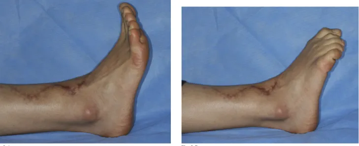
Fig. 5-A Fig. 5-B
At the one-year follow-up examination, the checkrein deformity of the great toe had disappeared and the patient was able to fully extend (Fig. 5-A) and flex (Fig. 5-B) the great toe.
215
TH EJO U R N A L O FBO N E& JO I N T SU R G E R YdJ B J S.O R G
VO L U M E9 2 - AdNU M B E R1dJA N UA R Y2 0 1 0
AR T E R I O V E N O U SMA L F O R M AT I O NIN F I LT R AT I N G T H E
Jae Kwang Kim, MD, PhD Jeong Suh Kim, MD
Department of Orthopedic Surgery, Ewha Womans Mokdong Hospital,
911-1, Mok-6-dong, Yangcheon-gu, Seoul 158-710,
South Korea. E-mail address for J.K. Kim: [email protected]
References
1. Konez O. Vascular anomalies (birthmarks) of the foot and ankle. J Am Podiatr Med Assoc. 2004;94:477-82.
2. Yu GV, Brarens RM, Vincent AL. Arteriovenous malformation of the foot: a case presentation. J Foot Ankle Surg. 2004;43:252-9.
3. Agir H, Sen C, Onyedi M. Extended lateral supramalleolar flap for very distal foot coverage: a case with arteriovenous malformation. J Foot Ankle Surg. 2007;46: 310-3.
4. Fayad LM, Hazirolan T, Bluemke D, Mitchell S. Vascular malformations in the extremities: emphasis on MR imaging features that guide treatment
options. Skeletal Radiol. 2006;35:127-37. Erratum in: Skeletal Radiol. 2006; 35:964.
5. Aub´a C, Hontanilla B. Prevention of a foot amputation: a large arteriovenous malformation reconstructed with a composite free flap. Plast Reconstr Surg. 2007;119:96e-100e.
6. Upton J, Mulliken JB, Murray JE. Classification and rationale for management of vascular anomalies in the upper extremity. J Hand Surg Am. 1985;10(6 Pt 2):970-5. 7. Carr MM, Mahoney JL, Bowen CV. Extremity arteriovenous malformations: review of a series. Can J Surg. 1994;37:293-9.