INTRODUCTION
The cornea and conjunctiva, which make up the ocular surface, are the tissues to interact with the external environment of the eye. The tear film of ocular surface is composed of multiple layers induced by tissues and glands.1 Each layer contains
specific molecules to protect surface cells from the environ-ment.2-4 A major protective mechanism is secretion of the
in-nermost layer of the tear film known as the mucous layer.2 Both
the cornea and conjunctiva epithelial cells generate insoluble mucins, whereas the conjunctiva epithelial cells produce solu-ble mucins.3-5 The mucins in the tear film maintain hydration
and provide lubrication between the cells of the ocular surface and conjunctiva during blinking,6,7 Additionally, mucins
pre-vent pathogens from binding to the ocular surface.8
Mucin production is regulated at multiple levels, including transcription, translation, post-translational modification, and cellular differentiation.9-11 Expression of membrane-spanning
insoluble mucin is dependent upon the cell differentiation st-age. Therefore, mucin production is dependent on the factors that regulate differentiation.12 In the cornea, MUC1, -4, and -16
are presented on the apical cells and mucin mRNA and protein have been shown to be upregulated by serum.13 Fibroblast
growth factor 10 increased MUC1 and MUC4,14 and retinoic
acid upregulated the protein expression of MUC16 in a conjunc-tival epithelial cell line.15 In addition, dexamethasone increased MUC16 expression in the corneal epithelial cells (CECs).16
Ex-pression of MUC7, a small secretory mucin, is induced by
lipo-Role of TGFBIp in Wound Healing and Mucin
Expression in Corneal Epithelial Cells
Yong-Sun Maeng
1, Ga-Hyun Lee
1, Boram Lee
1, Seung-Il Choi
1, Tae-im Kim
1, and Eung Kweon Kim
1,21Department of Ophthalmology, Corneal Dystrophy Research Institute, Yonsei University College of Medicine, Seoul; 2Institute of Vision Research, Severance Biomedical Science Institute, Yonsei University College of Medicine, Seoul, Korea.
Purpose: Transforming growth factor-β-induced protein (TGFBIp) is highly expressed in the cornea, and mutant TGFBIp induces corneal diseases. However, the function of TGFBIp in cornea epithelium is not fully investigated. Here, we tested the importance of TGFBIp in regulation of gene expression and corneal epithelial cell (CEC) activity.
Materials and Methods: The effect of TGFBIp on CEC activity was analyzed by cell migration, adhesion, proliferation and wound healing assay. Analysis of gene expression was examined by western blot and quantitative reverse transcription PCR.
Results: The results demonstrated that TGFBIp increased adhesion, migration, proliferation, and wound healing of CECs. Analysis of gene expression presented that TGFBIp-stimulated CECs exhibited increased expression of mucin family genes, such as MUC1, -4, -5AC, and -16. Furthermore, TGFBIp treatment increased the expression of MUC1, -4, -5AC, -7, and -16 in conjunctival epithelial cells. TGFBIp also increased the activity of intracellular signaling molecules ERK and AKT in CECs. Using pharmacologic inhibitors of ERK and AKT, we showed that the expression of mucin genes by TGFBIp is mediated by the activation of ERK and AKT signaling.
Conclusion: Our findings demonstrate that the locally generated TGFBIp in the cornea may contribute to wound healing of CECs by enhancing the migration, adhesion, and proliferation of CECs. In addition, our results suggest that TGFBIp has a protective effect on ocular surfaces by inducing the expression of mucin genes in corneal and conjunctival epithelial cells. These data suggest that TGF-BIp is a useful therapeutic target for patients with corneal wounds.
Key Words: TGFBIp, mucin, cornea, epithelial cells
pISSN: 0513-5796 · eISSN: 1976-2437
Received: March 9, 2016 Revised: July 1, 2016 Accepted: October 25, 2016
Corresponding author: Dr. Eung Kweon Kim, Department of Ophthalmology, Yonsei
University College of Medicine, 50-1 Yonsei-ro, Seodaemun-gu, Seoul 03722, Korea. Tel: 82-2-2228-0824, Fax: 82-2-2227-8129, E-mail: eungkkim@yuhs.ac
•The authors have no financial conflicts of interest. © Copyright: Yonsei University College of Medicine 2017
This is an Open Access article distributed under the terms of the Creative Com-mons Attribution Non-Commercial License (http://creativecomCom-mons.org/licenses/ by-nc/3.0) which permits unrestricted non-commercial use, distribution, and repro-duction in any medium, provided the original work is properly cited.
Yonsei Med J 2017 Mar;58(2):423-431 https://doi.org/10.3349/ymj.2017.58.2.423
polysaccharide interleukin (IL)-1β, IL-4, IL-13, tumor necrosis factor alpha, and epidermal growth factor (EGF) in airway ep-ithelial cells.17 However, the regulation of MUC7 production in
ocular tissues is limited. Expression of MUC5AC, a gel-forming mucin, is shown to be upregulated in the presence of retinoic acid,18 but little is known about the gene expression of
gel-form-ing mucins in the eye.
Transforming growth factor-β-induced protein (TGFBIp) is an extracellular matrix (ECM) protein and is expressed in vari-ous types of cells, including smooth muscle cells, chondrocytes, fibroblasts, tumor cells, and CECs.19-23 As a component of the
ECM, TGFBIp interacts with ECM proteins, such as laminin, collagen, fibronectin, and glycosaminoglycans,19 and increases
cell migration, differentiation, adhesion, wound healing, me-tastasis, spread, and proliferation through interactions with in-tergrins.24-29
In cornea, mutation of TGFBI gene is responsible for 5q31-linked autosomal dominant corneal dystrophies.30 These
dis-eases are characterized by accumulation of deposits in the cor-nea, resulting in a loss of transparency. Corneal dystrophy is ch-aracterized by a reduction in visual acuity and often culminates in blindness due to the accumulation of protein deposits in the cornea. Immunohistological studies demonstrated that mutant TGFBIp is abundant in the pathologic deposits in all TGFBIp-related corneal dystrophies,31 while wild-type TGFBIp exists
mainly in the extracellular space of CECs, below the corneal epithelial layer, and in the corneal stromal layer.31 TGFBIp
ap-pears to exist in both a covalently bound state and a free soluble form.32 The bound state TGFBIp may exhibit as anchors for
cells in the ECM, while the soluble TGFBIp may serve a regula-tory function. An sodium dodecyl sulfate (SDS)-insoluble frac-tion of TGFBIp is covalently bound to the type XII collagen, and its interaction provide anchoring for cells to the ECM.32
There-fore, interaction between TGFBIp and collagen is important for understanding the homeostasis of cornea and the pathobi-ology of TGFBI-linked corneal dystrophies. However, the role of wild-type TGFBIp in corneal/conjunctival epithelial cells is largely unknown, despite its clear expression in the cornea.
Therefore, the identification of the novel role of TGFBIp in healthy corneal/conjunctival epithelial cells is important for the understanding of the cornea pathophysiology. Here, we report a novel role and underlying signaling mechanism for the TGF-BIp in CEC and mucin expression.
MATERIALS AND METHODS
Cell cultureImmortalized SV40 human CEC line (p4) was obtained from Dr. Hitoshi Watanabe (Osaka University, Osaka, Japan).33 Cells
were cultured in Dulbeco’s modified ragle’s media (DMEM)/ F12 (Invitrogen, Carlsbad, CA, USA) supplemented with human recombinant EGF (10 ng/mL) (Upstate Biotechnology, Lake
Placid, NY, USA) and 10% fetal bovine serum. Cells were incu-bated in a CO2-regulated incubator and the medium was
re-newed every two days.
Human conjunctival epithelial cells were given as a gift by Dr. Tae-im Kim.34 Human conjunctival tissues were donated by the
YONSEI eye bank (Severance EYE & ENT Hospital, Seoul, Ko-rea). Conjunctiva was dissected 2-mm lateral to the limbus of the cornea and incubated with 1.4 U dispase (Sigma-Aldrich Corp., St. Louis, MO, USA) for 1 hour in a 37°C incubator. Con-junctival epithelial cells were then removed with a cell scraper and seeded on 60 mm plate. Cells were cultured in KGM-Gold medium (Lonza, Walkersville, MD, USA). The culture medium was changed every other day until the cells reached 60% to 70% confluence (~5–6 days). After reaching confluence, epithelial cells (1×10⁵ cells) were used to analyze gene expression.
Human corneal tissue was harvested from healthy corneas from the eye bank after penetrating or lamellar keratoplasty. Donor confidentiality was maintained in accordance with the Declaration of Helsinki and was approved by the Severance Hospital IRB Committee (CR04124), Yonsei University.
Corneas (described below) and epithelial tissue were collect-ed by scraping and then storcollect-ed at -80°C until RNA extraction and analysis of mucin expression by quantitative reverse tran-scription PCR (qRT-PCR).
Primary CECs were isolated from corneas using a method described by Maldonado and Furcht.35 Briefly, the cornea was
placed in a 35 mm plate containing dispase II (1.2 U/mL) and incubated for 3-hour at 37°C. The epithelial layer was gently scraped free with a no. 15 scalpel blade. The cell suspension was transferred to a 50-mL tube and centrifuged at 1000×g for 4 minutes. Collected cells were transferred to a flask coated with a mixture of fibronectin and collagen (Sigma-Aldrich Corp.), containing 5 mL of serum-free CnT-20 medium.
Cell migration assay
Cell migration was assayed using the Transwell system (Corning Costar, Acton, MA, USA) with 6.5 mm-diameter polycarbonate filters (8-μm pore size). Briefly, the lower surface of the filter was coated with 10 μg/mL fibronectin (Sigma-Aldrich Corp.), 10 μg/ mL recombinant human TGFBIp (rhTGFBIp) (Sino Biological Inc., Beijing, China), or bovine serum albumin at a concentra-tion of 3% (w/v) as a control for nonspecific binding. SV40-CECs or primary CECs (105) were seeded onto chemotaxis filters in
DMEM plus 1% fetal bovine serum. After the 5-hour migration period, non-migrating cells were removed and migrating cells were stained with hematoxylin and eosin (H&E) and quantified using Kodak 1D software (Eastman Kodak, Rochester, NY, USA). Results are representative of three different experiments in du-plicate.
Wound-healing assay
The wound-healing assay was performed by scratching con-fluent SV40-CECs or primary CECs on 35-mm dishes with
mi-cropipette tips. The cells were then washed to remove debris and treated with rhTGFBIp (10 μg/mL) for 16 hours. Images were captured at 0 and 16 hours after wounding. For quantita-tive analysis, five fields per plate were photographed, and dis-tances between the front lines were measured using ImageJ software (National Institutes of Health). Each assay was re-peated three times.
Cell-matrix adhesion
Cell-matrix adhesion assays were performed as described pre-viously.36 The 96-well plates were coated with 10 μg/mL human
fibronectin (Sigma-Aldrich Corp.) or 2.5–10 μg/mL rhTGFBIp. SV40-CECs or primary CECs in adhesion buffer (serum-free media: DMEM or CnT-20) were seeded at 105 cells/well in 100
μL volume and incubated for 30 minutes at 37°C. After two washes to remove non-adherent cells, adherent cells were stained with H&E and quantified in triplicate by counting ad-herent cells in five randomly selected fields per well (Axiovert 100; Carl Zeiss Micro-Imaging, Thornwood, NY, USA). Results are representative of three different experiments in duplicate. Cell proliferation assay
CEC proliferation was assessed using MTT assays (3-(4,5-di-methylthiazol-2yl)-2,5-diphenyltetrazolium bromide) accord-ing to the manufacturer’s protocol. Briefly, SV40-CECs or pri-mary CECs were seeded onto 96-well plates and incubated with different concentrations of rhTGFBIp (2.5–10 μg/mL) for 72 hours. Fifteen microliters of MTT solution and 200 μL di-methyl sulfoxide were successively added to each well and the optical density measurement (490 nm) was performed. All groups of experiments were performed in quintuplicate. Real-time quantitative reverse transcription PCR (Real-time qRT-PCR)
SV40-CECs, primary CECs or conjunctival epithelial cells (2×10⁵
cells/well) were seeded onto 60 mm plates and treated with TGFBIp (10 μg/mL) for 24 hours. Total RNA was isolated from cells by extraction in TRIZOL reagent (Invitrogen) at each time point (3, 6, 9, 12, and 24 hours). SV40-CECs (2×10⁵ cells/well)
were seeded onto 60 mm plates, After 24 hours, cells were pre-incubated for 40 minutes with or without PD98059 (10 μM) or Wortmannin (100 nM), then stimulated with TGFBIp (10 μg/ mL) for 6 hours. Total RNA was isolated from cells by extrac-tion in TRIZOL reagent (Invitrogen). Using the Power SYBR Green RNA-to-CTTM 1-Step kit (Applied Biosystems, Foster
City, CA, USA) and StepOnePlusTM (Applied Biosystems),
mRNA expression of the human glyceraldehyde 3-phosphate dehydrogenase (GAPDH), MUCIN1, MUCIN4, MUCIN5AC, MUCIN7, and MUCIN16 genes was measured according to the manufacturer’s instructions. The qRT-PCR conditions for all genes were as follows: 48°C for 30 minutes, 95°C for 10 minutes, then 40 cycles of 95°C for 15 seconds, and 60°C for 1 minute. The results are based on cycle threshold (Ct) values.
We calculated differences between the Ct values for experi-mental and reference genes (GAPDH) and graphed the results as the ratio of each RNA to the calibrator sample. Primers used for gene amplification were the following: MUCIN1 5’-AGCGTGAGTGATGTGCCATT-3’ (sense) and 5’-AGCGCA ACCAGAACACAGAC-3’ (antisense); MUCIN4 5’-GGTGGTG GAGGCGTTCTTAT-3’ (sense) and 5’-CTCACGTTCAGGGCT GTCAC-3’ (antisense); MUCIN5AC 5’-CGCTCAGCTGT TCTCTGGAC-3’ (sense) and 5’-GCACAGGTCGACTGGTTCT G-3’ (antisense); MUCIN7 5’-TGCCCCAATTACCACACCTA-3’ (sense) and 5’-TATTTTGGCCAGGAGCTGAA-3’ (antisense); MUCIN16 5’-CCAACTCTTCCGAAACAGCA-3’ (sense) and 5’-GCCAGTGGCGAGAAGTTACA-3’ (antisense); GAPDH 5’-A TGGGGAAGGTGAAGGTCG-3’ (sense), and 5’-GGGGTCATT GATGGCAACAATA-3’ (antisense). Three independent experi-ments were performed and statistical analysis was carried out using Newman-Keuls multiple comparison tests.
Western blotting
SV40-CECs (2×10⁵ cells/well) were seeded onto 60 mm plates.
After 24 hours, cells were pre-incubated for 40 minutes with or without PD98059 (10 μM) or Wortmannin (100 nM), and then stimulated with TGFBIp (10 μg/mL) for 8 hours. Growth medi-um was then removed, and the cells were rinsed twice with phosphate buffered saline prior to lysis with a radioimmuno-precipitation assay (RIPA) buffer. Cell lysates were electropho-resed on SDS-polyacrylamide gel electrophoresis (PAGE) and proteins were transferred onto polyvinylidene difluoride mem-branes. The blocked membranes were incubated with the ap-propriate antibody [anti-human MUCIN1, MUCIN4, or ACTIN (1:1000 dilution, Abcam Inc., Cambridge, MA, USA)], and the immunoreactive bands were visualized with a chemilumines-cent reagent, as recommended by Amersham Biosciences, Inc. (Piscataway, NJ, USA).
SV40-CECs (2×10⁵ cells/well) were seeded onto 60 mm plates.
After 24 hours, cells were treated with TGFBIp (10 μg/mL) for the indicated period of time (5, 10, 15, 30, and 60 minutes). Cells were harvested with RIPA buffer and cell lysates were electro-phoresed on SDS-PAGE and proteins were transferred onto polyvinylidene difluoride membranes. The blocked membranes were incubated with the appropriate antibody [anti-human pFAK, pSRC, pERK, pAKT, FAK, SRC, ERK, and AKT (1:1000 di-lution, Cell Signaling Technology Inc., Danvers, MA, USA)], and the immunoreactive bands were visualized with an enhanced chemiluminescence immunoblotting system (GE Healthcare, Buckinghamshire, UK).
Statistical analysis
Data are presented as mean±standard error and statistical comparisons between groups were performed by one-way ANOVA followed by Tukey’s test or Newman-Keuls multiple-comparison test when appropriate. All experiments were re-peated at least three times.
RESULTS
TGFBIp increases migration, adhesion, proliferation, and wound healing of corneal epithelial cells
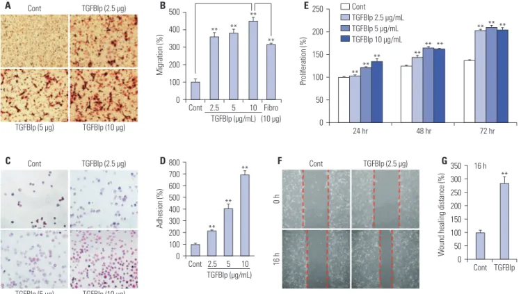
To assess the effect of TGFBIp on CEC activity, we first exam-ined the effect of TGFBIp on cell migration of CECs. As shown in Fig. 1A and B, TGFBIp increased CEC migration in a
dose-dependent manner. The magnitude of TGFBIp-induced CEC migration was higher than that induced by fibronectin, which is a known CEC migration-inducing factor (Fig. 1B).
Next, we analyzed the effect of TGFBIp on CEC adhesion and found that TGFBIp increased SV40-CEC adhesion in a dose-dependent manner (Fig. 1C and D). In addition, CECs stimu-lated with TGFBIp exhibited enhanced proliferation activity
Fig. 1. TGFBIp increases the migration, adhesion, proliferation, and wound healing of SV40-CECs. (A and B) Effect of TGFBIp on SV40-CECs migration. SV40-CECs were treated with TGFBIp or fibronectin and migration was quantified. (C and D) Effect of TGFBIp on SV40-CECs adhesion. SV40-CECs were treated with TGFBIp and cell adhesion was quantified. (E) Effect of TGFBIp on SV40-CECs proliferation. SV40-CECs were incubated with TGFBIp (2.5–10 μg/mL) for 72 hours and cell proliferation was assessed via MTT assay. (F and G) Effect of TGFBIp on SV40-CECs wound healing. SV40-CECs were scratched with micropipette tips and treated with TGFBIp. Images were captured at 0 and 16 hours after wounding. All data are presented as mean±SE from three independent experiments performed in duplicates or quintuplicate. **p<0.01 vs. control or fibronectin. TGFBIp, transforming growth factor-β-induced protein; CECs, corneal epithelial cells; SE, standard error; Cont, control.
Cont TGFBIp (5 μg) TGFBIp (2.5 μg) TGFBIp (10 μg) A Cont TGFBIp (5 μg) TGFBIp (2.5 μg) TGFBIp (10 μg) C Cont 0 h TGFBIp (2.5 μg) 16 h F Cont 2.5 5 10 D800 700 600 500 400 300 200 100 0 Adhesion (%) TGFBIp (μg/mL) ** ** ** Cont 2.5 5 10 Fibro (10 μg) B 500 400 300 200 100 0 Migration (%) TGFBIp (μg/mL) ** ** ** ** 24 hr 48 hr 72 hr Cont TGFBIp 2.5 μg/mL TGFBIp 5 μg/mL TGFBIp 10 μg/mL E250 200 150 100 50 0 Proliferation (%) ** ** ** ** ** ** ** ** ** G350 300 250 200 150 100 50 0 W
ound healing distance (%)
**
Cont 16 h
TGFBIp
Fig. 2. TGFBIp increases the migration, adhesion, proliferation, wound healing of primary CECs. (A) Effect of TGFBIp on primary CECs migration. Pri-mary CECs were treated with TGFBIp and migration was quantified. (B) Effect of TGFBIp on priPri-mary CECs adhesion. PriPri-mary CECs were treated with TGFBIp and cell adhesion was quantified. (C) Effect of TGFBIp on primary CECs proliferation. Primary CECs were incubated with TGFBIp (2.5–10 μg/ mL) for 72 hours and cell proliferation was assessed via MTT assay. (D) Effect of TGFBIp on primary CECs wound healing. Primary CECs were scratched with micropipette tips and treated with TGFBIp. For quantitative analysis, distances between front lines were measured using the ImageJ software. All data are presented as mean±SE from three independent experiments performed in duplicates or quintuplicate. **p<0.01 vs. control. TGFBIp, transforming growth factor-β-induced protein; CECs, corneal epithelial cells; SE, standard error; Cont, control.
A400 350 300 250 200 150 100 50 0 Migration (%) ** ** ** Cont 2.5 5 10 TGFBIp (μg/mL) B500 400 300 200 100 0 Adhesion (%) ** ** ** Cont 2.5 5 10 TGFBIp (μg/mL) D250 200 150 100 50 0 W ound healing distance (%) ** Cont TGFBIp 16 h C300 250 200 150 100 50 0 Proliferation (%) ** ** ** Cont 2.5 5 10 TGFBIp (μg/mL) (72 hr)
compared with the activity of unstimulated cells (Fig. 1E). This increased activity was replicated in the wound-healing assay, which showed that SV40-CECs migrated more into the scratch
wound after TGFBIp treatment (Fig. 1F and G). We cultured primary CECs from human corneal epithelial tissue and per-formed migration, adhesion, proliferation, and wound healing
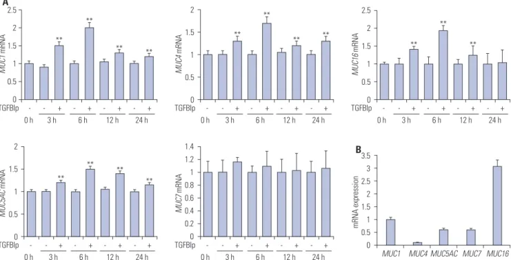
Fig. 3. TGFBIp increases the expression of mucins in SV40-CECs. (A) The temporal expression of each gene in SV40-CECs treated with TGFBIp was determined by qRT-PCR. (B) The mRNA level of each gene in SV40-CECs without TGFBIp treatment was determined by qRT-PCR. All results were nor-malized to GAPDH. **p<0.01 vs. control. TGFBIp, transforming growth factor-β-induced protein; CECs, corneal epithelial cells; qRT-PCR, quantitative reverse transcription PCR; GAPDH, glyceraldehyde 3-phosphate dehydrogenase.
A4 3 2 1 0 MUC1 mRNA ** ** - - + - + - + - + 3 h 6 h 12 h 24 h 0 h TGFBIp 2.5 2 1.5 1 0.5 0 MUC4 mRNA ** ** ** - - + - + - + - + 3 h 6 h 12 h 24 h 0 h TGFBIp 2.5 2 1.5 1 0.5 0 MUC16 mRNA ** ** ** - - + - + - + - + 3 h 6 h 12 h 24 h 0 h TGFBIp 3.5 3 2.5 2 1.5 1 0.5 0 MUC5AC mRNA ** ** - - + - + - + - + 3 h 6 h 12 h 24 h 0 h TGFBIp 2 1.5 1 0.5 0 MUC7 mRNA - - + - + - + - + 3 h 6 h 12 h 24 h 0 h TGFBIp 25 20 15 10 5 0 mRNA expression
MUC1 MUC4 MUC5AC MUC7 MUC16
B
Fig. 4. TGFBIp treatment induces the expression of mucins in primary CECs. (A) The temporal expression of each gene in primary CECs treated with TGFBIp was determined by qRT-PCR. (B) The mRNA level of each gene in primary CECs without TGFBIp treatment was determined by qRT-PCR. All results were normalized to those for GAPDH. **p<0.01 vs. control. TGFBIp, transforming growth factor-β-induced protein; CECs, corneal epithelial cells; qRT-PCR, quantitative reverse transcription PCR; GAPDH, glyceraldehyde 3-phosphate dehydrogenase.
A 2.5 2 1.5 1 0.5 0 MUC1 mRNA ** ** ** ** - - + - + - + - + 3 h 6 h 12 h 24 h 0 h TGFBIp 2 1.5 1 0.5 0 MUC4 mRNA ** ** ** ** - - + - + - + - + 3 h 6 h 12 h 24 h 0 h TGFBIp 2.5 2 1.5 1 0.5 0 MUC16 mRNA ** ** ** - - + - + - + - + 3 h 6 h 12 h 24 h 0 h TGFBIp 2 1.5 1 0.5 0 MUC5AC mRNA ** ** ** ** - - + - + - + - + 3 h 6 h 12 h 24 h 0 h TGFBIp 1.4 1.2 1 0.8 0.6 0.4 0.2 0 MUC7 mRNA - - + - + - + - + 3 h 6 h 12 h 24 h 0 h TGFBIp 3.5 3 2.5 2 1.5 1 0.5 0 mRNA expression
MUC1 MUC4 MUC5AC MUC7 MUC16
assays. As shown in Fig. 2, TGFBIp increased primary CEC mi-gration, adhesion, proliferation, and wound healing in a dose-dependent manner. Taken together, these results suggest that TGFBIp promotes migration, adhesion, and proliferation of CECs, which may enhance wound healing in these cells.
TGFBIp increases the expression of mucins in corneal and conjunctival epithelial cells
Next, we investigated whether TGFBIp promotes the expression of mucin genes in the corneal and conjunctival epithelial cells. As shown in Figs. 3A and 4A, TGFBIp treatment increased the expression of MUC1, -4, -5AC, and -16 in SV40-CECs and
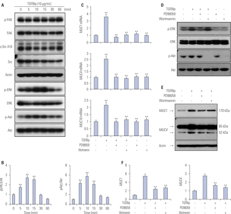
pri-Fig. 5. TGFBIp regulates the expression of mucins through the ERK and AKT signaling pathways in CECs. (A) SV40-CECs were treated with TGFBIp (10 μg/mL) for indicated period of time and cell lysates were subjected to Western blot analysis. (B) The relative ratios were normalized by arbitrarily set-ting the phosphorylation ratio at time 0 as 1. Similar results were obtained from three additional experiments (**p<0.01 vs. time 0). (C) SV40-CECs were pre-incubated with or without PD98059 (10 μM) or Wortmannin (100 nM), then stimulated with TGFBIp (10 μg/mL) for 6 hours. The mRNA level of each gene in CECs was determined by qRT-PCR. All results were normalized to GAPDH. **p<0.01 vs. control or TGFBIp. (D) SV40-CECs were pre-incubated with or without PD98059 (10 μM) or Wortmannin (100 nM) and then stimulated with TGFBIp (10 μg/mL) for 10 minutes. Cell lysates were subjected to Western blot. (E and F) SV40-CECs were pre-incubated with or without PD98059 (10 μM) or Wortmannin (100 nM) and then stimulated with TGFBIp (10 μg/mL) for 8 hours. Protein levels of mucin 1 and mucin 4 were analyzed by Western blot analysis. The relative ratios were normalized by arbitrarily setting the expression ratio for the control as 1. Similar results were obtained from three additional experiments (**p<0.01 vs. TGFBIp). TGFBIp, trans-forming growth factor-β-induced protein; CECs, corneal epithelial cells; qRT-PCR, quantitative reverse transcription PCR; GAPDH, glyceraldehyde 3-phosphate dehydrogenase. p-FAK FAK p-Src 418 Src Actin p-ERK ERK p-Akt Akt A 0 5 10 15 30 60 (min) TGFBIp (10 μg/mL) C 5 4 3 2 1 0 MUC1 mRNA ** ** ** ** ** 3 2.5 2 1.5 1 0.5 0 MUC4 mRNA ** ** ** ** ** - + + + + + -- -- -- + -- + TGFBIp PD98059 Wortmannin 2.5 2 1.5 1 0.5 0 MUC16 mRNA ** ** ** ** ** p-ERK p-Akt ERK Akt + + + + + - - - + - + TGFBIp PD98059 Wortmannin D Actin 52 kDa MUC1 → → → → 170 kDa MUC4 65 kDa - + + + - - + - - - + TGFBIp PD98059 Wortmannin E B4 3 2 1 0 pERK/ERK 0 5 10 15 30 60 Time (min) ** ** ** 8 6 4 2 0 pAkt/Akt 0 5 10 15 30 60 Time (min) ** ** ** F 8 6 4 2 0 MUC1 ** ** - + + + - - + - - - + TGFBIp PD98059 Wortmannin 4 3 2 1 0 MUC4 ** ** - + + + - - + - - - + TGFBIp PD98059 Wortmannin
mary CECs, and maximal expression was observed after 6 hours, whereas the mRNA levels of MUC7 were unchanged. Furthermore, TGFBIp also increased the expression of MUC1, -4, -5AC, -7, and -16 genes in conjunctival epithelial cells (Sup-plementary Fig. 1A, only online). Basal expression levels of MUC16 among the mucin genes were highest in SV40 immor-talized CECs, primary cultured human CECs, human corneal epithelial tissue, and conjunctival epithelial cells (Figs. 3B and 4B) (Supplementary Figs. 1B and 2, only online). These obser-vations provide evidence that TGFBIp may have a protective ef-fect on ocular surfaces by inducing the expression of mucin genes in corneal and conjunctival epithelial cells.
TGFBIp regulates the expression of mucins through the ERK and AKT signaling pathways in corneal epithelial cells
To determine which signaling pathways are involved in modu-lating expression of mucin genes in TGFBIp-stimulated SV40-CECs, we analyzed the activity of intracellular signaling mole-cules. As shown in Fig. 5A and B, phosphorylation of ERK and AKT signaling molecules was increased by TGFBIp stimulation in a time-dependent manner, whereas TGFBIp had no effect on FAK or SRC phosphorylation (Fig. 5A). To further investigate the significance of the ERK and AKT signaling pathway in regulating mucin expression in TGFBIp-stimulated CECs, CECs were pre-treated with PD98059, an ERK inhibitor, or Wortmannin, an AKT inhibitor, and then stimulated with TGFBIp. As shown in Fig. 5C, TGFBIp-stimulated CECs showed increased expression of MUC1, -4, and -16 mRNA, and this effect was effectively blocked by PD98059 or Wortmannin. Western blot analysis con-firmed the inhibition of TGFBIp-induced ERK and AKT phos-phorylation and of TGFBIp-induced MUC1 and -4 expression by PD98059 or Wortmannin (Fig. 5D, E, and F). These results suggest that ERK and AKT signaling are important in regulating the expression of mucin genes in TGFBIp-treated CECs.
DISCUSSION
In this study, we showed that TGFBIp promoted migration, ad-hesion, and proliferation of human CECs in a dose-dependent manner, and that these effects would mediate wound healing of CECs. These results suggest that TGFBIp secreted by CECs themselves may cause neighboring cells to activate wound-healing processes in an autocrine or paracrine manner.
Membrane-spanning and secretory mucins are critical to the health of the ocular surface and protection of this tissue from the external environment.6-8,37 In this study, we demonstrated
that TGFBIp increasesd the expression of MUC1, -4, -5AC, and -16 in CECs, whereas the expression of MUC7 was unchanged in response to TGFBIp. Furthermore, TGFBIp increased the ex-pression of MUC1, -4, -5AC, -7, and -16 in conjunctival epitheli-al cells. Interestingly, basepitheli-al expression of MUC16 was highest in
SV40 immortalized CECs, CECs from human tissue, and con-junctival epithelial cells. This finding suggests that MUC16 is predominantly expressed in the ocular surface and may con-tribute to the health of the ocular surface.
TGFBIp also significantly increased the phosphorylatoin of ERK and AKT in CECs, and inhibition of ERK and AKT signal-ing via pretreatment with PD98059 or Wortmannin markedly abrogated TGFBIp-mediated mucin expression. The phos-phorylation of FAK and Src remained unchanged with TGFBIp treatment. Previously, we reported that TGFBIp binds to inte-grin and activates the intracellular signaling molecules.38,39
However, the subtypes of integrin that mediate interactions with TGFBIp vary depending on the cell types. TGFBIp binds to integrin αVβ3 in corneal fibroblasts and activates the
endo-cytosis of TGFBIp itself,38 but in endothelial progenitor cells,
TGFBIp activates the intracellular signaling molecules via bind-ing to integrins a4 and a5 and induces differentiation of endo-thelial progenitor cells.39 Since TGFBIp-interacting proteins vary
depending on the cell type and downstream signaling pathway, the final outcome may also differ in varying cell types. In our study, we did not investigate whether TGFBIp activated ERK and AKT through integrin binding in the cell types studied. In addition, FAK and Src activation is not always induced by inte-grin binding of ligands.40 Therefore, we suggest that TGFBIp
may activate ERK and AKT with integrin binding and may not be able to induce the activation of FAK and Src independent of integrin binding in our cells.
Although TGFBIp is a secretory ECM protein, we found that the induction of mucins by TGFBIp is specifically ERK- and AKT-dependent. The relationship between TGFBIp signaling and mucin expression in CECs has been unclear until now. However, our present data indicate a direct link between TGF-BIp signaling and mucin expression in CECs, therefore, being the first to describe an interrelation between TGFBIp and mu-cins.
In summary, our findings clearly showed that TGFBIp was a critical factor for the health of the ocular surface. First, TGFBIp increased migration, adhesion, proliferation, and wound heal-ing of CECs. Second, TGFBIp activated the ERK and AKT sig-naling pathway, which in turn induced the expression of mu-cins in CECs. To the best of our knowledge, these data are the first to suggest that the locally generated TGFBIp in the cornea may contribute to wound healing of CECs by enhancing the migration, adhesion, and proliferation of CECs. In addition, TGFBIp may have a protective effect on the ocular surfaces by inducing the expression of mucin genes in corneal and con-junctival epithelial cells. Collectively, these results suggest that TGFBIp is a useful therapeutic target for patients with condi-tions such as corneal wounds.
ACKNOWLEDGMENTS
pro-gram through the National Research Foundation of Korea (NRF) funded by the Ministry of Education (NRF 2014R1A1A2057458, NRF-2016R1D1A1B03933337), and by a faculty research grant of Yonsei University College of Medicine for 2007 (6-2007-0173).
REFERENCES
1. Knop E, Knop N, Millar T, Obata H, Sullivan DA. The international workshop on meibomian gland dysfunction: report of the subcom-mittee on anatomy, physiology, and pathophysiology of the meibo-mian gland. Invest Ophthalmol Vis Sci 2011;52:1938-78.
2. Gipson IK, Argüeso P. Role of mucins in the function of the cor-neal and conjunctival epithelia. Int Rev Cytol 2003;231:1-49. 3. Dartt DA. Control of mucin production by ocular surface
epithe-lial cells. Exp Eye Res 2004;78:173-85.
4. Spurr-Michaud S, Argüeso P, Gipson I. Assay of mucins in human tear fluid. Exp Eye Res 2007;84:939-50.
5. Carraway KL, Perez A, Idris N, Jepson S, Arango M, Komatsu M, et al. MUC4/sialomucin complex, the intramembrane ErbB2 li-gand, in cancer and epithelia: to protect and to survive. Prog Nu-cleic Acid Res Mol Biol 2002;71:149-85.
6. Argüeso P. Glycobiology of the ocular surface: mucins and lectins. Jpn J Ophthalmol 2013;57:150-5.
7. Gipson IK. Distribution of mucins at the ocular surface. Exp Eye Res 2004;78:379-88.
8. Argüeso P, Guzman-Aranguez A, Mantelli F, Cao Z, Ricciuto J, Pan-jwani N. Association of cell surface mucins with galectin-3 con-tributes to the ocular surface epithelial barrier. J Biol Chem 2009; 284:23037-45.
9. Jonckheere N, Van Seuningen I. The membrane-bound mucins: from cell signalling to transcriptional regulation and expression in epithelial cancers. Biochimie 2010;92:1-11.
10. McGuckin MA, Quin RJ, Ward BG. Progesterone stimulates pro-duction and secretion of MUC1 epithelial mucin in steroid-re-sponsive breast cancer cell lines. Int J Oncol 1998;12:939-45. 11. Treon SP, Mollick JA, Urashima M, Teoh G, Chauhan D, Ogata A,
et al. MUC-1 core protein is expressed on multiple myeloma cells and is induced by dexamethasone. Blood 1999;93:1287-98. 12. Theodoropoulos G, Carraway KL. Molecular signaling in the
reg-ulation of mucins. J Cell Biochem 2007;102:1103-16.
13. Hori Y, Spurr-Michaud S, Russo CL, Argüeso P, Gipson IK. Differen-tial regulation of membrane-associated mucins in the human ocu-lar surface epithelium. Invest Ophthalmol Vis Sci 2004;45:114-22. 14. Ma M, Zhang Z, Niu W, Zheng W, Kelimu J, Ke B. Fibroblast
growth factor 10 upregulates the expression of mucins in rat con-junctival epithelial cells. Mol Vis 2011;17:2789-97.
15. Hori Y, Spurr-Michaud SJ, Russo CL, Argüeso P, Gipson IK. Effect of retinoic acid on gene expression in human conjunctival epithe-lium: secretory phospholipase A2 mediates retinoic acid induc-tion of MUC16. Invest Ophthalmol Vis Sci 2005;46:4050-61. 16. Seo KY, Chung SH, Lee JH, Park MY, Kim EK. Regulation of
mem-brane-associated mucins in the human corneal epithelial cells by dexamethasone. Cornea 2007;26:709-14.
17. Li S, Bobek LA. Functional analysis of human MUC7 mucin gene 5’-flanking region in lung epithelial cells. Am J Respir Cell Mol Biol 2006;35:593-601.
18. Tei M, Moccia R, Gipson IK. Developmental expression of mucin genes ASGP (rMuc4) and rMuc5ac by the rat ocular surface epi-thelium. Invest Ophthalmol Vis Sci 1999;40:1944-51.
19. Thapa N, Lee BH, Kim IS. TGFBIp/betaig-h3 protein: a versatile ma-trix molecule induced by TGF-beta. Int J Biochem Cell Biol 2007; 39:2183-94.
20. Gibson MA, Kumaratilake JS, Cleary EG. Immunohistochemical and ultrastructural localization of MP78/70 (betaig-h3) in extracel-lular matrix of developing and mature bovine tissues. J Histochem Cytochem 1997;45:1683-96.
21. Escribano J, Hernando N, Ghosh S, Crabb J, Coca-Prados M. cDNA from human ocular ciliary epithelium homologous to beta ig-h3 is preferentially expressed as an extracellular protein in the corneal epithelium. J Cell Physiol 1994;160:511-21.
22. Billings PC, Herrick DJ, Kucich U, Engelsberg BN, Abrams WR, Ma-carak EJ, et al. Extracellular matrix and nuclear localization of beta ig-h3 in human bladder smooth muscle and fibroblast cells. J Cell Biochem 2000;79:261-73.
23. Karring H, Runager K, Valnickova Z, Thøgersen IB, Møller-Peders-en T, Klintworth GK, et al. DifferMøller-Peders-ential expression and processing of transforming growth factor beta induced protein (TGFBIp) in the normal human cornea during postnatal development and aging. Exp Eye Res 2010;90:57-62.
24. Kim JE, Kim EH, Han EH, Park RW, Park IH, Jun SH, et al. A TGF-beta-inducible cell adhesion molecule, betaig-h3, is downregulat-ed in melorheostosis and involvdownregulat-ed in osteogenesis. J Cell Biochem 2000;77:169-78.
25. Bae JS, Lee SH, Kim JE, Choi JY, Park RW, Park JY, et al. Betaig-h3 supports keratinocyte adhesion, migration, and proliferation through alpha3beta1 integrin. Biochem Biophys Res Commun 2002;294:940-8.
26. Zhang Y, Wen G, Shao G, Wang C, Lin C, Fang H, et al. TGFBI defi-ciency predisposes mice to spontaneous tumor development. Can-cer Res 2009;69:37-44.
27. Maeng YS, Aguilar B, Choi SI, Kim EK. Inhibition of TGFBIp ex-pression reduces lymphangiogenesis and tumor metastasis. On-cogene 2016;35:196-205.
28. Park SW, Bae JS, Kim KS, Park SH, Lee BH, Choi JY, et al. Beta ig-h3 promotes renal proximal tubular epithelial cell adhesion, mi-gration and proliferation through the interaction with alpha3be-ta1 integrin. Exp Mol Med 2004;36:211-9.
29. Kim YH, Kwon HJ, Kim DS. Matrix metalloproteinase 9 (MMP-9)-dependent processing of βig-h3 protein regulates cell migration, invasion, and adhesion. J Biol Chem 2012;287:38957-69.
30. Fujiki K, Hotta Y, Nakayasu K, Yokoyama T, Takano T, Yamaguchi T, et al. A new L527R mutation of the betaIGH3 gene in patients with lattice corneal dystrophy with deep stromal opacities. Hum Genet 1998;103:286-9.
31. Streeten BW, Qi Y, Klintworth GK, Eagle RC Jr, Strauss JA, Bennett K. Immunolocalization of beta ig-h3 protein in 5q31-linked corneal dystrophies and normal corneas. Arch Ophthalmol 1999;117:67-75. 32. Runager K, Klintworth GK, Karring H, Enghild JJ. The insoluble
TGFBIp fraction of the cornea is covalently linked via a disulfide bond to type XII collagen. Biochemistry 2013;52:2821-7.
33. Araki-Sasaki K, Ohashi Y, Sasabe T, Hayashi K, Watanabe H, Tano Y, et al. An SV40-immortalized human corneal epithelial cell line and its characterization. Invest Ophthalmol Vis Sci 1995;36:614-21. 34. Lee H, Kim EK, Kim JY, Yang YM, Shin DM, Kang KK, et al.
DA-6034-induced mucin secretion via Ca2+-dependent pathways through P2Y receptor stimulation. Invest Ophthalmol Vis Sci 2014; 55:6565-74.
35. Maldonado BA, Furcht LT. Epidermal growth factor stimulates in-tegrin-mediated cell migration of cultured human corneal epithe-lial cells on fibronectin and arginine-glycine-aspartic acid peptide. Invest Ophthalmol Vis Sci 1995;36:2120-6.
36. Maeng YS, Choi HJ, Kwon JY, Park YW, Choi KS, Min JK, et al. En-dothelial progenitor cell homing: prominent role of the IGF2-IG-F2R-PLCbeta2 axis. Blood 2009;113:233-43.
po-tential of salivary histatin-5, two variants of histatin-5, and salivary mucin (MUC7) domain 1. Antimicrob Agents Chemother 2000; 44:1485-93.
38. Choi SI, Maeng YS, Kim TI, Lee Y, Kim YS, Kim EK. Lysosomal trafficking of TGFBIp via caveolae-mediated endocytosis. PLoS One 2015;10:e0119561.
39. Maeng YS, Choi YJ, Kim EK. TGFBIp regulates differentiation of EPC (CD133(+) C-kit(+) Lin(-) cells) to EC through activation of the Notch signaling pathway. Stem Cells 2015;33:2052-62. 40. Huveneers S, Danen EH. Adhesion signaling-crosstalk between